Download
1 / 59
610 likes | 804 Views
Sarcoidosis. Dr.Adil Al Sulami KAUH. Sarcoidosis is a multisystem inflammatory disease of unknown etiology that predominantly affects the lungs and intrathoracic lymph nodes. Sarcoidosis is manifested by the presence of noncaseating granulomas (NCGs) in affected organ tissues.

E N D
Sarcoidosis Dr.Adil Al Sulami KAUH
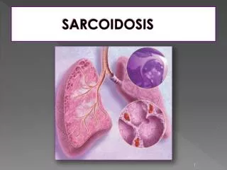
Sarcoidosis is a multisystem inflammatory disease of unknown etiology that predominantly affects the lungs and intrathoracic lymph nodes.
Sarcoidosis is manifested by the presence of noncaseating granulomas (NCGs) in affected organ tissues.
The modern history of sarcoidosis • In 1899, the pioneering Norwegian dermatologistCaesar Boeck describe skin nodules characterizedby compact, sharply defined foci of "epithelioid cells withlarge pale nuclei and also a few giant cells . • Thinking thisresembled sarcoma, he called the condition "multiple benignsarcoid of the skin.
Epidemiology • All racial . • All ethnic groups. • All ages (with the incidence peaking at 20 to 39 years). • M-F ratio 2:1.
The incidence • The highest annual incidence in northern European countries 5 - 40 / 100,000. • In Japan, the annual incidence 1 - 2 / 100,000. • Among black Americansis roughly 3 times that among white Americans (35.5 / 100,000, as compared with 10.9 / 100,000.
Pathophysiology T cells play a central role in the development of sarcoidosis, as they likely propagate an excessive cellular immune reaction.
The cause of sarcoidosis is unknown. Efforts to identify a possible infectious etiology have been unsuccessful.
Genetic and environmental factors seem to play a role. • As yet, no bacterial, fungal, or viral antigen has been consistently isolated from the sarcoidosis lesions. • Sarcoidosis is neither a malignant nor an autoimmune disease.
The following have been suggested as possible candidates that might play a role in causing sarcoidosis: • Mycobacteria, such as Mycobacterium tuberculosis, and atypical pathogens have been suggested. • Fungi and viruses, particularly Mycoplasma, Chlamydia, and Epstein-Barr virus, have been unconvincingly implicated.
Environmental Causes • Some of the earliest studiesof sarcoidosis reported associations with exposures to irritantsfound in rural settings, such as emissions from wood-burningstoves and tree pollen. • More recently, associations with sarcoidosisand exposure to inorganic particles, insecticides, and moldyenvironmentshave been reported. • Occupational studies haveshown positive associations with service in the U.S. Navy, metalworking, firefighting, and the handling of buildingsupplies.
Genetic Features • Familial sarcoidosis was first reported in 1923 in two affectedsisters . • No formal twin study has been reported, but the concordanceappears to be higher in monozygotic twins than in dizygotictwins. • In A Case-Control Study, patientswith sarcoidosis stated 5 times as often as control subjectsthat they had siblings or parents with sarcoidosis.
Presentation depends on the extent and severity of the organ involved. • Approximately 5% of cases are asymptomatic and incidentally detected by CXR. • Systemic symptoms occur in 45% of cases such as : • Fever. • anorexia • Fatigue. • Night sweats . • Weight loss . • Pulmonary, dyspnea on exertion, cough, chest pain, and hemoptysis (rare) occur in 50% of cases.
Löfgren's syndrome, an acutepresentation consisting of: • Fever. • Arthralgia. • erythema nodosum. • bilateral hilar adenopathy. • occurs in 9 to 34% of patients. Heerford's syndrome : • Anterior Uveitis • Fever • Parotid enlargment • Facial palsy
Pulmonary findings. • Dermatological manifestations. • Ocular manifestations . • Cardiac manifestations • Neurologic manifestations (rare)
Organ Involvement • Sarcoidal granulomas can involve any organ, but in more than 90% of patients, clinical sarcoidosis is manifested as intrathoracic LN enlargement, pulmonary involvement, skin or ocular signs and symptoms, or some combination of these findings.
Pulmonary Involvement • dyspnea, cough, vague chest discomfort, and wheezing. • Chest radiographs in patients with sarcoidosis have been classified into four stages: • stage 1, bilateral hilar lymphadenopathy without infiltration. • stage 2, bilateral hilar lymphadenopathy with infiltration. • stage 3, infiltration alone. • stage 4, fibrotic bands, bullae, hilar retraction, bronchiectasis, and diaphragmatic tenting. • These so-called stages represent radiographic patterns and do not indicate disease chronicity or correlate with changes in pulmonary function.
Cutaneous Involvement Although not life-threatening, but can be emotionally devastating. • Erythema nodosum may occur. • Lupus pernio is the most specific associated cutaneous lesion. • Violaceous rash is often seen on the cheeks or nose. • Osseous involvement may be present. • Maculopapular plaques are possible. • Lupus pernio is more common in women than in men and is associated with chronic disease and extrapulmonary involvement. • Erythema nodosum occurs in about 10% of patients with sarcoidosis and usually lasts for about 3 weeks. • Biopsy specimens of erythema nodosum lesions show nonspecific septal panniculitis, which neither confirms nor negates the diagnosis of sarcoidosis.
Liver and Spleen Involvement • 10% of all patients with sarcoidosis have elevated serum aminotransferase and alkaline phosphatase levels. • A cholestatic syndrome characterized by pruritus and jaundice, hepatic failure, or portal hypertension can develop (liver involvement is usually clinically silent). • Detection of hepatic and splenic lesions on CT is described in 5% and 15% of patients. • 60% of patients with hepatic manifestations of sarcoidosis have constitutional symptoms such as fever, night sweats, anorexia, and weight loss. • Portal hypertension with variceal bleeding, a hepatopulmonary syndrome with refractory hypoxemia, and cirrhosis leading to liver failure occur in only 1% of patients with sarcoidosis.
Neurologic Involvement • CNS is involved in up to 25% of patients with sarcoidosis who undergo autopsy, but only 10% of all patients with sarcoidosis present with neurologic symptoms. • The most common problems: • cranial-nerve palsies. • Headache. • Ataxia. • cognitive dysfunction. • Weakness. • seizures. • CSF Analysis : • nonspecific lymphocytic inflammation. • measuring ACE levels . • oligoclonal immunoglobulin bands in the CSF are elevated, making it difficult to differentiate sarcoidosis from multiple sclerosis. • Magnetic resonance imaging (MRI)
Ophthalmologic Complications • The eye and adnexa are involved in 25 -80% of patients with sarcoidosis,this necessitating routine slit-lamp and funduscopic examination. • Anterior or posterior granulomatous uveitis . • Conjunctival lesions and scleral plaques may also be noted. • Ocular involvement may lead to blindness if untreated. • Anterior uveitis (is the most common manifestation) chronic anterior uveitis, with insidious symptoms leading to glaucoma and vision loss, is more common than acute anterior uveitis.
Cardiac manifestations • Heart failure from cardiomyopathy rarely occurs. • Heart block and sudden death may occur. • Approximately 25% of patients may have NCGs at autopsy, but fewer than 5% have clinical cardiac disease. • Okada et al reported on cardiac infiltration associated with a novel heterogenous mutation (G481D in CARD15) in early-onset sarcoidosis.
Differential Diagnosis Hilar infiltrates: • Tuberculosis. • Lymphoma • Eosinophilic granuloma • Fungal infection • Lung cancer NCG on a biopsy : • Berylliosis • Catscratch disease • Fungal infection • Hypersensitivity pneumonitis • Leprosy • Primary biliary cirrhosis • Tuberculosis.
Diagnosis The diagnosis is established on the basis of: • Clinical finding. • Radiologic findings. • Supported by histologicevidence in one or more organs of noncaseating epithelioid-cellgranulomas in the absence of organisms or particles.
A diagnosisof sarcoidosis is reasonably certain without biopsy in patientswho present with Löfgren's syndrome.
Laboratory Studies • Routine lab evaluation often is unrevealing. • Hypercalcemia or hypercalciuria may occur (NCGs secrete 1,25 vitamin D). • Hypercalcemia is seen in about 10-13% of patients, whereas hypercalciuria is 3 times more common. • An elevated alkaline phosphatase level suggests hepatic involvement. • Angiotensin converting enzyme (ACE) levels may be elevated.
NCGs secrete ACE, which may function as a cytokine. • Serum ACE levels are elevated in 60% of patients at the time of diagnosis. • Levels may be increased in fluid from bronchoalveolar lavage or in CSF. • Sensitivity and specificity as a diagnostic test is limited (60 and 70%, respectively). • There is no clear prognostic value. • Serum ACE levels may decline in response to therapy. • Decisions on treatment should not be based on the ACE level alone.
Imaging Studies • A chest radiograph is central to evaluation. • Routine chest CT scan adds little. • HRCT of the chest may be helpful.
Biopsy specimen • A biopsy specimen should be obtained from the involved organthat is most easily accessed, such as the skin, peripheral LN, lacrimal glands, or conjunctiva. • If diagnosis requirespulmonary tissue, transbronchial biopsy by means of bronchoscopyhas a diagnostic yield of at least 85% when multiple lung segmentsare sampled.
The central histologic finding is the presence of NCGs with special stains negative for fungus and mycobacteria.
Sarcoidal granulomas have no unique histologic features to differentiatethem from other granulomas. • Special stains for acid-fast bacilliand fungi, as well as cultures of such organisms, are essential. • If the results of lung biopsy with bronchoscopy are negativeand other organs are not obviously involved, biopsy of intrathoraciclymph nodes, which are often enlarged in patients with sarcoidosis, may be necessary to confirm the diagnosis.
Most patients (>75%) require only symptomatic therapy NSAID. • Approximately 10% of patients need treatment for extrapulmonary disease. • 15% of patients require treatment for persistent pulmonary disease.
Corticosteroids are the mainstay of therapy • prednisone given daily and then tapered over a 6-month course is adequate for pulmonary disease. • Earlier recommendations suggested an initial dose of 1 mg/kg/d of prednisone; however, more recent expert opinions endorse a lower dose (eg, 40 mg/d), which is tapered to every other day long-term therapy over several weeks. • Most patients who require long-term steroids can be treated using 10-15 mg of prednisone every other day. • High-dose inhaled corticosteroids may be an option, but conclusive data are lacking.
Data suggest that corticosteroid use may be associated with increased relapse rates. • Occasionally, certain patients cannot tolerate or do not respond to corticosteroids.
Noncorticosteroid agents Used more frequently. Common indications : • Steroid-resistant disease. • Intolerable adverse effects. • patient desire not to take corticosteroids.
Methotrexate (MTX) has been a successful alternative to prednisone and is a steroid-sparing agent. • Chloroquine and hydroxychloroquine are antimalarial drugs with immunomodulating properties, which have been used for cutaneous lesions, hypercalcemia, neurological sarcoidosis, and bone lesions. • Chloroquine has also been shown to be efficacious for the treatment and maintenance of chronic pulmonary sarcoidosis.
Cyclophosphamide has been rarely used with modest success as a steroid-sparing treatment in patients with refractory sarcoidosis. • Azathioprine is another second-line therapy, which is best used as a steroid-sparing agent rather than as a single-drug treatment for sarcoidosis. • Chlorambucil is an alkylating agent that may be beneficial in patients with progressive disease unresponsive to corticosteroids or when corticosteroids are contraindicated. • Cyclosporine is a fungal cyclic polypeptide with lymphocyte-suppressive properties that may be of limited benefit in skin sarcoidosis or in progressive sarcoid resistant to conventional therapy.
Infliximab and thalidomide have been used for refractory sarcoidosis, particularly for cutaneous disease. • Infliximab appears to be an effective treatment for patients with systemic manifestations such as lupus pernio, uveitis, hepatic sarcoidosis, and neurosarcoidosis. • Tetracyclines have shown promise for the treatment of cutaneous sarcoidosis.
For pulmonary disease • Asymptomatic PFT and/or CXR abnormalities are not an indication for treatment. • In patients with minimal symptoms, serial reevaluation is prudent. • Significant respiratory symptoms associated with PFT and CXR abnormalities likely require therapy. • For such patients, treatment is indicated if objective evidence of recent deterioration in lung function exists. • Corticosteroids can result in small improvements in the functional vital capacity and in the radiographic appearance in patients with more severe stage II and III disease.