Download
1 / 1
10 likes | 491 Views
Abstract. Results. Comparison of macrophage in myostatin null mice and wild-type Aracely Acevedo REU Program: Iowa State University, Faculty Advisor: Dr. Max Rothschild Mentor: Dr. James Reecy. C. B. A. Introduction.

E N D
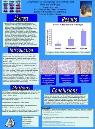
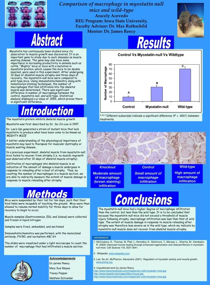
Abstract Results Comparison of macrophage in myostatin null mice and wild-typeAracely Acevedo REU Program: Iowa State University, Faculty Advisor: Dr. Max RothschildMentor: Dr. James Reecy C B A Introduction A, B, C Different subscripts indicate a significant difference (P < .0001) between treatments. The myostatin protein inhibits skeletal muscle growth Myostatin was first described by Dr. Se-Jin Lee in 1997. Dr. Lee’s lab generated a strain of mutant mice that lack myostatin to produce what have been come to be known as ‘MIGHTY MICE’ A better understanding of the physiological importance of myostatin may lead to therapies for muscular dystrophy or muscle wasting disease. In a previous experiment, skeletal muscle from myostatin-null mice failed to recover from atrophy (i.e. no muscle regrowth was observed after 10 days of skeletal muscle atrophy). Infiltration of macrophages into skeletal muscle is an indication of the amount of damage a muscle undergoes in response to reloading after a bout of atrophy. Thus, by counting the number of macrophages in a muscle section, we are able to indirectly measure the extent of muscle damage in response to muscle reloading after atrophy Myostatin has continuously been studied since its association to muscle growth was discovered. It is an important gene to study due to such diseases as muscle wasting disease. The gene may also have some importance in increasing productivity in animals such as cattle. “Mighty” mice or mice with a mutation in myostatin protein, which causes the mice to be double muscled, were used in this experiment. After undergoing 10 days of skeletal muscle atrophy and three days of recovery, the myostatin-null mice were compared to wild-type mice. Using immunohistochemistry along with hematoxilyn staining techniques, the number of macrophages that had infiltrated into the skeletal muscle was determined. There was significant difference in number of macrophage between the control, myostatin-null, and wild type. Statistical analysis displayed a p value of .0001, which proves there is significant difference. Wild-type High amount of macrophage infiltration Knockout Moderate amount of macrophage (brown staining) infiltration Control Small amount of macrophage infiltration Methods Conclusions Mice were suspended by their tail for ten days, such that their hind limbs were incapable of touching the ground. Mice were then allowed to resume normal mobility for three days to allow for recovery to begin to occur. Muscle samples (Gastrocnemius, EDL and Soleus) were collected and frozen in liquid nitrogen Samples were fixed, embedded, and sectioned Immunohistochemistry was performed, with the monoclonal antibody, F4/80, and vectastain ABC kit The slides were visualized under a light microscope to count the number of macrophage that had infiltrated a muscle section. The myostatin-null mice had a higher degree of macrophage infiltration than the control, but less than the wild type. It is to be concluded that because the myostatin-null mice did not exceed a threshold of muscle injury following atrophy, macrophage infiltration was less than that of wild type. The extent of muscle damage in response to muscle reloading after atrophy was therefore less severe as in the wild type, which my indicate by myostatin-null muscle does not recover from skeletal muscle atrophy. REFERENCES 1. McCroskery, S., Thomas, M, Platt, L, Hennebry, A., Nishimura, T., McLeay, L., Sharma, M., Kambadur, R. (2005) Improved muscle healing through enhanced regeneration and reduced fibrosis in myostatin-null mice. Cell Science 118, 3531-3541. 2. Wikipedia: www.wikipedia.com 3. Lee, Se-Jin, McPherron, Alexandria (2001). Regulation of myostatin activity and muscle growth. www.pnhas.org 4. Unpublished work by James Reecy http://www.mesomorphosis.com/images/elzi-volk/myostatin-mice.jpg http://www.hogwild.net/images/Misc/mouse.jpg http://www.mesomorphosis.com/images/elzi-volk/myostatin-mice.jpg Acknowledgements Dr.James Reecy Mary Sue Mayes Tracey Pepper Matthew Schneider