Download
1 / 23
240 likes | 417 Views
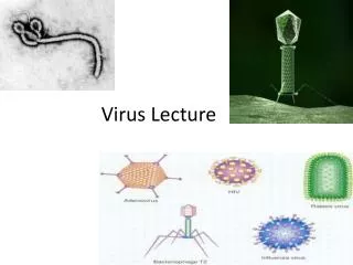
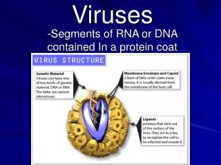
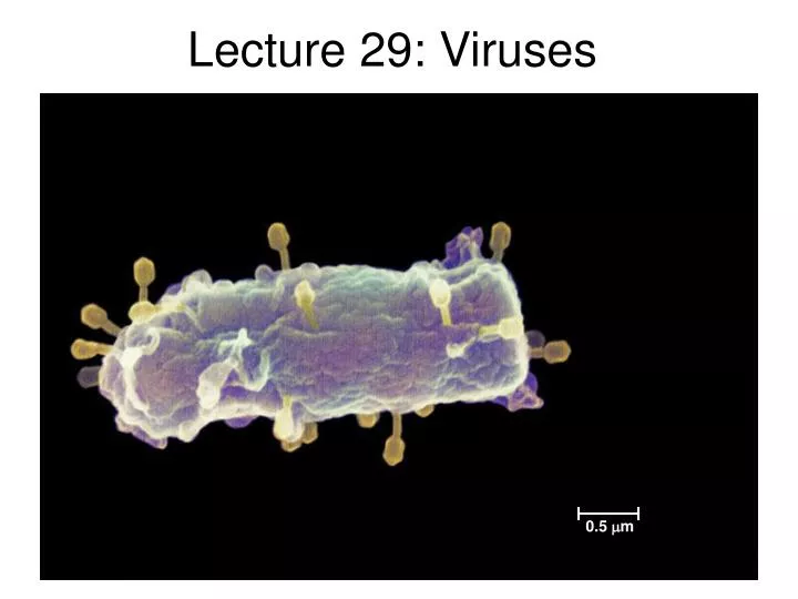
0.5 m. Lecture 29: Viruses. Lecture outline 11/11/05. Types of viruses Bacteriophage Lytic and lysogenic life cycles DNA viruses RNA viruses Influenza HIV Prions Mad cow disease. Figure 18.4 Viral structure. Capsomere of capsid. Membranous envelope. RNA. Capsomere. DNA. Head.

E N D
0.5 m Lecture 29: Viruses
Lecture outline 11/11/05 • Types of viruses • Bacteriophage • Lytic and lysogenic life cycles • DNA viruses • RNA viruses • Influenza • HIV • Prions • Mad cow disease
Figure 18.4 Viral structure Capsomereof capsid Membranousenvelope RNA Capsomere DNA Head Capsid Tail sheath DNA RNA Tail fiber Glycoprotein Glycoprotein 80 225 nm 18 250 mm 80–200 nm (diameter) 70–90 nm (diameter) 50 nm 20 nm 50 nm 50 nm (d) Bacteriophage T4 (a) Tobacco mosaic virus (b) Adenoviruses (c) Influenza viruses
VIRUS Entry into cell and uncoating of DNA Transcription DNA Capsid Replication HOST CELL Viral DNA mRNA Viral DNA Capsid proteins Self-assembly of new virus particles and their exit from cell Figure 18.5 Viral reproductive cycle
Head Capsomereof capsid Tail sheath DNA RNA DNA Capsomere Tail fiber 80 225 nm Glycoprotein 70–90 nm (diameter) 18 250 mm 50 nm 20 nm 50 nm (d) Bacteriophage T4 (b) Adenoviruses Figure 18.4d Figure 18.4a, b (a) Tobacco mosaic virus A capsidis the protein shell that encloses the viral genome
Membranousenvelope Capsid RNA Glycoprotein 80–200 nm (diameter) 50 nm (c) Influenza viruses Figure 18.4c Viral Envelopes are derived from the membrane of the host cell
0.5 m Bacteriophage • Viruses of bacteria have been studied for decades • T1, T2, T4 • “virulent” • Lambda • “temperate” See the animation
The lytic cycle of T4 Attachment. binds to specificreceptor sites on cell surface. 1 2 Entry of phage DNA and degradation of host DNA. 5 Release (lysis) Phage assembly Synthesis of viral genomes and proteins. 3 4 Assembly of phage capsid Head Tail fibers Tails
Bacterial chromosome The lytic and lysogenic cycles of phage This is a “temperate” phage Attachment and injection of DNA. Phage DNA Many cell divisions produce a large population of bacteria infected with the prophage. Phage DNA circularizes Phage Occasionally, a prophage exits the bacterial chromosome, initiating a lytic cycle. Lysogenic cycle Lytic cycle Replicates with host DNA Certain factors determine whether Lysis and release Prophage or Integrated into host chromosome. New phage particles synthesized
Classes of Animal Viruses DNA Viruses RNA Viruses
Smallpox nmhm.washingtondc.museum
Influenza One of the few viruses with genome in segments (8) “H5N1” Spikes of hemagglutanin And neuraminidase
Glycoproteins on the viral envelope bind to specific receptor molecules(not shown) on the host cell, promoting viral entry into the cell. 1 Capsid RNA Envelope (with glycoproteins) Capsid and viral genome enter cell 2 HOST CELL The viral genome (red) functions as a template forsynthesis of complementary RNA strands (pink) by a viral enzyme. 3 Viral genome (RNA) Template mRNA Complementary RNA strands also function as mRNA, which is translated into both capsid proteins (in the cytosol)and glycoproteins for the viral envelope (in the ER). 5 Capsid proteins New copies of viral genome RNA are made using complementary RNA strands as templates. 4 ER Copy of genome (RNA) Glyco- proteins Vesicles transport envelope glycoproteins to the plasma membrane. 6 New virus 8 7 A capsid assembles around each viral genome molecule. The reproductive cycle of an enveloped RNA virus
Why are flu vaccines so hard to make? • Flu strains are highly variable • Recombination among the viral gene segments • RNA polymerase has high mutation rate • Now have some antiviral drugs (e.g. Tamiflu) • blocks the neuramidase enzyme so virus isn’t released from cell
HIV www.who.int/hiv/facts/en/
Glycoprotein Viral envelope Capsid RNA(two identicalstrands) Reversetranscriptase The structure of HIV, the retrovirus that causes AIDS Only 9 genes in HIV: Viral coat proteins Reverse transcriptase Integrase Protease
1 Viral RNA enters cell 8 New capsids are assembled Virus particles bud off. 9 HIV reproduction Reverse transcriptase synthesizes DNA from RNA template . HIV Membrane of white blood cell 2 HOST CELL 3 Reverse transcriptase Makes second DNA strand. Viral RNA RNA-DNAhybrid Incorporated into host chromosome. 4 0.25 µm HIV entering a cell DNA NUCLEUS Provirus ChromosomalDNA 5 New viral RNA is transcribed. RNA genomefor the nextviral generation mRNA New viral proteins are produced. 6 New HIV leaving a cell
Reverse transcriptase is a special DNA polymerase 1. Copies DNA from an RNA template 2. Removes RNA template
thymidine azt AZT • Azidothymidine • a modified thymidine • The first anti-retroviral drug • Stops DNA synthesis because it does not have a 3’OH • Originally developed as an anti cancer drug, but too many side effects
Protease inhibitors- another class of drugs for HIV Inhibitor in active site Protein in active site HIV initially produces one long polypeptide. Protease is necessary to cut the polypeptide into individual enzymes www.chemistry.wustl.edu/~edudev/LabTutorials/HIV/
Originalprion Prion Many prions Normalprotein Newprion Prions are infectious mis-folded proteins Starts a slow chain reaction, causing regular proteins to assume the new shape Altered PRP proteins in nerve cells cause Mad Cow Disease