Download
1 / 49
510 likes | 729 Views
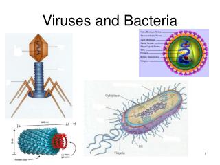
Viruses and Bacteria. Ch 19 & 27 Campbells. Structure of Viruses. Viruses are not cells Viruses are very small infectious particles consisting of nucleic acid enclosed in a protein coat and, in some cases, a membranous envelope. Capsids and Envelopes.

E N D
Viruses and Bacteria Ch 19 & 27 Campbells
Structure of Viruses • Viruses are not cells • Viruses are very small infectious particles consisting of nucleic acid enclosed in a protein coat and, in some cases, a membranous envelope
Capsids and Envelopes • A capsid is the protein shell that encloses the viral genome • Capsids are built from protein subunits called capsomeres • A capsid can have various structures
Fig. 19-3 RNA Membranous envelope Head DNA RNA DNA Capsomere Capsid Tail sheath Tail fiber Capsomere of capsid Glycoproteins Glycoprotein 18 250 nm 70–90 nm (diameter) 80 225 nm 80–200 nm (diameter) 20 nm 50 nm 50 nm 50 nm (a) Tobacco mosaic virus (b) Adenoviruses (d) Bacteriophage T4 (c) Influenza viruses
Some viruses have membranous envelopes that help them infect hosts • These viral envelopes surround the capsids of influenza viruses and many other viruses found in animals • Viral envelopes, which are derived from the host cell’s membrane, contain a combination of viral and host cell molecules
Bacteriophages, also called phages, are viruses that infect bacteria • They have the most complex capsids found among viruses • Phages have an elongated capsid head that encloses their DNA • A protein tail piece attaches the phage to the host and injects the phage DNA inside
General Features of Viral Reproductive Cycles • Once a viral genome has entered a cell, the cell begins to manufacture viral proteins • The virus makes use of host enzymes, ribosomes, tRNAs, amino acids, ATP, and other molecules • Viral nucleic acid molecules and capsomeres spontaneously self-assemble into new viruses Animation: Simplified Viral Reproductive Cycle
Fig. 19-4 VIRUS Entry and uncoating 1 DNA Capsid Transcription and manufacture of capsid proteins 3 Replication 2 HOST CELL Viral DNA mRNA Capsid proteins Viral DNA Self-assembly of new virus particles and their exit from the cell 4
The Lytic Cycle • The lytic cycle is a phage reproductive cycle that culminates in the death of the host cell • The lytic cycle produces new phages and digests the host’s cell wall, releasing the progeny viruses • A phage that reproduces only by the lytic cycle is called a virulent phage • Bacteria have defenses against phages, including restriction enzymes that recognize and cut up certain phage DNA Animation: Phage T4 Lytic Cycle
Fig. 19-5-5 Attachment 1 2 Entry of phage DNA and degradation of host DNA 5 Release Phage assembly 4 Assembly 3 Synthesis of viral genomes and proteins Head Tail Tail fibers
The Lysogenic Cycle • The lysogenic cycle replicates the phage genome without destroying the host • The viral DNA molecule is incorporated into the host cell’s chromosome • This integrated viral DNA is known as a prophage • Every time the host divides, it copies the phage DNA and passes the copies to daughter cells Animation: Phage Lambda Lysogenic and Lytic Cycles
Fig. 19-6 Daughter cell with prophage Phage DNA The phage injects its DNA. Cell divisions produce population of bacteria infected with the prophage. Phage DNA circularizes. Phage Bacterial chromosome Occasionally, a prophage exits the bacterial chromosome, initiating a lytic cycle. Lytic cycle Lysogenic cycle The bacterium reproduces, copying the prophage and transmitting it to daughter cells. The cell lyses, releasing phages. Lytic cycle is induced Lysogenic cycle is entered or Prophage Phage DNA integrates into the bacterial chromosome, becoming a prophage. New phage DNA and proteins are synthesized and assembled into phages.
RNA as Viral Genetic Material • The broadest variety of RNA genomes is found in viruses that infect animals • Retroviruses use reverse transcriptase to copy their RNA genome into DNA • HIV (human immunodeficiency virus) is the retrovirus that causes AIDS (acquired immunodeficiency syndrome)
Fig. 19-8a Viral envelope Glycoprotein Capsid RNA (two identical strands) HOST CELL Reverse transcriptase HIV Reverse transcriptase Viral RNA RNA-DNA hybrid DNA NUCLEUS Provirus Chromosomal DNA RNA genome for the next viral generation mRNA New virus
Vaccines are harmless derivatives of pathogenic microbes that stimulate the immune system to mount defenses against the actual pathogen • Vaccines can prevent certain viral illnesses • Viral infections cannot be treated by antibiotics • Antiviral drugs can help to treat, though not cure, viral infections
Fig. 19-9c (c) Vaccinating ducks
Viral Diseases in Plants • More than 2,000 types of viral diseases of plants are known and cause spots on leaves and fruits, stunted growth, and damaged flowers or roots • Most plant viruses have an RNA genome
Viroids and Prions: The Simplest Infectious Agents • Viroids are circular RNA molecules that infect plants and disrupt their growth • Prions are slow-acting, virtually indestructible infectious proteins that cause brain diseases in mammals • Prions propagate by converting normal proteins into the prion version • Scrapie in sheep, mad cow disease, and Creutzfeldt-Jakob disease in humans are all caused by prions
Fig. 27-2 2 µm 5 µm 1 µm (a) Spherical (cocci) (b) Rod-shaped (bacilli) (c) Spiral
Cell-Surface Structures • An important feature of nearly all prokaryotic cells is their cell wall, which maintains cell shape, provides physical protection, and prevents the cell from bursting in a hypotonic environment • Eukaryote cell walls are made of cellulose or chitin • Bacterial cell walls contain peptidoglycan, a network of sugar polymers cross-linked by polypeptides
Archaea contain polysaccharides and proteins but lack peptidoglycan • Using the Gram stain, scientists classify many bacterial species into Gram-positive and Gram-negative groups based on cell wall composition • Gram-negative bacteria have less peptidoglycan and an outer membrane that can be toxic, and they are more likely to be antibiotic resistant
Fig. 27-3 Carbohydrate portion of lipopolysaccharide Outer membrane Peptidoglycan layer Cell wall Cell wall Peptidoglycan layer Plasma membrane Plasma membrane Protein Protein Gram- positive bacteria Gram- negative bacteria 20 µm (b) Gram-negative: crystal violet is easily rinsed away, revealing red dye. (a) Gram-positive: peptidoglycan traps crystal violet.
Fig. 27-3c Gram- positive bacteria Gram- negative bacteria 20 µm
Fig. 27-7 1 µm 0.2 µm Respiratory membrane Thylakoid membranes (a) Aerobic prokaryote (b) Photosynthetic prokaryote
Fig. 27-8 Chromosome Plasmids 1 µm
The typical prokaryotic genome is a ring of DNA that is not surrounded by a membrane and that is located in a nucleoid region
Reproduction and Adaptation • Prokaryotes reproduce quickly by binary fission and can divide every 1–3 hours • Many prokaryotes form metabolically inactive endospores, which can remain viable in harsh conditions for centuries
Fig. 27-9 Endospore 0.3 µm
Transformation and Transduction • A prokaryotic cell can take up and incorporate foreign DNA from the surrounding environment in a process called transformation • Transduction is the movement of genes between bacteria by bacteriophages (viruses that infect bacteria)
Fig. 27-11-4 Phage DNA A+ B+ A+ B+ Donor cell A+ Recombination A+ A– B– Recipient cell A+ B– Recombinant cell
Conjugation and Plasmids • Conjugation is the process where genetic material is transferred between bacterial cells • Sex pili allow cells to connect and pull together for DNA transfer • A piece of DNA called the F factor is required for the production of sex pili • The F factor can exist as a separate plasmid or as DNA within the bacterial chromosome
Fig. 27-12 1 µm Sex pilus
Concept 27.3: Diverse nutritional and metabolic adaptations have evolved in prokaryotes • Phototrophs obtain energy from light • Chemotrophs obtain energy from chemicals • Autotrophs require CO2 as a carbon source • Heterotrophs require an organic nutrient to make organic compounds • These factors can be combined to give the four major modes of nutrition: photoautotrophy, chemoautotrophy, photoheterotrophy, and chemoheterotrophy
The Role of Oxygen in Metabolism • Prokaryotic metabolism varies with respect to O2: • Obligate aerobes require O2 for cellular respiration • Obligate anaerobes are poisoned by O2 and use fermentation or anaerobic respiration • Facultative anaerobes can survive with or without O2
Nitrogen Metabolism • Prokaryotes can metabolize nitrogen in a variety of ways • In nitrogen fixation, some prokaryotes convert atmospheric nitrogen (N2) to ammonia (NH3)
Fig. 27-16 Domain Eukarya Eukaryotes Korarcheotes Euryarchaeotes Domain Archaea Crenarchaeotes UNIVERSAL ANCESTOR Nanoarchaeotes Proteobacteria Chlamydias Spirochetes Domain Bacteria Cyanobacteria Gram-positive bacteria
Some archaea live in extreme environments and are called extremophiles • Extreme halophiles live in highly saline environments • Extreme thermophiles thrive in very hot environments
Methanogens live in swamps and marshes and produce methane as a waste product • Methanogens are strict anaerobes and are poisoned by O2 • In recent years, genetic prospecting has revealed many new groups of archaea • Some of these may offer clues to the early evolution of life on Earth
Example: Rhizobium,which forms root nodules in legumes and fixes atmospheric N2 • Example: Agrobacterium,which produces tumors in plants and is used in genetic engineering
Fig. 27-18c 2.5 µm Rhizobium (arrows) inside a root cell of a legume (TEM)
Chlamydias • These bacteria are parasites that live within animal cells • Chlamydia trachomatis causes blindness and nongonococcal urethritis by sexual transmission
Chemical Cycling • Prokaryotes play a major role in the recycling of chemical elements between the living and nonliving components of ecosystems • Chemoheterotrophic prokaryotes function as decomposers, breaking down corpses, dead vegetation, and waste products • Nitrogen-fixing prokaryotes add usable nitrogen to the environment
In mutualism, both symbiotic organisms benefit • In commensalism, one organism benefits while neither harming nor helping the other in any significant way • In parasitism, an organism called a parasite harms but does not kill its host • Parasites that cause disease are called pathogens
Pathogenic prokaryotes typically cause disease by releasing exotoxins or endotoxins • Exotoxins cause disease even if the prokaryotes that produce them are not present • Endotoxins are released only when bacteria die and their cell walls break down • Many pathogenic bacteria are potential weapons of bioterrorism