Download
1 / 126
1.26k likes | 1.27k Views
A Anemia. Blood formation. 1-Intrautrine life: Blood cells are formed in the liver and spleen up to the fifth months. After 5th month : bone marrow share in the formation of these cells. 2-After birth: Formation of these cells will be restricted to the bone morrow. SITES. STEPS.

E N D
A • Anemia
Blood formation • 1-Intrautrine life: • Blood cells are formed in the liver and spleen up to the fifth months. • After 5th month : bone marrow share in the formation of these cells. • 2-After birth: • Formation of these cells will be restricted to the bone morrow. SITES STEPS
Blood formation • 3-Adulthood • The active bone morrow will be: • Restricted to axial skeleton : flat bones, vertebrae, ribs, sternum, ilia . • Some extension to the proximal ends of long bone mainly femur. • Extramedullary haemopoiesis:When demand for blood formation is increased: • The active red BM extends into the shafts of the long bones. • The spleen and liver will regain their ability to produce blood elements when bone morrow is affected by some diseases. SITES
Pleuri-potent cells Blood formation • BFU-E • CFU-E • Proerythroblast • Basophilic erythroblast • Polychromatic erythroblast • Othrochromatic erythroblast • Reticulocytes • CFU-G • Myleloblast • Promyelocyte • Myelocyte • Metamgelocyte • Juvenil • Granulocyte (PML) • CFU-Mega • Megakaryoblast • Megadaryocyte • Platelet SITES STEPS Erthrocyto-poiesis Granulocyto- poiesis Thrambocytopoiesis
Blood formation STEPS
Blood formation Blood Constituents • Organelles (formed elements): • RBCs • WBCs • Thrombocytes • Fluid components: plasma in which the above elements are suspended and contain fibrinogen.
Blood formation Erythrocytopoiesis • Normal erythrocytopoiesis depends on: • Healthy BM: normal stem cells and architecture. • Elements essential for erythropoies: (Maturation factors ) • Regulatory hormones: GPO, Androgen thyroxin, cortisol and ACT. • Nutritional elements: Protein (high biological value), • Mineral (Iron, cupper, znic, selenium ) • Vitamins (B, Folic acid, vit c)
Anemia Anaemia = reduction of O2 carrying capacity of the blood with inadequate O2 supply to tissue . Anaemia is diagnosed when there is a reduction of (RBCs number) below the reference range for age and sex of the individual . More accurately is defined as reduction in the red cell mass Definition Types C/P
Anemia AETIOLOGICAL CLASSIFICATION I- Decrease red cell production. • A-Dyshaemopoietic anaemia: (Decreased maturation due to deficiency of maturation factors essential for erythropoiesis). • Mineral deficiency: iron, zinc, selenium, cupper • Vitamin deficiency: B12, folic acid ; Vit C& pyridoxine • Hormonal deficiency: anaemia of renal diseases, pituitary, thyroid or suprarenal deficiency. • Protein deficiency : high class Definition Types C/P
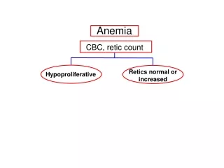
Anemia AETIOLOGICAL CLASSIFICATION I- Decrease red cell production. • B- Hypoproliferative anaemias (BM failure ): • Aplastic anaemia. • Myelophthisic anaemia (BM replacement anaemia). • Anaemia of chronic diseases. Types
Anemia AETIOLOGICAL CLASSIFICATION • II- Haemolytic anaemia: • Short life-span of RBCs • III- Acute post haemorrhagic anaemia: • Loss of RBCs • IV- Mixed anaemia. • eg. Megalobastosis associated with haemolysis • V: Dilutconal anaemia: (raised plasma volume) • Pregnancy • Oliguric RF • Volume-overload Types
Anemia MORPHOLOGICAL CLASSIFICATION • A. Microcytic-hypochromic anaemias: • Thalassaemia. • Iron deficiency anaemia. • Anaemia of chronic disease. • Sideroblastic anaemia: • Hereditary • Chronic lead poisoning. Types
Anemia MORPHOLOGICAL CLASSIFICATION • B-Normocytic-normochromic anaemias: • Acute post –haemorrhagic anaemia. • Hemolytic anaemia. • Aplastic anaemia. • Myelophthisic anaemia. • Anaemia of chronic diseases . Types
Anemia MORPHOLOGICAL CLASSIFICATION • C- Macrocytic- normochromic anaemias: • Megaloblastic anaemia. • Marked reticulocytosis. • Myelodysplastic syndromes. • Myxoedema. • Acquired sideroblastic anaemia. Types
I- PATHOPHYSIOLOGY Anemia The clinical feature of the anaemia could be explained by the following factors. Definition • 1-Tissue Hypoxia • Impaired functions of the tissues, the degree of impairment depends an the need of the tissue to O2 so CVS, CNS and skeletal muscles are much affected. Types C/P
I- PATHOPHYSIOLOGY Anemia The clinical feature of the anaemia could be explained by the following factors. • 2- Compensatory mechanisms • Increased COP. • Increased O2 delivery from HB to the tissue. • Increased erythropoietin production with stimulation of erythropoiesis • Increased plasma volume. • Redistribution of the blood from less to more vital organs. C/P
I- PATHOPHYSIOLOGY Anemia The clinical feature of the anaemia could be explained by the following factors. • 3-Rate of blood loss: • The rapid the rate of blood loss, the more the severe symptoms will occur especially in elderly. While the slowly falling HB allows for haemodynamic compensation with less symptom. • 4- Cause: C/P
II- GENERAL SYMPTOMS OF ANAEMIA: Anemia • 1-Neurological • Dizziness, fainting, lack of concentration • Blurred or diminished vision • Headache, tinnitus • Paraesthesia in the fingers and toes • Insomnia, irritability. • 2-CVS: • Angina, dyspnea, palpitation and intermittent claudication by exertion • HF in severe cases or presence of other organic cardiac disease, it is high COP failure. • 3-Musculo skeletal: • Easy fatigability. • Tiredness and lassitude. C/P
II- GENERAL SYMPTOMS OF ANAEMIA: Anemia • 4-GIT: • Dyspepsia and anorexia • 5-Gental • Loss of libido & impotence • Menstrual abnormalities as amenorrhea. • 6-May be polyuria. C/P
III- PHYSICAL SIGNS: Anemia • Pallor of the skin and mucous membranes • The colour of the skin is unreliable because it depends upon the degree of skin pigmentation and the amount of fluid in the subcutaneous tissues. • Best examined : palmer creases, nail bed, and mucous membrane . • Peripheral oedema : • Slight oedema of the legs probably due to increase in the capillary permeability secondary to hypoxia. • High COP failure. • Fever : • Mild fever may occur in sever anaemia but other causes should be excluded C/P
III- PHYSICAL SIGNS: Anemia • Fundal changes: • Retinal hemorrhages of the flame shape type, exudates and rarely papilloedema. • The cardiovascular system: • Increased velocity with decreased viscosity of the blood in addition to capillary dilation: • Pulse; tachycardia, bounding pulse • Cardiac examination : • Loud HS • S3 over mitral or tricuspid area • Haemic murmur: ejection systolic, hearted allover the precordium • Raised jugular venous pressure. C/P
III- PHYSICAL SIGNS: Anemia • Proteinuria and impairment of the concentrating power of kidneys due to anoxia of renal tubules. IV: SPECIAL MANIFESTATIONS RELATED TO THE CAUSE C/P
Microcytic Anemia Basic nutritional features and metabolism of iron • Dietary sources : • Red meat and liver, bread, eggs and green vegetables, mainly in ferric form, • Daily minimum requirement 10-12mg of which about 1mg is absorbed.
Microcytic Anemia Basic nutritional features and metabolism of iron Absorption • In the stomach the iron is released from its complex form and is reduced to ferrous form (action of gastrin and Hcl). • Iron absorption takes place in the duodenum and proximal jejunum. • Factor controlling iron absorption • Iron absorption is under regulatory system (Apoferritin-Transferrin system ) present in the intestinal mucosa and regulate absorption of the iron according to body requirement .
Microcytic Anemia Basic nutritional features and metabolism of iron Absorption • Factors enhancing iron absorption • Pregnancy • Iron deficiency anaemia • Increased erythropoiesis • Vit. C. • Factors decrease iron absorption : • Excess phosphate, tannates, phytate in diet • Iron overload haemochromatosis • Decreased erythropoies eg a plastic anaemia • Malabsorption syndrome • Decreased HCI atrophic gastritis .
Microcytic Anemia Basic nutritional features and metabolism of iron Transport • There is a diurnal rhythm with higher level in the morning, iron is transported in the plasma bound to transferrin. • Utilization : incorporation into Hb, myoglobin and tissue enzymes.
Microcytic Anemia Basic nutritional features and metabolism of iron • Storage : • About two-thirds of the total body iron is in the circulation as haemoglobin. • Iron is stored in reticulo-endothelial cells, hepatocytes, spleen, bone marrow and skeletal muscle about two thirds as ferritin and one-third as haemosiderin. • Iron loss: (0.5 mg/day). Via • Hair and nail growth • Epithelium of the skin and mucus membrane. • body excreta : stool and urine
Microcytic Anemia • True deficiency (defective intake) Rare: • Prolonged starvation and famines. • Infancy (milk is poor in iron) • Conditioneddeficiency (normal demand and intake but there are defective absorption and utilization) Not common: • Ferric form cause. • Decreased HCL • Iron binder: phosphate, phytate, tannates • Malobsorption syndrome. • Relative deficiency :(increased requirement ) common cause. • Menstruating females. • Pregnancy, labor. • Growing children . • Convalescence from disease. Aetiology C/P Lab DD TTT
Microcytic Anemia • Chronic blood loss : the commonest cause • Frank blood loss • Menorrhagia • Repeated GI bleeding, haemeptysis, epistaxis, haematuria • Bleeding tendencies • Repeated blood donation • Occult blood loss via GIT: • Anckylstoma & schistomiasis • Oozing OV, PU. • Neoplasm. • Inflammatory bowel disease ulcerative colitis • TB enteritis. Aetiology
Microcytic Anemia • General manifestations of anaemia • Especial manifestations of iron deficiency anaemia . • Epithelial changes: • GIT manifestations • Angular stomatitis. • Atrophic glossitis (red, glazed, smooth tongue). • Atrophy of the gastric mucosa • Small splenomegaly • Dermatological changes • Brittle nails thinning and ridging and loss of luster. • Koilonychia; spoon-shaped nails in severe cases: • Brittle hair Aetiology C/P Lab DD TTT
Microcytic Anemia • Features of special types: • 1- Plummer-Vinson –syndrome: • Common in middle aged female • Postcricoid esophageal web cause dysphagia 2- Ankylostoma infestation: • Perverted appetite: (pica) eating mud, stones and chalk. • Epigastric pain (DD= D. ulcer ), altering bowel habits • Endemic parasites (trapezoid face) • The symptoms are mild and to variable. • Investigations: • Stool: Ova. • CBC: eosinphilia. C/P
Microcytic Anemia • To diagnose iron deficiency anaemia Aetiology • CBC: • The red cells are microcytic, (MCV<80f1) and hypochromic (MCH < 27pg) anisocytosis and poikilocytosis (variation in size and shape). • Eosinophilia is present in cases of ankylostoma infestation. • Bone Marrow: • Absent iron store, • Normoplastic hyperplasia, • Hb in maturely erythroblasts. C/P Lab DD TTT
Microcytic Anemia • To diagnose iron deficiency anaemia • Serum iron study • Decreased serum iron (n=70-170 mg%). • Increased total iron binding capacity (TIBC).(n=250-450mg%). • Decreased serum ferritin levels . • Reflect iron stores. • The normal values are 30-300 mg/L in males and 15-200 mg/L in females and investigation of gastrointestinal tract are often required. • Falsely raised value in cases of acute phase reactant e.g malignancy . • Decreased transferrin saturation (n=25-50%) Lab
Microcytic Anemia B-Investigation for the causes • Stool analysis: • Occult blood , ankylstoma & Bilharizasis . • GI cause: • Imaging : barium swallow, meal or enema. • Endoscopic studies: upper and lower. • Achlorhydria • Haemostatic profile. Lab
Microcytic Anemia Aetiology C/P • Microcytic anaemia • Anaemia of chronic disease Lab DD TTT
Microcytic Anemia • The aim of the treatment is to correct the anaemia and build up iron stores • Correction of underlying cause if possible • Iron replacement • Oral iron: • 200 mg anhydrous ferrous sulphate three times daily. • The response is a rise in Hb concentration of 1 gm/dl per week, • Side effects nausea, abdominal pain, diarrhea or constipation. Aetiology C/P Lab DD TTT
Microcytic Anemia • Parenteral iron: • Indication : • General intolerance of oral preparation even at low dose. • Severe malabsorption and those who have gastrointestional diseases. • Rapid iron loss. • Preparation • Iron sucrose ( ferrosac – venofer) • Good safety and efficacy profile. • Given IV or slowly IV infusion. • Amp: 5ml = 100 mg. TTT
Microcytic Anemia • Iron dextran (cosmofer - imferon) • Good efficacy but more side effects. • Giving IM, or IV infusion. • Amp 2 ml = 100mg. • Side effects: • Local pain, staining inflammation, abscess formation • General: hypersensitivity reaction, fever, rigor, hypotension • N.B: Test dose is required prior to the use of iron therapy. TTT
Microcytic Anemia • Transfusion therapy : • It should be packed RBCs. • Indications: • Sever (Hb < 7 gm/dl) andsymptomatizing anemia/dl) • Complicated anaemia:-HF TTT
Microcytic Anemia Aetiology Type of anaemia may be microcytic hypochromic or more common normocytic normochromic . Endocrinal disorders : hypothyroidism, hypocorticalisin, hypogonadism in male . Inflammatory diseases : collagenosis, chron’s disease. Chronic infection: as TB, sarcoidosis, oestomylitis . Neoplastic diseases . Malabsorption. Organ failure, liver and kidney Mechanism Lab TTT
Microcytic Anemia Aetiology • Decreased serum iron and TIBC. • Normal or raised serum ferritin • Normal BM iron. Mechanism Lab TTT
Microcytic Anemia • It is a refractory microcytic hypochromic anaemia (peripheral blood) characterized by the presence of sidroblasts in the bone marrow. • Sidroblasts are: • Erythroblasts inside which iron accumulate into the mitochondria of erythroblasts owing to disordered haeme synthesis . • A ring of iron granules is formed around the nucleus. Definition Aetiology
Microcytic Anemia • Inherited: X-linked disease transmitted by females. • Acquired : • Primary: one of the myelodysplastic syndromes • Secondary: Other types of myelodysplasia Myeloproliferative disorders Myeloid leukemia Drugs as isoniazid, alcohol, lead Definition Aetiology
Microcytic Anemia Differential diagnosis of Microcytic anaemia:
Macrocytic Anemia MACROCYTIC ANAEMIAS This can be divided into: 1-Megaloblastic type. 2-Non megaloblastic types depending on bone marrow findings.
Macrocytic Anemia MEGALOBLASTIC ANAEMIA Megaloblastic anaemia This anaemia in which DNA syntheses is impaired with delayed nuclear maturation. This will lead to formation of megaloblast i.e. large cells with large immature nucleic ( cellular gigantism). The cells affected are those rapidly developing: haematopoietic cells, GI mucosa, and skin. Megaloblastic changes occur in 1- Vitamin B12 deficiency or abnormal vit. B12 metabolism 2- Folic acid deficiency or abnormal folate metabolism 3- Congenital enzyme deficiencies in DNA synthesis or drugs interfering with DNA synthesis (hydroxyurea, azathioprine). 4-Myelodysplasia due to dyserythropoiesis.
Macrocytic Anemia Hematological values 1-MCV>96 Fl unless there is a coexisting cause of microcytosis. 2-The peripheral blood film shows macrocytes with hypersegmented polymorphs with six or more lobes in the nucleus. If severe there may be leucopenia and thrombocytopenia.
Macrocytic Anemia • (COBLAMIN)12 VITAMIN B • *Vit B12 is synthesized by certain micro-organism. • *Sources: meat, fish, eggs, and milk but not plants • *It is not destroyed by cooking . • *Daily requirement is 1-2 mg / day. • *Storage : the average adult stores 2-3 mg in the liver, it may take two years or more before B12 deficiency, develops as the daily losses are small (1-2mg).
Macrocytic Anemia • Absorption: Vit. B12 is liberated in the stomach, bound by intrinsic factor (IF) and absorbed through the terminal ileum, transported by transcobalamin I and to lesser extend by transcobalamin II and III. • It is essential for: • 1-haematopoiesis • 2-GIT mucosa integration • 3-Formation of myelin of nervous system.
Macrocytic Anemia CAUSES OF VIT. B12 DEFICIENCY I- True deficiency: (Decreased intake) -Poor socioeconomic status. -Vegetarians, alcoholic II-condition deficiency A-Decreased absorption . 1-Decreased intrinsic factors commonest cause. *Pernicious anaemia. *Total or partial gastrectomy *Atrophic gastritis. *Gastric cancer. *Non- Addisonian pernicious anaemia a- In association with hypogammaglobulinemia, gastric atrophy, achlorhydria and absent intrinsic factor but no antibodies b-In infancy: Selective failure of IF, normal mucosa. C- Juvenil PA failure of IF secretion with gastric atrophy rarely with antibodies