Download
1 / 82
830 likes | 1.04k Views
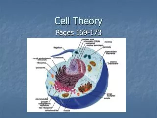
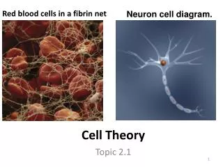
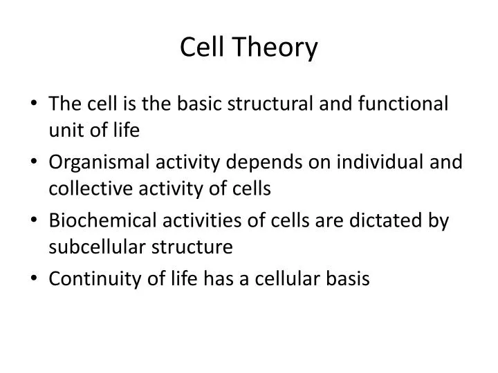
Cell Theory. The cell is the basic structural and functional unit of life Organismal activity depends on individual and collective activity of cells Biochemical activities of cells are dictated by subcellular structure Continuity of life has a cellular basis. Chromatin. Nuclear envelope.

E N D
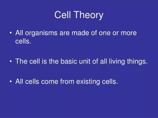
Cell Theory • The cell is the basic structural and functional unit of life • Organismal activity depends on individual and collective activity of cells • Biochemical activities of cells are dictated by subcellular structure • Continuity of life has a cellular basis
Chromatin Nuclear envelope Nucleus Nucleolus Plasma membrane Smooth endoplasmic reticulum Cytosol Lysosome Mitochondrion Centrioles Rough endoplasmic reticulum Centrosome matrix Ribosomes Golgi apparatus Microvilli Secretion being released from cell by exocytosis Microfilament Microtubule Intermediate filaments Peroxisome Figure 3.2
Plasma Membrane • Separates intracellular fluids from extracellular fluids • Plays a dynamic role in cellular activity • Glycocalyx is a glycoprotein area abutting the cell that provides highly specific biological markers by which cells recognize one another
Fluid Mosaic Model • Double bilayer of lipids with imbedded, dispersed proteins • Bilayer consists of phospholipids, cholesterol, and glycolipids • Glycolipids are lipids with bound carbohydrate • Phospholipids have hydrophobic and hydrophilic bipoles PLAY Membrane Structure
Fluid Mosaic Model Figure 3.3
Functions of Membrane Proteins PLAY • Transport • Enzymatic activity • Receptors for signal transduction Transport Protein PLAY Enzymes PLAY Receptor Proteins Figure 3.4.1
Functions of Membrane Proteins • Intercellular adhesion • Cell-cell recognition • Attachment to cytoskeleton and extracellular matrix PLAY Structural Proteins Figure 3.4.2
Plasma Membrane Surfaces • Differ in the kind and amount of lipids they contain • Glycolipids are found only in the outer membrane surface • 20% of all membrane lipid is cholesterol
Lipid Rafts • Make up 20% of the outer membrane surface • Composed of sphingolipids and cholesterol • Are concentrating platforms for cell-signaling molecules
Membrane Junctions • Tight junction – impermeable junction that encircles the cell • Desmosome – anchoring junction scattered along the sides of cells • Gap junction – a nexus that allows chemical substances to pass between cells
Membrane Junctions: Tight Junction Figure 3.5a
Membrane Junctions: Desmosome Figure 3.5b
Membrane Junctions: Gap Junction Figure 3.5c
Membrane Potential • Voltage across a membrane • Resting membrane potential – the point where K+ potential is balanced by the membrane potential • Ranges from –20 to –200 mV • Results from Na+ and K+ concentration gradients across the membrane • Differential permeability of the plasma membrane to Na+ and K+ • Steady state – potential maintained by active transport of ions
Generation and Maintenance of Membrane Potential PLAY InterActive Physiology ®: Nervous System I: The Membrane Potential Figure 3.15
Cell Adhesion Molecules (CAMs) • Anchor cells to the extracellular matrix • Assist in movement of cells past one another • Rally protective white blood cells to injured or infected areas
Roles of Membrane Receptors • Contact signaling – important in normal development and immunity • Electrical signaling – voltage-regulated “ion gates” in nerve and muscle tissue • Chemical signaling – neurotransmitters bind to chemically gated channel-linked receptors in nerve and muscle tissue • G protein-linked receptors – ligands bind to a receptor which activates a G protein, causing the release of a second messenger, such as cyclic AMP
Operation of a G Protein • An extracellular ligand (first messenger), binds to a specific plasma membrane protein • The receptor activates a G protein that relays the message to an effector protein
Operation of a G Protein • The effector is an enzyme that produces a second messenger inside the cell • The second messenger activates a kinase • The activated kinase can trigger a variety of cellular responses
Operation of a G Protein Extracellular fluid First messenger (ligand) Effector (e.g., enzyme) 1 Active second messenger (e.g., cyclic AMP) 4 3 G protein 2 5 Membrane receptor Inactive second messenger Activated (phosphorylated) kinases 6 Cascade of cellular responses (metabolic and structural changes) Cytoplasm Figure 3.16
Operation of a G Protein Extracellular fluid First messenger (ligand) 1 Membrane receptor Cytoplasm Figure 3.16
Operation of a G Protein Extracellular fluid First messenger (ligand) 1 G protein 2 Membrane receptor Cytoplasm Figure 3.16
Operation of a G Protein Extracellular fluid First messenger (ligand) Effector (e.g., enzyme) 1 3 G protein 2 Membrane receptor Cytoplasm Figure 3.16
Operation of a G Protein Extracellular fluid First messenger (ligand) Effector (e.g., enzyme) 1 Active second messenger (e.g., cyclic AMP) 4 3 G protein 2 Membrane receptor Inactive second messenger Cytoplasm Figure 3.16
Operation of a G Protein Extracellular fluid First messenger (ligand) Effector (e.g., enzyme) 1 Active second messenger (e.g., cyclic AMP) 4 3 G protein 2 5 Membrane receptor Inactive second messenger Activated (phosphorylated) kinases Cytoplasm Figure 3.16
Operation of a G Protein Extracellular fluid First messenger (ligand) Effector (e.g., enzyme) 1 Active second messenger (e.g., cyclic AMP) 4 3 G protein 2 5 Membrane receptor Inactive second messenger Activated (phosphorylated) kinases 6 Cascade of cellular responses (metabolic and structural changes) Cytoplasm Figure 3.16
Cytoplasm • Cytoplasm – material between plasma membrane and the nucleus • Cytosol – largely water with dissolved protein, salts, sugars, and other solutes
Cytoplasm • Cytoplasmic organelles – metabolic machinery of the cell • Inclusions – chemical substances such as glycosomes, glycogen granules, and pigment
Cytoplasmic Organelles • Specialized cellular compartments • Membranous • Mitochondria, peroxisomes, lysosomes, endoplasmic reticulum, and Golgi apparatus • Nonmembranous • Cytoskeleton, centrioles, and ribosomes
Mitochondria • Double membrane structure with shelf-like cristae • Provide most of the cell’s ATP via aerobic cellular respiration • Contain their own DNA and RNA
Mitochondria Figure 3.17a, b
Ribosomes • Granules containing protein and rRNA • Site of protein synthesis • Free ribosomes synthesize soluble proteins • Membrane-bound ribosomes synthesize proteins to be incorporated into membranes
Endoplasmic Reticulum (ER) • Interconnected tubes and parallel membranes enclosing cisternae • Continuous with the nuclear membrane • Two varieties – rough ER and smooth ER
Endoplasmic Reticulum (ER) Figure 3.18a, c
Rough (ER) • External surface studded with ribosomes • Manufactures all secreted proteins • Responsible for the synthesis of integral membrane proteins and phospholipids for cell membranes
Signal Mechanism of Protein Synthesis • mRNA – ribosome complex is directed to rough ER by a signal-recognition particle (SRP) • SRP is released and polypeptide grows into cisternae • The protein is released into the cisternae and sugar groups are added
Signal Mechanism of Protein Synthesis • The protein folds into a three-dimensional conformation • The protein is enclosed in a transport vesicle and moves toward the Golgi apparatus
Cytosol Transport vesicle budding off Coatomer- coated transport vesicle 5 Ribosomes mRNA 3 4 Sugar group 2 1 Released glycoprotein Signal sequence removed Signal sequence Receptor site Growing polypeptide Signal- recognition particle (SRP) ER cisterna ER membrane Signal Mechanism of Protein Synthesis Figure 3.19
Cytosol mRNA 1 Signal sequence Receptor site Signal- recognition particle (SRP) ER cisterna ER membrane Signal Mechanism of Protein Synthesis Figure 3.19
Cytosol mRNA 2 1 Signal sequence Receptor site Growing polypeptide Signal- recognition particle (SRP) ER cisterna ER membrane Signal Mechanism of Protein Synthesis Figure 3.19
Cytosol Ribosomes mRNA 3 2 1 Signal sequence removed Signal sequence Receptor site Growing polypeptide Signal- recognition particle (SRP) ER cisterna ER membrane Signal Mechanism of Protein Synthesis Figure 3.19
Cytosol Ribosomes mRNA 3 4 2 1 Released glycoprotein Signal sequence removed Signal sequence Receptor site Growing polypeptide Signal- recognition particle (SRP) ER cisterna ER membrane Signal Mechanism of Protein Synthesis Figure 3.19
Cytosol Transport vesicle budding off 5 Ribosomes mRNA 3 4 Sugar group 2 1 Released glycoprotein Signal sequence removed Signal sequence Receptor site Growing polypeptide Signal- recognition particle (SRP) ER cisterna ER membrane Signal Mechanism of Protein Synthesis Figure 3.19
Cytosol Transport vesicle budding off Coatomer- coated transport vesicle 5 Ribosomes mRNA 3 4 Sugar group 2 1 Released glycoprotein Signal sequence removed Signal sequence Receptor site Growing polypeptide Signal- recognition particle (SRP) ER cisterna ER membrane Signal Mechanism of Protein Synthesis Figure 3.19
Smooth ER • Tubules arranged in a looping network • Catalyzes the following reactions in various organs of the body • In the liver – lipid and cholesterol metabolism, breakdown of glycogen and, along with the kidneys, detoxification of drugs • In the testes – synthesis of steroid-based hormones
Smooth ER • Catalyzes the following reactions in various organs of the body (continued) • In the intestinal cells – absorption, synthesis, and transport of fats • In skeletal and cardiac muscle – storage and release of calcium
Golgi Apparatus • Stacked and flattened membranous sacs • Functions in modification, concentration, and packaging of proteins • Transport vessels from the ER fuse with the cis face of the Golgi apparatus
Golgi Apparatus • Proteins then pass through the Golgi apparatus to the trans face • Secretory vesicles leave the trans face of the Golgi stack and move to designated parts of the cell
Golgi Apparatus Figure 3.20a
Rough ER Cisterna Proteins in cisterna Phagosome Membrane Vesicle Lysosomes containing acid hydrolase enzymes Vesicle incorporated into plasma membrane Pathway 3 Coatomer coat Golgi apparatus Pathway 2 Secretory vesicles Pathway 1 Plasma membrane Proteins Secretion by exocytosis Extracellular fluid Role of the Golgi Apparatus Figure 3.21