Download
1 / 54
580 likes | 1.13k Views
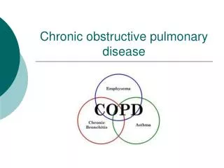
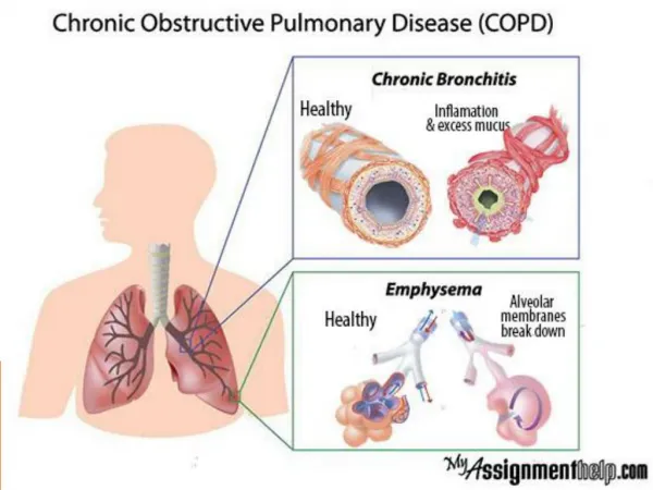
Chronic Obstructive Pulmonary Disease (COPD). COPD Description. Characterized by presence of airflow obstruction Caused by emphysema or chronic bronchitis Generally progressive May be accompanied by airway hyperreactivity May be partially reversible. Emphysema Description.

E N D
COPDDescription • Characterized by presence of airflow obstruction • Caused by emphysema or chronic bronchitis • Generally progressive • May be accompanied by airway hyperreactivity • May be partially reversible
EmphysemaDescription • Abnormal permanent enlargement of the air space distal to the terminal bronchioles • Accompanied by destruction of bronchioles
Chronic BronchitisDescription • Presence of chronic productive cough for 3 or more months in each of 2 successive years in a patient whom other causes of chronic cough have been excluded
COPDCauses • Cigarette smoking • Primary cause of COPD*** • Clinically significant airway obstruction develops in 15% of smokers • 80% to 90% of COPD deaths are related to tobacco smoking • > 1 in 5 deaths is result of cigarette smoking
COPDCauses • Cigarette smoking • Nicotine stimulates sympathetic nervous system resulting in: • HR • Peripheral vasoconstriction • BP and cardiac workload
COPDCauses • Cigarette smoking • Compounds problems in a person with CAD • Ciliary activity • Possible loss of ciliated cells • Abnormal dilation of the distal air space • Alveolar wall destruction • Carbon monoxide • O2 carrying capacity • Impairs psychomotor performance and judgment • Cellular hyperplasia • Production of mucus • Reduction in airway diameter • Increased difficulty in clearing secretions
COPDCauses • Secondhand smoke exposure associated with: • Pulmonary function • Risk of lung cancer • Mortality rates from ischemic heart disease
COPDCauses • Infection • Major contributing factor to the aggravation and progression of COPD • Heredity • -Antitrypsin (AAT) deficiency (produced by liver and found in lungs); accounts for < 1% of COPD cases • Emphysema results from lysis of lung tissues by proteolytic enzymes from neutrophils and macrophages
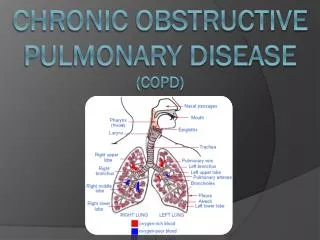
Pathophysiology of Chronic Bronchitis and Emphysema Fig. 28-7
EmphysemaPathophysiology • Hyperinflation of alveoli • Destruction of alveolar walls • Destruction of alveolar capillary walls • Narrowed airways • Loss of lung elasticity
Emphysema Pathophysiology • Two types: • Centrilobular (central part of lobule) • Most common • Panlobular (destruction of whole lobule) • Usually associated with AAT deficiency
Emphysema Pathophysiology • Structural changes are: • Hyperinflation of alveoli • Destruction of alveolar capillary walls • Narrowed, tortuous small airways • Loss of lung elasticity
Emphysema Pathophysiology • Small bronchioles become obstructed as a result of • Mucus • Smooth muscle spasm • Inflammatory process • Collapse of bronchiolar walls • Recurrent infections production/stimulation of neutrophils and macrophages release proteolytic enzymes alveolar destruction inflammation, exudate, and edema
Emphysema Pathophysiology • Elastin and collagen are destroyed • Air goes into the lungs but is unable to come out on its own and remains in the lung • Causes bronchioles to collapse
Emphysema Pathophysiology • Trapped air hyperinflation and overdistention • As more alveoli coalesce, blebs and bullae may develop • Destruction of alveolar walls and capillaries reduced surface area for O2 diffusion • Compensation is done by increasing respiratory rate to increase alveolar ventilation • Hypoxemia usually develops late in disease
EmphysemaClinical Manifestations • Dyspnea • Progresses in severity • Patient will first complain of dyspnea on exertion and progress to interfering with ADLs and rest
Emphysema Clinical Manifestations • Minimal coughing with no to small amounts of sputum • Overdistention of alveoli causes diaphragm to flatten and AP diameter to increase
Emphysema Clinical Manifestations • Patient becomes chest breather, relying on accessory muscles • Ribs become fixed in inspiratory position
EmphysemaClinical Manifestations • Patient is underweight (despite adequate calorie intake)
Chronic BronchitisPathophysiology Pathologic lung changes are: • Hyperplasia of mucus-secreting glands in trachea and bronchi • Increase in goblet cells • Disappearance of cilia • Chronic inflammatory changes and narrrowing of small airways • Altered fxn of alveolar macrophages infections
Chronic BronchitisPathophysiology Chronic inflammation • Primary pathologic mechanism causing changes • Narrow airway lumen and reduced airflow d/t • hyperplasia of mucus glands • Inflammatory swelling • Excess, thick mucus
Chronic BronchitisPathophysiology • Greater resistance to airflow increases work of breathing • Hypoxemia and hypercapnia develop more frequently in chronic bronchitis than emphysema
Chronic BronchitisPathophysiology • Bronchioles are clogged with mucus and pose a physical barrier to ventilation • Hypoxemia and hypercapnia d/t lack of ventilation and O2 diffusion • Tendency to hypoventilate and retain CO2 • Frequently patients require O2 both at rest and during exercise
Chronic BronchitisPathophysiology • Cough is often ineffective to remove secretions because the person cannot breathe deeply enough to cause air flow distal to the secretions • Bronchospasm frequently develops • More common with history of smoking or asthma
Chronic BronchitisClinical Manifestations • Earliest symptoms: • Frequent, productive cough during winter • Frequent respiratory infections
Chronic BronchitisClinical Manifestations • Bronchospasm at end of paroxysms of coughing • Cough • Dyspnea on exertion • History of smoking • Normal weight or heavyset • Ruddy (bluish-red) appearance d/t • polycythemia (increased Hgb d/t chronic hypoxemia)) • cyanosis
Chronic BronchitisClinical Manifestations • Hypoxemia and hypercapnia • Results from hypoventilation and airway resistance + problems with alveolar gas exchange
COPDComplications • Pulmonary hypertension (pulmonary vessel constriction d/t alveolar hypoxia & acidosis) • Cor pulmonale (Rt heart hypertrophy + RV failure) • Pneumonia • Acute Respiratory Failure
COPDDiagnostic Studies • Chest x-rays early in the disease may not show abnormalities • History and physical exam • Pulmonary function studies • reduced FEV1/FVC and residual volume and total lung capacity
COPDDiagnostic Studies • ABGs • PaO2 • PaCO2 (especially in chronic bronchitis) • pH (especially in chronic bronchitis) • Bicarbonate level found in late stages COPD
COPDCollaborative Care • Smoking cessation • Most significant factor in slowing the progression of the disease
COPDCollaborative Care:Drug Therapy • Bronchodilators – as maintenance therapy • -adrenergic agonists (e.g. Ventolin) • MDI or nebulizer preferred • Anticholinergics (e.g. Atrovent)
COPDCollaborative Care: Oxygen Therapy • O2 therapy • Raises PO2 in inspired air • Treats hypoxemia • Titrate to lowest effective dose
COPDCollaborative Care: Oxygen Therapy • Chronic O2 therapy at home • Improved prognosis • Improved neuropsychologic function • Increased exercise tolerance • Decreased hematocrit • Reduced pulmonary hypertension
COPDCollaborative Care: Respiratory Therapy • Breathing retraining • Pursed-lip breathing • Prolongs exhalation and prevents bronchiolar collapse and air trapping • Diaphragmatic breathing • Focuseson using diaphragm instead of accessory muscles to achieve maximum inhalation and slow respiratory rate • See text re how to teach
COPDCollaborative Care: Respiratory Therapy • Huff coughing (Table 28-21) • Chest physiotherapy – to bring secretions into larger, more central airways • Postural drainage • Percussion • Vibration
Positions for Postural Drainage Positions for Postural Drainage Fig. 28-16
COPDCollaborative Care • Encourage patient to remain as active as possible
COPDCollaborative Care • Surgical Therapy • Lung volume reduction surgery • Lung transplant
COPDCollaborative Care • Nutritional therapy • Full stomachs press on diaphragm causing dyspnea and discomfort • Difficulty eating and breathing at the same time leads to inadequate amounts being eaten
COPDCollaborative Care • Nutritional therapy • To decrease dyspnea and conserve energy • Rest at least 30 minutes prior to eating • Use bronchodilator before meals • Select foods that can be prepared in advance • 5-6 small meals to avoid bloating • Avoid foods that require a great deal of chewing • Avoid exercises and treatments 1 hour before and after eating
COPDCollaborative Care • Nutritional therapy • Avoid gas-forming foods • High-calorie, high-protein diet is recommended • Supplements • Avoid high carbohydrate diet to prevent increase in CO2 load
Nursing ManagementNursing Diagnoses • Ineffective airway clearance • Impaired gas exchange • Imbalanced nutrition: less than body requirements • Disturbed sleep pattern • Risk for infection
Nursing ManagementNursing Implementation Health Promotion • STOP SMOKING!!! • Avoid or control exposure to occupational and environmental pollutants and irritants • Early detection of small-airway disease • Early diagnosis of respiratory tract infections
Nursing ManagementNursing Implementation Acute Intervention • Required for complications like pneumonia, cor pulmonale, and acute respiratory failure
Nursing ManagementNursing Implementation Ambulatory and Home Care • Pulmonary rehabilitation • Control and alleviate symptoms of pathophysiologic complications of respiratory impairment
Nursing ManagementNursing Implementation Ambulatory and Home Care • Teach patient how to achieve optimal capability in carrying out ADLs • Physical therapy • Nutrition • Education • Activity considerations • Exercise trainingof upper extremities to help improve function and relieve dyspnea
Nursing ManagementNursing Implementation • Ambulatory and Home Care • Explore alternative methods of ADLs • Encourage patient to sit while performing activities • Coordinated walking
Nursing ManagementNursing Implementation Ambulatory and Home Care • Slow, pursed-lip breathing • After exercise, wait 5 minutes before using -adrenergic agonist MDI