Download
1 / 49
541 likes | 1.31k Views
Neuroanatomical Techniques. General avenues in neuroanatomy. Descriptive neuroanatomy: What does a structure (cell/cell group/nucleus) look like? Where is a structure localized? Which neuron connects with what? Functional neuroanatomy What structure is associated with what function?

E N D
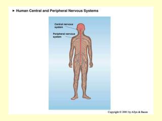
General avenues in neuroanatomy • Descriptive neuroanatomy: • What does a structure (cell/cell group/nucleus) look like? • Where is a structure localized? • Which neuron connects with what? • Functional neuroanatomy • What structure is associated with what function? • How does manipulation, injury, disease and experience influence the structure and connectivity of the nervous system?
Objectives • Short history of modern neuroanatomy • Histochemical stains • Neuronal and axonal tracing • Immmunohistochemistry & in situ hybridization • Genetic labeling of cells and connections
History of modern neuroanatomy Rudolf Albert von Kölliker (1817-1905) nucleus of Kölliker (Rexed lamina X), continuity of axon and neuron Heinrich Wilhelm Gottfried Waldeyer (1837-1921) Introduced the term “neuron” and “chromosome” Camilio Golgi (1843-1926) Golgi method; Golgi cells; Golgi apparatus; Golgi tendon organ; Golgi-Mazzoni corpuscle Santiago Ramon y Cajal (1852-1934) Cajal's gold-sublimate method for astrocytes horizontal cell of Cajal (Retzius-Cajal cell in cortex) interstitial nucleus of Cajal
Common immunohistochemical stains • Golgi: sparse, but random • Hematoxylin/Eosin: cell stain • Nissl (thionin): cell body stain • Kluver Barrera: mixed cell fiber stain • Weil: myelinated fiber stain • Acetycholine-esterase; cytochrome oxidase
Golgi Stain Jim Conner, UCSD
Nissl (thionin) stain Brainmaps.org Brain-map.org
Cytochrome oxidase Adams, Sincich and Horton, J Neuroscience 2007 “Metabolic marker” Mitochondria in dendrites and somata
Anterograde and Retrograde Tracing • Anterograde tracing: identification of projections • Uptake of the tracer by cell body • Transport along axon • Axon is labeled • Retrograde tracing: identification of the cells that give rise to afferent projections • Injection of tracer in fiber tract, terminal field or peripheral target • Uptake of the tracer by axons • Cell body is labeled
Anterograde tracing: identification of projections • Uptake of the tracer by cell bodies (1 or many) • Transport along axon/s • Axon/s labeled
Retrograde tracing: identification of the cells that give rise to afferent projections • Injection of tracer in fiber tract, terminal field or peripheral target • Uptake of the tracer by axons • Cell bodies labeled (1 or many)
Retrograde labeling with HRP First introduced by Kristensson & Olsson (1971) LaVail & LaVail (1972) Spinal cord motor neurons 1: 40 µm (TMB) 2: 1 µm (TMB) 3: 7 µm (TMB) 4: 7 µm (DAB) Van der Want et al.1997
Brief History of Tracing • Degeneration techniques: • Anterograde: Wallerian degeneration Silver impregnation methods: Nauta 1950, Nauta and Gygax 1954, Fink and Heimer 1967 • Retrograde chromatolysis (disintegration of Nissl bodies as a result of injury/disease) • Autoradiography: anterograde transport of radioactive amino acids (Grafstein, 1967) • Retrograde transport of HRP (horseradish peroxidase) (Kristensson & Olsson, 1971) Fink-Heimer stain (Heimer 1999)
Chromatolysis http://cclcm.ccf.org/vm/VM_cases/neuro_cases_PNS_muscle.htm Normal (10x) Diseased (20x) Anterior horn motor neurons
Anterograde tracing with radioactive amino acids First introduced by Grafstein (1967) A: terminal field B: white matter tract Edwards and Hendrickson in: Neuroanatomical tract tracing
(Transneuronal) anterograde tracing with radioactive amino acids
Uptake Mechanisms • Active uptake: • Lectins bind to sugar moieties of membrane glycoproteins • Uptake at nerve terminals • Uptake by fibers of passage • Passive incorporation: lipophilic substances • Intracellular injection
Types of tracers • Lipophilic dyes: DiI, DiO, DiA • Dextran conjugates: BDA, fluororuby… • Lectins: WGA(wheat germ agglutinin), PHA-L (Phaseolus vulgaris leuco-agglutinin) • Bacterial toxins: CTB (cholera toxin beta subunit) • Biocyctin • Viruses: pseudorabies, GFP recombinant viruses… • Retrograde tracers: FB, DiY, Fluorogold, Microspheres • (Transgenic animals)
Application of tracers • Pressure injection: glass micropipette Hamilton syringe • Iontophorestic injection: charged tracers • Extracellular and intracellular application • Electrophysiological measurements can be taken before tracer application • Dye Crystals: Carbocyanic dyes, WGA-HRP
Transport • Diffusion in membrane: • DiI, DiO, DiA • Slow, dependent on temperature, fixation • Active transport through vesicles • Faster, up to 2 cm/day • HRP & CTB stay in vesicles-granular appearance • PHA-L, FB better cell morphology • Intracellular diffusion
Detection • Fluorescence • Enzyme reaction: HRP (WGA-HRP, CTB-HRP) • Antibodies • Streptavidin-HRP conjugate for biotinylated tracers e.g. BDA, biocytin
Lectins and Toxins • High affinity to specific sugars • Bind to glycoproteins on membrane and are internalized • WGA: wheat germ agglutinin • PHA-L: Phaseolus vulgaris leuco-agglutinin • Concavalin A, agglutinins from soy bean, lens, rhicinus… • CTB: cholera toxin beta subunit • Tetanus toxin fragment C • Unmodified, biotinylated or conjugated to HRP or fluorophors
WGA-HRP • Retrograde, anterograde and transneuronal transport • Very fast transport: • retrograde: 100 mm/day • anterograde: 300 mm/day • Disadvantages: • wide diffusion • artefact • Tissue is fragile due to need of weak fixation
Cholera Toxin beta subunit (CTB) • Retrograde, anterograde and transganglionic • Detection: antibody, HRP conjugate, conjugated to fluorophor • Application: 1 % aqueous solution, iontophoresis or pressure injection • Different efficiency in labeling among different neuronal populations and species
PHA-L • Mostly anterograde • Application: 2.5%, iontophoresis • Detection: immunohistochemically • Highly sensitive • Long transport times (2-7 weeks) • Not very effective in old animals
Anterograde tracing with PHA-L Nigrostriatal projections Gerfen et al. in: Neuroanatomical tract tracing
Lipophilic Carbocyanine Dyes • DiI, DiO, DiA: differ in exc/ems wavelengths • Anterograde and retrograde transport • Can be used in vivo (DiI & DiA) and in fixed tissue (DiI & DiO) for post-mortem labeling • Best choice for fixed tissue: slow diffusion (2 mm/month) • Non-toxic • Slice cultures, cell labeling in vitro, time lapse videomicroscopy
Lipophilic Carbocyanine Dyes DiI label from corpus callosum, Hoechst counterstain DiI (orange) callosal & DiA (green) striatal projection neurons From: Vercelli et al. 2000
Labeling of radial glia Thanos et al. 2000
Dextran amines • Polysaccharides • Soluble in water • Molecular weights from 3,000 -100,000 kD • Anterograde and retrograde transport: uptake by lesioned fibers and cells • One of the best tracers • Conjugated either to biotin or Fluorophores • BDA (biotinylated dextran amine) • FR: Fluororuby (tetramethyl rhodamine DA) • Fluoro-emerald (fluorescein conjugated DA) • Alexa-dye conjugated DA (488, 594, 632...)
Biotinylated dextran amine (BDA) • Anterograde and retrograde transport • Highly sensitive and detailed • Iontophoretic and pressure injection • Visualization using ABC and DAB • Anterograde: MW 10,000 kD • Retrograde: MW 3,000 kD (in sodium citrate -HCl pH 3)
BDA Reiner et al. 2000
Biocytin/Neurobiotin • Application: 5% solution, pressure injection or iontophoresis • Fast degradation-short survival time 2-3 days • Mostly anterograde transport • Requires glutaraldehyde fixation
Fluorogold • Application : 1-10%, pressure injection or iontophoresis • Retrogradely transported • Often granular appearance of labeled cell somata • Antibodies against Fluorogold available • Exc.: 325 nm, emm.:440 nm • Labeling for extended time: several months • Long-term toxicity
Fluorogold Fluorescence Immunolabeling Naumann et al. 2000
Cell Filling with Lucifer Yellow Layer V Corticospinal neurons Ling Wang, UCSD
Choosing the Right Tracer • Points to consider: • Anterograde or retrograde tracing • Transport time • Efficient transport in investigated system: • Age of animal, species and neuronal population • Complete cell filling necessary • Compatibility with double labeling/ electrophysiology • Stability of labeling • Spread of tracer at the injection site • Cost?
In situ Hybridization Method of localizing, either mRNA within the cytoplasm or DNA within the chromosomes, by hybridizing the sequence of interest to a complimentary strand of a nucleotide probe.
In situ Hybridization Karin Loew, UCSD Dark grains= mRNA; blue= counterstain
In situ Hybridization (bright field detection methods)
Multiplex mRNA detectionDave Kosman (Ethan Bier and Bill McGinnis labs, UC San Diego) http://superfly.ucsd.edu/%7Edavek/images/quad.html
Controls • Specificity of probe • Sequence analysis • Testing by Northern blot • Negative controls: • RNase treatment pre-hybridization • Addition of an excess of unlabeled probe • Hybridization with sense probe • Tissue known not to express the gene of interest • Positive Controls: • Comparison with protein product • Comparison to probes hybridizing to different part of the same mRNA • Tissue known to express the gene of interest • Poly dT probe or housekeeping gene to check RNA integrity
Immunohistochemistry • Fixation: formalin, paraformaldehyde, glutaraldehyde • ± parafinn embedding • Tissue cutting: cryostat, sliding microtome, vibratome • Tissue penetration: mild detergents • Blocking of unspecific binding • Primary antibody binding • Secondary antibody for detection
Detection Methods • Horseradisch peroxidase: • PAP (peroxidase anti peroxidase) • ABC (avidin-biotin-complex) method: • secondary antibody is biotinylated, • detection with streptavidin-HRP complex • Alkaline phosphatase • APAAP (alkaline phosphatase anti-alkaline phosphatase • TSA (tyramide signal amplification) method =CSA (catalyzed signal amplification) • Fluorescence
BAC-transgenic mice expressing GFP or CRE under the control of a gene specific promoter Expression patterns can be due to promoter and/or ‘positional effects’
Transgenic “Golgi” stains • Crossing of YFP mice with transgenics or KO or conditional KO
Combining cell type specificity with tracing and molecular ‘anatomy’ Example: DRD4…’experiment’
Viruses • Replication in/competent neurotropic viruses: • HSV • Pseudorabies • Multisynaptic retrograde tracing • Highly sensitive as viruses can replicate after infection • Pathways over several orders of synapses can be followed depending on the survival time • Cell type specific promoters