Download
1 / 1
10 likes | 66 Views
No. 105. Relationship with changes in hormonal oligospermia, anthropometric parameters and sperm morphology. A. ERKOVICH 1 , N . TEMNIKOV 1 , L . OSADCHUK 2. Introduction

E N D
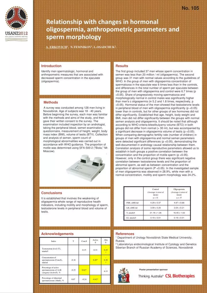
No. 105 Relationship with changes in hormonal oligospermia, anthropometric parameters and sperm morphology A. ERKOVICH1, N.TEMNIKOV1, L.OSADCHUK2. Introduction Identify men spermatologic, hormonal and anthropometric measures that are associated with decreased sperm concentration in the ejaculate (oligospermia). Results The first group included 37 men whose sperm concentration in semen was less than 20 million / ml (oligospermia). The second group was 31 men with normal values according to the guidelines of WHO. In the group of men with oligospermia concentration of spermatozoa in the ejaculate was 6 times less than in the controls, and differences in the total number of sperm per ejaculate between the group of men with oligospermia and control were 6.7 times (p <0.05). Share of progressively moving spermatozoa and morphologically normal in control males was significantly higher than men's c oligospermia (in 5.2 and 1.8 times, respectively, p <0.05). Hormonal status of the men showed that testosterone levels in peripheral blood of men with oligospermia significantly (p <0.05) lower than in controls, but for other groups of sex hormones did not differ significantly. Established that age, height, body weight and BMI, men did not differ significantly between the groups with normal semen analysis and oligospermia. It should be noted that although according to WHO criteria bitestikulyarny volume (BTO) in both groups did not differ from normal (> 30 ml), but was accompanied by a significant decrease in oligospermia volume of testis (p <0.05). When comparing demographic fertility rate (number of children) in groups of men with oligospermia and normal semen parameters were detected significant differences (p <0.05), demonstrating the well-documented in andrology causal relationship between them. Correlation analysis of some reproductive parameters allowed us to establish in both groups a positive correlation between the concentration and the proportion of motile sperm (p <0.05). However, only in the control group there was significant negative correlation between testosterone levels and the proportion of abnormal sperm, as well as between concentration and the proportion of abnormal sperm (P <0.05). In the investigated sample of men oligospermia was observed in 28.9%, while men with a normal concentration, motility and sperm morphology was 24.2%. Methods A survey was conducted among 128 men living in Novosibirsk. Age of subjects was 18 - 40 years. Before beginning the survey, each man was familiar with the methods and aims of the study, and then gave their written consent to the survey. The examination included inspection by an andrologist, taking the peripheral blood, semen examination, questionnaire, measurement of height, weight, body mass index (BMI), volume of testis (BTV). Collection and analysis of semen, sperm count of morphological abnormalities was carried out in accordance with WHO guidance. The proportion of motile was determined using SFA-500-2 ("Biola," Mr. Moscow). Conclusions It is established that involves the weakening of oligospermia whole range of reproductive health indicators, including motility and morphology of sperm, testosterone levels in peripheral blood and volume of testis. Acknowledgements References 1Department of Urology Novosibirsk State Medical University, Russia; 2Laboratoriya endocrinological Institute of Cytology and Genetics Siberian Branch of Russian Academy ofSciences,Novosibirsk Poster presentation sponsor