Download
1 / 38
410 likes | 972 Views
* KNEE JOINT * ANKLE JOINT * HIP JOINT. Prof . Ahmed Fathalla Ibrahim Professor of Anatomy College of Medicine King Saud University E-mail: ahmedfathala@hotmail.com. KNEE JOINT. OBJECTIVES. At the end of the lecture, students should be able to:

E N D
* KNEE JOINT * ANKLE JOINT* HIP JOINT Prof. Ahmed FathallaIbrahim Professor of Anatomy College of Medicine King Saud University E-mail: ahmedfathala@hotmail.com
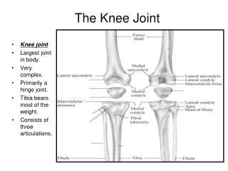
OBJECTIVES At the end of the lecture, students should be able to: • List the type & articular surfaces of knee joint. • Describe the capsule of knee joint, its extra- & intra-capsular ligaments. • List important bursae in relation to knee joint. • Describe movements of knee joint. • Apply Hilton’s law about nerve supply of joints.
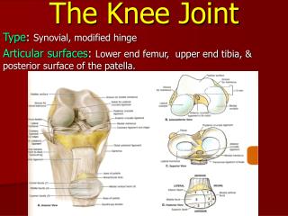
TYPES & ARTICULAR SURFACES Knee joint is formed of: Three bones. Three articulations. Femoro-tibial articulations: between the 2 femoral condyles & upper surfaces of the 2 tibialcondyles(Type: synovial, modified hinge). Femoro-patellar articulations: between posterior surface of patella & patellar surface of femur (Type: synovial, plane).
CAPSULE • Is deficient anteriorly& is replaced by: quadriceps femoris tendon, patella & ligamentum patellae. • Possesses 2 openings: one for popliteus tendon & one for communication with suprapatellar bursa.
EXTRA-CAPSULAR LIGAMENTS Ligamentum patellae (patellar ligament): from patella to tibialtuberosity. Medial (tibial) collateral ligament: from medial epicondyle of femur to upper part of medial surface of tibia (firmly attached to medial meniscus). Lateral (fibular) collateral ligament:from lateral epicondyle of femur to head of fibula (separated from lateral meniscus by popliteus tendon). Oblique popliteal ligament: extension of semimembranosus tendon.
INTRA-CAPSULAR LIGAMENTS MENISCI ATTACHMENTS: Each meniscus is attached by anterior & posterior horns into upper surface of tibia. The outer surface of medial meniscus is also attached to capsule & medial collateral ligament: medial meniscus is less mobile & more liable to be injured. FUNCTIONS: They deepen articular surfaces of tibialcondyles. They serve as cushions between tibia & femur. They are 2 C-shaped plates of fibro-cartilage. The medial meniscus is large & oval. The lateral meniscus is small & circular.
INTRA-CAPSULAR LIGAMENTS ANTERIOR & POSTERIOR CRUCIATE LIGAMENTS ATTACHMENTS: Anterior cruciate: from anterior part of intercondylar area of tibia to posterior part of lateral condyle of femur. Posterior cruciate: from posterior part of intercondylar area of tibia to anterior part of medial condyle of femur. FUNCTIONS: Anterior cruciate: prevents posterior displacement of femur on tibia. Posterior cruciate: prevents anterior displacement of femur on tibia.
IMPORTANT BURSAE RELATED TO KNEE • Suprapatellar bursa: between femur & quadriceps tendon, communicates with • synovial membrane of knee joint (Clinical importance?) • Prepatellar bursa: between patella & skin. • Deep infrapatellar bursa: between tibia & ligamentum patella. • Subcutaneous infrapatellar bursa: between tibialtuberosity & skin. • Popliteal bursa (not shown): between popliteus tendon & capsule, communicates with synovial membrane of knee joint.
MOVEMENTS • FLEXION: • Mainly by hamstring muscles: biceps femoris , semitendinosus & semimembranosus. • Assisted by sartorius , gracilis & popliteus. • EXTENSION: Quadriceps femoris. • ACTIVE ROTATION (PERFORMED WHEN KNEE IS FLEXED): A) MEDIAL ROTATION: • Mainly by semitendinosus & semimembranosus. • Assisted by sartorius & gracilis. B) LATERAL ROTATION: Biceps femoris.
MOVEMENTS (cont’d) • INACTIVE (DEPENDANT) ROTATION: A) LOCKING OF KNEE: • Lateral rotation of tibia, at the end of extension • Results mainly by tension of anterior cruciate ligament. • In locked knee, all ligaments become tight. B) UNLOCKING OF KNEE: • Medial rotation of tibia, at the beginning of flexion. • Performed by popliteus to relax ligaments & allow easy flexion.
NERVE SUPPLY REMEMBER HILTON’S LAW: “The joint is supplied by branches from nerves supplying muscles acting on it”.
OBJECTIVES At the end of the lecture, students should be able to: • List the type & articular surfaces of ankle joint. • Describe the ligaments of ankle joints. • Describe movements of ankle joint.
TYPES & ARTICULAR SURFACES • TYPE: • It is a synovial, hinge joint. • ARTICULAR SURFACES: UPPER: • A socket formed by: the lower end of tibia, medial malleolus & lateral malleolus. LOWER: • Body of talus.
LIGAMENTS MEDIAL (DELTOID) LIGAMENT: A strong triangular ligament. Apex: attached to medial malleolus. Base: subdivided into 4 parts: Anterior tibiotalar part. Tibionavicular part. Tibiocalcaneal part. Posterior tibiotalar part. LATERAL LIGAMENT: Composed of 3 separate ligaments (WHY?). Anterior talofibular ligament. Calcaneofibular ligament. Posterior talofibular ligament.
MOVEMENTS DORSIFLEXION: • Performed by muscles of anterior compartment of leg (tibialis anterior, extensor hallucislongus, extensor digitorumlongus & peroneustertius). PLANTERFLEXION: • Initiated by soleus. • Maintained by gastrocnemius. • Assisted by other muscles in posterior compartment of leg (tibialis posterior, flexor digitorumlongus & flexor hallucislongus) + muscles of lateral compartment of leg (peroneuslongus & peroneusbrevis).
N.B. • INVERSION & EVERSION MOVEMENTS occur at the talo-calcaneo-navicular joint.
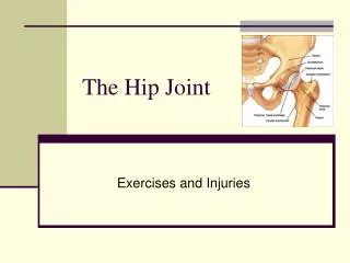
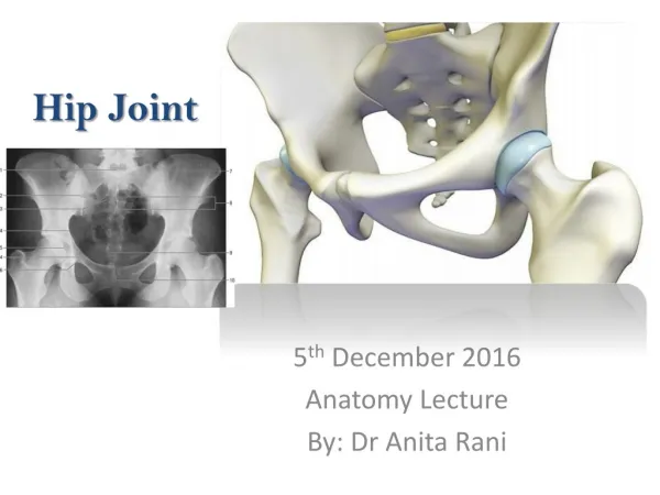
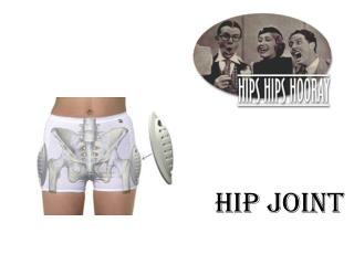
OBJECTIVES At the end of the lecture, students should be able to: • List the type & articular surfaces of hip joint. • Describe the ligaments of hip joints. • Describe movements of hip joint.
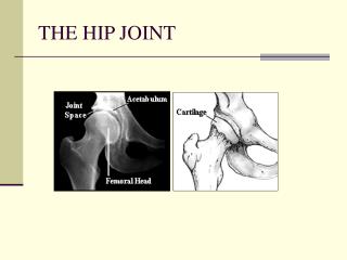
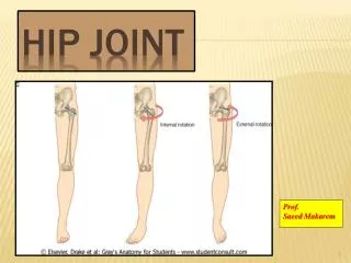
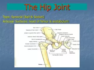
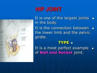
TYPES & ARTICULAR SURFACES • TYPE: • It is a synovial, ball & socketjoint. • ARTICULAR SURFACES: • Acetabulum of hip (pelvic) bone • Head of femur
LIGAMENTS(3 Extracapsular) Intertrochanteric line • Iliofemoral ligament: Y-shaped, anterior to joint, limits extension • Pubofemoral ligament: antero-inferior to joint, limits abduction & lateral rotation • Ischiofemoral ligament: posterior to joint, limits medial rotation
LIGAMENTS(3 Intracapsular) • Acetabular labrum: fibro-cartilaginous collar attached to margins of acetabulum • to increase its depth for better retaining of head of femur. • Transverse acetabular ligament: converts acetabular notch into foramen through • which pass acetabular vessels • Ligament of femoral head: carries vessels to head of femur
MOVEMENTS • FLEXION:Iliopsoas (mainly), sartorius, pectineus, rectus femoris. • EXTENSION: Hamstrings (mainly), gluteus maximus (powerful extensor). • ABDUCTION: Gluteus medius & minimus, sartorius. • ADDUCTION: Adductors, gracilis. • MEDIAL ROTATION: Gluteus medius & minimus. • LATERAL ROTATION: Gluteus maximus, quadratusfemoris, piriformis, obturatorexternus & internus.
QUESTION 1 • The muscle that extends the hip & flexes the knee joint is: • Gluteus maximus. • Quadriceps femoris. • Sartorius. • Semitendinosus.
QUESTION 2 • The bursa that communicates with the synovial membrane of knee joint is: • Suprapatellar. • Prepatellar. • Subcutaneous infrapatellar. • Deep infrapatellar.
QUESTION 3 • The muscle that dorsiflexes the ankle is: • Flexor digitorumlongus. • Tibialis anterior. • Peroneusbrevis. • Gastrocnemius.