Download
1 / 7
70 likes | 97 Views
Surgical pathology deals with examination of fluids, tissues and even foreign objects extracted from the patient for clinical evaluations, diagnosis and further correlations. Journal of Surgical Pathology and Diagnosis aims to provide integrated knowledge and a new perspective on current practices in surgical pathology. The Journal encourages novel insights, technological developments and critical observations while providing an expanded coverage of the relevant topics ranging right from fundamental experimental results to advanced diagnostic procedures. The Journal focuses on improved approaches in biopsies and resections, organ systems, immune-staining techniques, use of markers in identification of tumors and correlation with radiological findings. The Journal publishes different forms of scholarly communications including case studies, commentaries, letter to the editor and expert opinions. The Journal was established in the year 2018 and since then has successfully produced two volumes of peer-reviewed articles. The recently published literature of the Journal include key research areas such as immunohistrochemistry for HPV infection, structural polymorphisms, mitochondrial functioning in specimens, and comparative account of colposcopy and histo and cytopathology. With open access model of publication, the contributing authors have the advantage of greater outreach and recognition. Interested authors can submit their manuscripts at https://www.scholarscentral.org/submissions/surgical-pathology-diagnosis.html or they can opt to send as an e-mail attachment to the Editorial Office at manuscripts@hilarispublisher.com. Upon submission, the authors get manuscript ID within 72 hours and the online editorial tracking and review system allows faster manuscript processing with average turnaround time being 3 weeks.

E N D
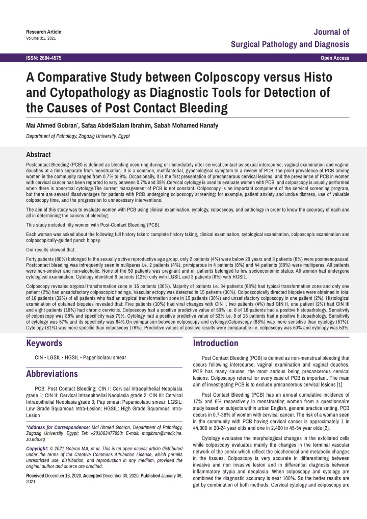
Journal of Research Article Volume 3:1, 2021 Surgical Pathology and Diagnosis ISSN: 2684-4575 Open Access A Comparative Study between Colposcopy versus Histo and Cytopathology as Diagnostic Tools for Detection of the Causes of Post Contact Bleeding Mai Ahmed Gobran*, Safaa AbdelSalam Ibrahim, Sabah Mohamed Hanafy Department of Pathology, Zagazig University, Egypt Abstract Postcontact Bleeding (PCB) is defined as bleeding occurring during or immediately after cervical contact as sexual intercourse, vaginal examination and vaginal douches at a time separate from menstruation. It is a common, multifactorial, gynecological symptom.In a review of PCB, the point prevalence of PCB among women in the community ranged from 0.7% to 9%. Occasionally, it is the first presentation of precancerous cervical lesions, and the prevalence of PCB in women with cervical cancer has been reported to vary between 0.7% and 39%.Cervical cytology is used to evaluate women with PCB, and colposcopy is usually performed when there is abnormal cytology.The current management of PCB is not constant. Colposcopy is an important component of the cervical screening program, but there are several disadvantages for patients with PCB undergoing colposcopy screening; for example, patient anxiety and undue distress, use of valuable colposcopy time, and the progression to unnecessary interventions. The aim of this study was to evaluate women with PCB using clinical examination, cytology, colposcopy, and pathology in order to know the accuracy of each and all in determining the causes of bleeding. This study included fifty women with Post-Contact Bleeding (PCB). Each woman was asked about the following full history taken: complete history taking, clinical examination, cytological examination, colposcopic examination and colposcopically-guided punch biopsy. Our results showed that: Forty patients (80%) belonged to the sexually active reproductive age group, only 2 patients (4%) were below 20 years and 3 patients (6%) were postmenopausal. Postcontact bleeding was infrequently seen in nulliparas i.e. 2 patients (4%), primiparous in 4 patients (8%) and 44 patients (88%) were multiparas. All patients were non-smoker and non-alcoholic. None of the 50 patients was pregnant and all patients belonged to low socioeconomic status. All women had undergone cytological examination. Cytology identified 6 patients (12%) only with LGSIL and 3 patients (6%) with HGSIL. Colposcopy revealed atypical transformation zone in 15 patients (30%). Majority of patients i.e. 34 patients (68%) had typical transformation zone and only one patient (2%) had unsatisfactory colposcopic findings. Vascular ectopy was detected in 15 patients (30%). Colposcopically directed biopsies were obtained in total of 16 patients (32%) of all patients who had an atypical transformation zone in 15 patients (30%) and unsatisfactory colposcopy in one patient (2%). Histological examination of obtained biopsies revealed that: Five patients (10%) had viral changes with CIN I, two patients (4%) had CIN II, one patient (2%) had CIN III and eight patients (16%) had chronic cervicitis. Colposcopy had a positive predictive value of 50% i.e. 8 of 16 patients had a positive histopathology. Sensitivity of colposcopy was 88% and specificity was 79%. Cytology had a positive predictive value of 53% i.e. 8 of 15 patients had a positive histopathology. Sensitivity of cytology was 57% and its specificity was 84%.On comparison between colposcopy and cytology:Colposcopy (88%) was more sensitive than cytology (57%). Cytology (81%) was more specific than colposcopy (79%). Predictive values of positive results were comparable i.e. colposcopy was 50% and cytology was 53%. Keywords Introduction CIN • LGSIL • HGSIL • Papanicolaou smear Post Contact Bleeding (PCB) is defined as non-menstrual bleeding that occurs following intercourse, vaginal examination and vaginal douches. PCB has many causes, the most serious being precancerous cervical lesions. Colposcopy referral for every case of PCB is important. The main aim of investigating PCB is to exclude precancerous cervical lesions [1]. Abbreviations PCB: Post Contact Bleeding; CIN I: Cervical Intraepithelial Neoplasia grade 1; CIN II: Cervical Intraepithelial Neoplasia grade 2; CIN III: Cervical Intraepithelial Neoplasia grade 3; Pap smear: Papanicolaou smear; LGSIL: Low Grade Squamous Intra-Lesion; HGSIL: High Grade Squamous Intra- Lesion Post Contact Bleeding (PCB) has an annual cumulative incidence of 17% and 6% respectively in menstruating women from a questionnaire study based on subjects within urban English, general practice setting. PCB occurs in 0.7-39% of women with cervical cancer. The risk of a woman seen in the community with PCB having cervical cancer is approximately 1 in 44,000 in 20-24 year olds and one in 2,400 in 45-54 year olds [2]. *Address for Correspondence: Mai Ahmed Gobran, Department of Pathology, Zagazig University, Egypt; Tel: +201063477990; E-mail: magibran@medicine. zu.edu.eg Cytology evaluates the morphological changes in the exfoliated cells while colposcopy evaluates mainly the changes in the terminal vascular network of the cervix which reflect the biochemical and metabolic changes in the tissues. Colposcopy is very accurate in differentiating between invasive and non invasive lesion and in differential diagnosis between inflammatory atypia and neoplasia. When colposcopy and cytology are combined the diagnostic accuracy is near 100%. So the better results are got by combination of both methods. Cervical cytology and colposcopy are Copyright: © 2021 Gobran MA, et al. This is an open-access article distributed under the terms of the Creative Commons Attribution License, which permits unrestricted use, distribution, and reproduction in any medium, provided the original author and source are credited. Received December 16, 2020; Accepted December 30, 2020; Published January 06, 2021
Gobran MA, et al. J Surg Path Diag, Volume 3:1, 2021 used to evaluate women with PCB, and histopathological examination is the final route of diagnosis of precancerous cervical lesions [3]. Aim of the work The aim of this study is to evaluate women with postcontact bleeding using clinical examination, cytology, colposcopy, and pathology in order to know the accuracy of each and all in determining the causes of bleeding. Patients and Methods This study was carried in Outpatient Obstetric and Gynecologic Clinic, Zigzag university hospitals during the period from 2018 to 2020. The research team was made up of gynecologists and pathologists. The study included fifty women with Post-Contact Bleeding (PCB) who met all the inclusion criteria, recruiting with their age or parity limit. Inclusion criteria Figure 1. (a): histopathology picture of normal cervical epithelium (× 100'H&E);1. (b): histopathological image of CIN Low grade;with atypical changes with nuclear pleomorphism and hyperchromatism affecting lower one third of cervical epithelium(× 100,H&E);1(c):histopathological image of CIN High grade;with marked atypical changes with nuclear pleomorphism and hyperchromatism affecting more than two third of cervical epithelium(× 100,H&E);1. (d):histopathological image of well differentiated squamous cell carcinoma with formation of cell nests of malignant squamous epithelial cells showing extensive nuclear pleomorphism and hyperchromatism & central keratin whorls invading basement membrane of epithelium & infiltrating the cervical submucosa(× 100,H&E); • All women presented at Gynecology Outpatient Clinic with post contact bleeding. • Aged 20-65 years. • Any parity. • Sexually active women. Exclusion criteria Cytological examination: The smears were taken by cervical scraping using Ayres spatula in the majority of participant, while cytobrushes were reserved for use in women with nulliparous os. The specimen was applied on the slide and immersed immediately in 95% ethyl alcohol for at least 30 minutes. Thereafter, the slide was sent to the pathologist for staining and examination. II- • Pregnant women. • Women with bleeding due to vulvovagintis, acute cervicitis or polyps. • Women unwilling to cooperate in the study. Papanicolaou smears showing atypia with viral changes, mild, moderate or severe dyskaryosis and carcinoma in situ were considered positive cytology. Metaplastic changes, inflammatory smears and inflammatory atypia without viral changes were considered negative cytology. • Women with frank evidence of invasive cervical carcinoma. • Women with prior total hysterectomy. Methods Using the Bethesda system, patients with viral changes and mild dyskaryosis were be graded as Low-Grade Squamous Intraepithelial Lesion (LG-SIL) and those with moderate to severe dyskaryosis and carcinoma in situ were graded as High-Grade Squamous Intraepithelial Lesion (HG-SIL). Each woman was asked about the following full history taken: 1. Personal history, including: age of the patient, age of marriage and its duration. 2. Main complaint which is post contact bleeding and its duration. III- Colposcopic examination: Patients were evaluated colposcopically. The abnormal colposcopic evidence of lesions is acetowhite areas, abnormal angioarchitecture, punctuations, mosaic pattern and gland openings. 3. Menstrual history. 4. Obstetric history. 5. Contraceptive history including the type and duration of use. Colposcopy is indicated in all cases with postcontact bleeding and positive papsmear as: 6. Past history of medical problems and surgical procedures she had, especially cervical surgery as previous cauterization. • Atypical Squamous Cells of Undetermined Significance (ASCUS). 7. Family history of similar condition or cancer cervix. • Human Papilloma Virus (HPV). 8. Husband history for smoking and venereal diseases. • Low-Grade Squamous Intraepithelial Lesion (LGSIL). 9. History of sexually transmitted diseases Methodology • High-Grade Squamous Intraepithelial Lesion (HGSIL). • Malignant cells. Clinical examination: After obtaining a history, the participant moved to the examination table to be examined by the researcher, a bivalve disposable speculum was inserted into the vagina, and the vulva, vaginal walls and the cervix were inspected with comment on the morphology and any apparent lesion seen Figures 1a-1d. I- According to colposcopic examination, the patients were classified into two groups: 1) Normal colposcopic findings. 2) Abnormal colposcopic findings: a. Significant colposcopy, which showed different colposcopic lesions, Page 2 of 7
Gobran MA, et al. J Surg Path Diag, Volume 3:1, 2021 vascular changes and gave us the possibility that CIN lesions I, II and III were found in biopsies. Mean ± SD 2.2 ± 2.1 Range Patient characteristics Occupation 0-11 b. Highly significant colposcopy, which showed suspicious colposcopic lesion usually with vascular changes and gave us the possibility that an early precancerous cervical lesion was found in biopsies. No % c. Unsatisfactory colposcopy, which showed colposcopic changes showing an atypical transformation zone grades 1-3 (according to grade of clarity of demarcation), and suspect invasive cancer was considered positive colposcopy. Unemployed 46 92 Employed Education 4 8 In cases of unsatisfactory colposcopy, two tablets of 200 μg misoprostol (400 μg Cytotec) were inserted into the posterior vaginal fornix and the patients were observed in the hospital for 6 hours and the side effects of the drug were noted such as abdominal pain, perception of increase in body temperature, vaginal bleeding, headache or vomiting before a colposcopic re-examination . Illiterate 10 20 Read and write 21 42 Moderate education 19 38 High education Residence 0 0 Colposcopically-guided punch biopsy: It was obtained by punch biopsy forceps from suspicious areas wherever these were located. One or more biopsies were obtained from each patient. Specimens were preserved with 10% formalin solution and were sent for examination by pathologist. I- Rural 24 48 Urban Smoking 26 52 The results were obtained, tabulated and statistically analyzed. In the aim of evaluating and comparing the accuracy of cytology and colposcopy, the sensitivity and specificity, the predictive value for positive test results and for negative test results, and the percentages of false-positive results and false-negative results were calculated for Pap smears and colposcopy, with histopathologic results as the gold standard. Yes 0 0 No Types of contraception Oral contraception Injected contraception Tubal ligation Condom IUCD Duration of symptoms 0-3 months 4-6 months 7-9 months 10-12 months > 12 months 50 No 20 20 2 2 6 No 23 11 2 7 7 100 % 40 40 4 4 12 % 46 22 4 14 14 Statistical analysis: Data were entered checked and analyzed using Epi-Info version 6 and SPP for Windows version 8. Results In this prospective controlled study, 50 patients with post contact bleeding presented to Outpatient Clinic of Obstetrics and Gynecology Department. Age was shown in Table 1, forty patients (80%) belonged to the sexually active reproductive age group, only 2 patients (4%) were below 20 years and 3 patients (6%) were postmenopausal. Age < 20 years No 2 % 4 Table 1. Demographic data of patients. Postcontact bleeding was infrequently seen in nulliparas i.e. 2 patients (4%), primiparous in 4 patients (8%) and 44 patients (88%) were multiparas. All patients were non-smoker and non-alcoholic. None of the 50 patients was pregnant and all patients belonged to low socioeconomic status. Approximately, two thirds of the patients i.e. 34 patients (68%) reported the symptoms early i.e. within 6 months of onset and 7 patients (14%) waited for more than one year. All women had undergone cytological examination. Cytology identified 6 patients (12%) only with LGSIL and 3 patients (6%) with HGSIL.The majority of patients i.e. 34 patients (68%) had nonspecific inflammation and 6 smears (12%) had metaplasia Table 2. Colposcopically directed biopsies were obtained in total of 16 patients (32%) of all patients who had an atypical transformation zone in 15 patients (30%) and unsatisfactory colposcopy in one patient (2%) Table 3.Histological examination of obtained biopsies revealed that: Five patients (10%) had viral changes with CIN I, two patients (4%) had CIN II, one patient (2%) had CIN III and eight patients (16%) had chronic cervicitis, as shown in Table 4. AS showen in Tables 5 and 6. Colposcopy had a positive predictive value of 50% i.e. 8 of 16 patients had a positive histopathology. Sensitivity of colposcopy was 88% and specificity was 79%. 21-30 years 18 36 31-40 years 22 44 41-50 years 7 14 > 50 years Mean ± SD 1 2 36.3 ± 10.1 Range PARITY Nullipara 19-55 No 2 % 4 Para 1 4 8 Para 2 17 34 Para 3 14 28 Para 4 13 26 Page 3 of 7
Gobran MA, et al. J Surg Path Diag, Volume 3:1, 2021 I. Colposcopy (88%) was more sensitive than cytology (57%). No % II. Cytology (81%) was more specific than colposcopy (79%). Normal 1 2 Predictive values of positive results were comparable i.e. colposcopy was 50% and cytology was 53%. Abnormal LGSIL 6 12 Discussion HGSIL 3 6 Postcontact Bleeding (PCB) is defined as non-menstrual bleeding that occurs following sexual intercourse, vaginal examination and vaginal douches. PBC has many causes the most serious being precancerous cervical lesion. Colposcopy done for every case of PCB, the main aim of investigation of PCB is to exclude precancerous cervical lesion [4]. Non-specific inflammation 34 68 6 12 Metaplasia Total 50 100 In this study, we used cervical cytology to evaluate women with unhealthy cervix, and colposcopy is performed to all cases. Histopathological examination is done for positive cytological and colposcopic cases. This study icnluded 50 women. The wide range of age (mean, 36.3 years), parity (mean, 2.2), occupation, levels of education and residence of the womenere included in this study making them more or less represented the general population. Table 2. Results of cytological examination. No % Typical transformation zone 34 68 Atypical transformation zone 15 30 This study included 50 women only who referred for cytopathology and colposcopy because there was logistic difficulty in performing cytopathology and colposcopy for all women due to the economic cost, man hours and difficulty in convincing the women. The wide range of age (mean, 36.3 years), parity (mean, 2.2), occupation, levels of education and residence of the women were included in this study making them more or less represented the general population. Grade I 11 22 Grade II 3 6 Grade III 1 2 Unsatisfactory 1 2 Another study included 200 women (100 cases and 100 controls) with the mean age of both cases and controls was 47 years, almost all women were Muslems (97%) and about a third had been born in Bamako. They found that there was no statistical difference as regard the result of cervical lesions and age (p value=0.95) [5]. Table 3. Colposcopic findings. No % Chronic cervicitis CIN I CIN II CIN III 8 5 2 1 16 10 4 2 On the other hand, In other study it was found that cervical cancer in women less than 19 years of age is rare compared with older women. The highest reported prevalence (3.77%) of Squamous Intraepithelial Lesions (SIL) among 10,296 cytology smears from patients aged 10 to 19 years, 18 percent of SILs werehigh grade [6]. Table 4. Histopathological findings. Ries et al. reported that the incidence rate of invasive cervical cancer was 0/100,000 year for ages 10 to 14 years; 0/100,000/year for ages 15 to 19 years and 1.7/100,000/year for ages 20 to 24 years from 1995 to 1999 [7]. False Positive (FP) True Positive (TP) True Negative (TN) False Negative (FN) Other study found that only three percent of adolescents with LSIL progressed to HSIL within three years. On the other hand, five percent of adolescents who developed an HPV infection developed a HSIL, half of whom did not have a prceding detectable LSIL [8]. 8 8 31 1 Table 5. Application of sensitivity and specificity tests and calculation of predictive value of positive results as regard colposcopy. In our study Incidence of unhealthy cervix among the 50 women did not reflect the true incidence in Egypt. Women were showing mild cytological changes. Cytology identified 6 patients only with LGSIL and 3 patients (6%). The majority of patients i.e. 34 patients (68%) had nonspecific inflammation and 6 smears (12%) had metaplasia. So, we preferred immediate referral for cytopathology and colposcopy for those patients at risk of not being treated in a timely manner, of not returning for a second appointment. False Positive (FP) True Positive (TP) True Negative (TN) False Negative (FN) 7 8 37 6 Table 6. Application of sensitivity and specificity tests and calculation of predictive value of positive results as regard cytology. According to Jeffcoate, cervicitis is a common finding in nearly all multiparous women and in many nulliparous women as well. This could be the reason for inflammatory report in most of the Pap smear test in the study as majority of women had at least one child Figures 2a-2f. Cytology had a positive predictive value of 53% i.e. 8 of 15 patients had a positive histopathology. Sensitivity of cytology was 57% and its specificity was 84%. On comparison between colposcopy and cytology: Page 4 of 7
Gobran MA, et al. J Surg Path Diag, Volume 3:1, 2021 Figure 1. (a):Pap smear of normal cervix(× 100,Papnucleai stain); 2.(b):Pap smear of ASCUS(Atypical squamous epithelial Cells of Undetermined Significance) with presence of scattered atypical cells with mildly enlarged &hyperchromatic nuclei(× 100,Pap); 2.(c):Pap smear of CIN Low grade/Grade I showing mild atypical changes with mildly irregular memberne and hyperchromatic nuclei(× 100,Pap); 2.(d):Pap smear of CIN High grade/Grade II showing moderate atypical changes with moderate irregular memberne and hyperchromatic nuclei(× 100,Pap); 2.(e):Pap smear of CIN High grade/Grade III showing marked atypical changes with severe irregular memberne and enlarged hyperchromatic nuclei(×100,Pap); 2.(f):Pap smear of Squamous cell carcinoma showing marked hypercellularity .The malignant cells arranged in sheets showed marked atypia with irregularly hyperchromatic nuclei with marked pleomorphism(× 100,Pap). In the present study, women who were married and had begun their sexual activity before 20 years of age were more likely to have an unhealthy cervix. Unhealthy cervix was also more common among the women with 3 or more children. Repeated childbirth may cause injury to cervix and erosion and cervicitis are also common in multipara, which can give cervix an abnormal appearance as remarked by Jeffcoate [9]. overlap and reduced debris, inflammatory cells and blood, thus simplifying screening. In this group of women with unhealthy cervix that the index of suspicion of malignancy is high, investigation work up through smears and colposcpoy is mandatory in all cases. The commonest cause of unhealthy cervix was a vascular ectopy. In the absence of neoplasia and infection, a vascular ectopy which is persistently symptomatic, may be treated by conservative measures, such as cauterization, cryotherapy or laser vaporization. Colposcopy revealed atypical transformation zone in 15 patients (30%). Majority of patients i.e. 34 patients (68%) had typical transformation zone and only one patient (2%) had unsatisfactory colposcopic findings. Vascular ectopy was the commonest benign lesion seen in colposcopy and it is a term cioned for an erosion or ectopy surrounded by a well- vascularized, metaplastic epithelium. It was detected in 15 patients (30%). Singh, et al. commented that visual screening was inferior to cytology or colposcopy as it detected only 63% of abnormality in comparison to 71% by cytology or colposcopy and concluded that visual screening would still be useful where cytological screening is not feasible [11]. Sensitivity of colposcopy was 88% and specificity was 79%. Sensitivity of cytology was 57% and its specificity was 84%; so, colposcopy was more sensitive and less specific than cytology in this study and previous study [10] that was done in the early cancer detection unit. Similarly, Misra, et al. reported 11.2% cervical dysplasia and 1.9% maliganncy in cytology of unhealthy cervix compared to 3.3% dysplasia and 0.02% malignancy in healthy cervix [12]. The positive predictive value of colposcopy in this study was 50%. The predictive value of positive results of cytology and colposcopy were almost comparable i.e. 53% and 50%, respectively. Similarly in a small study of Pap test done in 100 women, dysplasia was seen in 7 women with clinically normal looking cervix, 6 in cervicitis and one each in atrophic uterus and cervical polyp. Dysplasia occurs in majority of healthy cervices [13]. In this study, false positive rate of colposcopy was 50% which weas comparable to that of pap smear (46%). The false negative results of colposcopy was markedly low (3%) as compared to 13% of cytology. big study was conducted in 6 different countries with total number of women participating 56, 939 women which adds to the strength of the results. There was similar or comparable level of sensitivty and specificity to our study [14]. Although this is time consuming and expensive in screening program, it may prove worthwhile in women with unhealthy cervix. This was designed to produce a thin layer smear from cells collected in fluid medium. Other 2 studies had lower sensitivity than this stdy but still high indicating the good performance of the tests [15,16]. Slides prepared by this method show optimal cell preservation, minimal Page 5 of 7
Gobran MA, et al. J Surg Path Diag, Volume 3:1, 2021 Conclusion Pradhan, et al. compared cervical cytology in women with health/ unhealthy cervix. They derived that dysplasia was slightly higher among the women with unhealthy cervix in comparison to healthy cervix, but the difference was not statistically significance. Hence, they emphasized on the importance of universal screening of both the healthy and unhealthy looking cervix [17]. All women at high risk for development of cervical precancerous lesions should be screened annually with special attention to women married at teenage, smokers, immunosuppressed and those with postcontact bleedi. Colposcopy fills the gap between cytology and histopathology. So, we recommend the widespread use of colposcopy in all university hospitals and general hospitals to detect cervical lesions. As cytology detects a crime, the colposcopy locates the culprit. Workshops and training programs for colposcopy are also recommended. Tehranian, et al. Evaluated women with Postcontact Bleeding (PCB) using clinical examination, cytology, colposcopy, and pathologic findings. Because of its higher sensitivity, colposcopy can be recommended for the investigation of persistent PCB, even if women with persistent PCB have a normal physical examination and negative cytologic result [18]. References In our patients, fourteen patients (28%) had satisfactory re- examination. The conversion from unsatisfactory to satisfactory colposcopy was not statistically significnat different (p=0.2). All these patients in our study with successful conversion from an unsatisfactory to a satisfactory re-examination after they were treated by inserting vaginal misoprostol had BMI<25 kg/m2. Ten out of fourteen patients were parous and pre- menopause, and eleven out of ten patients had LSIL indication. 1. Lee T, Dresang “Evidence-based Clinical Medicine: Colposcopy: An Evidence-based Update.” J Am Board Fam Pract 18(2005): 383-392. 2. Ephraim, Shorr “New Techniques for Staining Vaginal Smears: III. A simple Differential Stain.” Science 94(1941):545-546. 3. Jin, L , M Qi, D Z Chen and A Anderson, et.al “Indole-3-Carbinol Prevents Cervical Cancer in Human Papillomavirus Type (HPV16) Transgenic Mice.” Cancer Res 59(2009): 3991-3997. Three patients of the fourteen patients had side effects before the re- examination. The side effects included abdomial pain (13.3%), perception of increase in body temperature (10%) and vomiting in one patient (3.3%). Only one patient had mild abdominal pain at 2 weeks after the re- examination. There were no severe or life-threatening side effects. 4. JT, Shen, Naliet RH and Schlaerth JB “Efficiency of cotton tipped applicators for obtaining cells from the uterine cervix for Pap smears.” Acta Cytol 28(2004): 541. 5. Michael, Sindos, Ndisang D and Pisal N “Measurement of Brn-3a levels in Pap Smear Provides a Novel Diagnostic Marker for the Detection of Cervical Neoplasia.” Gynaecol Oncol 90(2003): 366-371. The Ameican Cancer society reported that the overall age-adjusted incidence rates of cervical squamous cell carcinoma declined by 51%, from 13.39 per 100,000 women in 1970-1972 to 6.56 per 100,000 women in 1994-1996. Conversely, incidence rates of adenocarcinoma and adenosquamous carcinoma increased considerably, from 1.3 and 0.15 per 100,000 women respectively, in 1970-1972, to 1.83 and 0.41 per 100,000 women respectively, in 1994-1996. The observed increase in overall incidence rates of adenocarcinoma and adenosquamous carcinoma was mainly observed in women aged 20-49 years. The risk of developing such invsaive carcinomas of the cervix I nthe 20-34 year and 35-49 age groups trebled or doubled respectively, over that period. The icnidence rates of cervical adenocarcinoma for older woemn decreased slightly [19]. 6. FJ, Montz “Impact of Iincreasing Papanicolaou Test Sensitivity and Compliance: A Modeled Cost and Outcomes Analysis.” Obstet Gynecol 97(2002): 781-800. 7. LAG, Ries, Eisner MP and Kosary CL Cancer statistics review. National Cancer Institute, Bethesda, MD, (2010) 8. H, Mithcell and Meldley G “Differences between Papanicolaou Smears with Correct and Incorrect Diagnosis.” Cytopathology 6(1995): 368-375. 9. N, Jeffcoate Tumours of the cervix uteri, In: Jeffcoate's Principles of Gynaecology. London,(2001) Masaad studied 2131 women with AIDS (1661 HIV-positive and 470 HIV- negative) and observed that 62.7% of the HIV-positive women and 31.7% of the HIV-negative women had evidence of HPV infection; 13.6% and 3.6%, respectively, had oncogenic HPV strains associated with cervical cancer. At baseline, 37.7% of the HIV positive women and 17.3% of the HIV negative women had abnormal cervical cytology of any grade, mostly ASCUS (19.5% and 10.8%), AGCUS (1.9% and 2.7%), or low-grade SIL (14.1% and 2.5%). High-grade SIL was seen in 32 (2.1%) of the HIV-positive and 6 (1.4%) of the HIV-negative women, while only one HIV-positive woman and no HIV- negative women had cervical carcinoma [20]. 10. Ian J, Etherington “Telecolposcopy: A Feasibility Study in Primary Care.” J Telemed Telecare 8(2003): 22-24. 11. Veena, Singh, Schgal A and Luthra UK “Screening for Cervical Cancer by Direct Inspection. BMJ 304(1992): 534-35. 12. Jata S, Misra, Das K and Chandrawati R “Results of Clinically Downstaging Cervical Cancer in a Cytological Screening Programme.” Diagn Cytopathol 19(1998): 344-348. 13. V, Shrivastava and Bhanot UK “Prospective Study of 100 cases of Pap Smear.” J Nep Med Assoc 37(1998): 635-640. Tam, et al. Found that the rate of abnormal Pap smears in women with Systemic Lupus Erythematosus (SLE) was 16.5% comapred to just 5.7% in the health population. They also found that the rate of squamous intraepithelial lesiosn was almost 6 times higher in lupus patients compared to those without the disease A(11.8% versus 2%). So, they proved that SLE itself remained an independent risk factor for abnormal Pap smears [21]. 14. Jeff, Susman “Telemedicine Marches on: The Efficacy of Remote Telecolposcopy.” J Fam Pract 52(2003): 264. 15. R, Snoeck, Noel JC, Muller C and M. Bossens, et al. “Incidence of Human Papilloma Virus in Patients with Invasive Cervical Carcinoma.” Cancer Genet Cytogenet 7(2010): 118-123. 16. NY, Suffern “Pap Net Testing System. Package insert.” Neuromedical system (2006) Thanapprapasr et al. Assessed the effectiveness of misoprostol in overcoming an unsatisfactory colposcopy in the patients with an abnormal cervical cytology and stated that four hundred micrograms of vaginal misoprostol were not proved to be effective in converting an unsatisfactory to a satisfactory colposcopy [22]. 17. Neelam, Pradhan, Giri K and Rana A “Cervical cytological study in unhealthy and healthy looking cervix.” N J Obstet. Gynaecol. 2(2007): 42-47. 18. Afsaneh, Tehranian, Rezaii N, Mohit M and Bita Eslami, et al. “Evaluation of Women Presenting with Postcoital Bleeding by Cytology and Colposcopy.” Int J Gynecol Obstet 105(2009):18-20. Page 6 of 7
Gobran MA, et al. J Surg Path Diag, Volume 3:1, 2021 22. Duangmani, Thanapprapasr, Wilailak S, Israngura N and Nathpong Israngura Na Ayudhya et al. “Can vaginal misoprostol effectively increase rate of a satisfactory colposcopy? A randomized double-blind placebo-controlled trial.” Jpn J Clin Oncol. 40(2009): 203-207 19. American Cancer Society: Cancer facts and figures. Atlanta, American Cancer Society (2002) 20. L. Stewart, Massad, Lonky NM, Mutch DG and J S Blanco, et al. “Use of Speculoscopy in The Evaluation of Women with Atypical Papanicolaou Smears.” J Reprod Med 38(1993): 153-169. How to cite this article: Gobran, Mai Ahmed, Safaa AbdelSalam Ibrahim, and Sabah Mohamed Hanafy. “A Comparative Study between Colposcopy versus Histo and Cytopathology as Diagnostic Tools for Detection of the Causes of Post Contact Bleeding.” J Surg Path Diag 3 (2021). 21. Tuuminen, Tamara, Pekka P and Jorma P “The use of serologic tests for the diagnosis of chlamydial infections.” J Microbiol Meth 42(2004): 265-279. Page 7 of 7