Download
1 / 39
600 likes | 1.76k Views
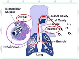
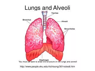
Oxygen Concentration and Partial Pressure in the Alveoli. The oxygen concentration in the alveoli, and its partial pressure is controlled by: The rate of absorption of oxygen into the blood

E N D
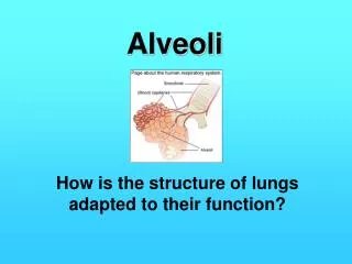
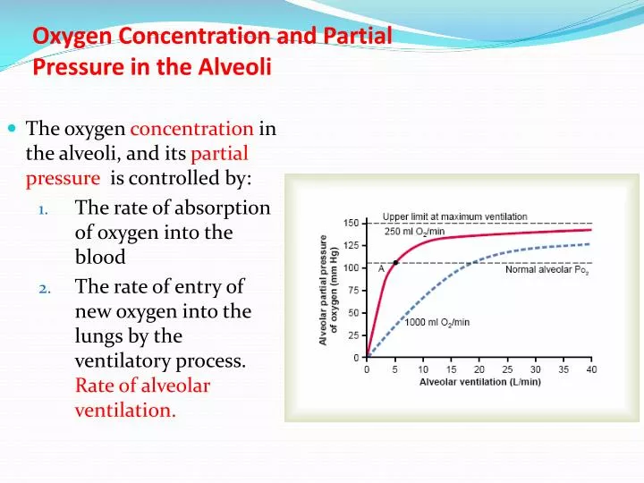
Oxygen Concentration and PartialPressure in the Alveoli • The oxygen concentration in the alveoli, and its partial pressure is controlled by: • The rate of absorption of oxygen into the blood • The rate of entry of new oxygen into the lungs by the ventilatory process. Rate of alveolar ventilation.
CO2 Concentration and PartialPressure in the Alveoli • Determined by two factors: • First, the alveolar PCO2 increases directly in proportion to the rate of carbon dioxide excretion • Second, the alveolar PCO2 decreases in inverse proportion to alveolar ventilation.
REGULATION OF RESPIRATION By Dr. Mudassar Ali Roomi (MBBS, M.Phil.) Assist. Prof. Physiology
Control of respiration Two types: • Nervous control of respiration • Chemical control of respiration
Control of repiration Components: • Sensors • gather information • Central controller • integrate signals • Effectors • muscles
Respiratory centre • Located bilaterally in medulla oblongata and pons. • Composed of 1. Dorsal Respiratory Group (DRG) 2. Ventral Respiratory Group (VRG) 3. Pneumotaxic center 4. Apneustic center
Pre-Botzinger complex (pre-BOTC) • A collection of pace-maker cells at the upper end of Dorsal Respiratory Group (DRG) • Synaptic connection with DRG • Function: Discharges rhythmic respiratory signals
Dorsal Respiratory Group (DRG) • Extends most of the length of M. oblongata • LOCATION: Neurons located in nucleus of tractus solitarius and additional neurons in reticular substance of medulla • vagus and glossopharyngeal nerve terminates at Nucleus of tractussolitarius • Both nerves – afferent nerves for resp. signals to center • Pace maker neurons send ramp signals to inspiratory muscles in a Rhythmic fashion
Ramp signals controlled by • Pneumotaxic center • Stretch receptors in the lungs Significance of ramp signals • No gasping • Smooth inflation of lungs Full cycle of respiration 5 seconds • 2sec inspiration • 3 sec expiration
Fibers from respiratory center (DRG) reach the motor neurons in spinal cord between C3 & C5 to form phrenic nerve • Complete lesion of spinal cord above C3 will stop the breathing • Lesion after C5 will not affect the respiration
The Hering-Breuer Inflation Reflex • Muscular portions of the walls of the bronchi and bronchioles throughout the lungs have stretch receptors • Transmit signals through the vagi into the dorsal respiratory group of neurons when the lungs become overstretched. • Switches Off the inspiratory ramp and thus stops further inspiration • These signals affect inspiration in much the same way as signals from the pneumotaxic center • It also increases rate of respiration
The Hering-Breuer Inflation Reflex • This reflex is activated when tidal volume increases to more than three times normal • Therefore, this reflex appears to be mainly a protective mechanism for preventing excess lung inflation
Lung “J Receptors.” • Location: In the alveolar walls in juxtaposition to the pulmonary capillaries • Stimalation: Stimulated especially when the pulmonary capillaries become engorged with blood or • Example: When pulmonary edema occurs in such conditions as congestive heart failure. • Their excitation may give the person a feeling of dyspnea.
Ventral Respiratory Group (VRG) • LOCATION: Ventral part of medulla • Two nuclei • (1) Nucleus Ambiguusrostrally • (2) Nucleus Retroambiguus caudally • Both types of neurons – INSPIRATORY & EXPIRATORY • Center remain inactive during quite breathing • Active only in increased pulmonary ventilation, during which signal from DRG spill over to VRG • Stimulation of accessory inspiratory muscles & expiratory muscles
Pneumotaxic Center • Location: Upper part of Pons • Function: Switches off Ramp Signal • Controls rate and duration of Inspiratory ramp signals • Strong stimulation may reduce Inspiratory phase to 0.5 sec respiratory rate ↑ to 30 – 40/min • Weak stimulation may ↑ Inspiratory phase to 5sec or more respiratory rate ↓ to 3-5/ min
Apneustic Center • Located in lower part of pons • Function: Prevent inspiratory neurons from being switched off → prolonged inspiration • Shortens expiration • Such Respiration called – apneusis
CHEMICAL CONTROL OF RESPIRATION Following chemical stimuli stimulate the respiration: • Excess CO2 • Excess Hydrogen ion • Decreased Oxygen
Central chemosensitivearea • Stimulated by CO2 & H+ .Oxygen have no effect Peripheral chemoreceptors • Stimulated by O2. CO2 & H+ has little effect
Location of Chemosenstive area • Located bilaterally beneath the ventral surface of medulla • Hydrogen ions are only the main direct stimulus for these group of neurons
Decreased Stimulatory Effect of Carbon Dioxide After the First 1 to 2 Days • CO2 has a potent acute effect on controlling respiratory drive but only a weak chronic effect after a few days of adaptation. • Mechanism of adaptation: Renal readjustment of the hydrogen ion by increasing the blood bicarbonate, which binds with the hydrogen ions in the blood and cerebrospinal fluid to reduce their concentrations
Acclimatization of chemoreceptors • Mountain climbers have found that when they ascend a mountain slowly • Over a period of days rather than a period of hours • They breathe much more deeply and therefore can withstand far lower atmospheric oxygen concentrations than when they ascend rapidly
The reason is within 2 to 3 days, the respiratory center in the brain stem loses about four fifths of its sensitivity to changes in Pco2 and hydrogen ions. • Therefore, the excess ventilatory blow-off of carbon dioxide that normally would inhibit an increase in respiration fails to occur • Low oxygen can drive the respiratory system to a much higher level of alveolar ventilation than under acute condition • The alveolar ventilation often increases 400 to 500 per cent after 2 to 3 days of low oxygen
Peripheral Chemoreceptor • Carotid bodies through Hering N to Glossopharyngeal N • Aortic Bodies through Vagus N to DRG • Both bodies are supplied by special minute arteries direct from the arterial trunk
Stimulation of the Chemoreceptors by Decreased Arterial Oxygen
Effect of Carbon Dioxide and Hydrogen Ion Concentration onChemoreceptor Activity They have a weak effect but stimulation by way of the peripheral chemoreceptors occurs as much as five times as rapidly as central stimulation
PERIODIC BREATHING An abnormality of respiration • CHEYNE-STOKES BREATHING is characterized by slowly waxing and waning respiration occurring about every 40 to 60 seconds
CHEYNE-STOKES BREATHING mechanism : • Overbreathes decrease CO2 & increase O2 in pulmonary blood • It takes several seconds before the changed pulmonary blood can be transported to the brain and inhibit the excess ventilation • Overventilated for an extra few seconds. • Therefore, when the overventilated blood finally reaches the brain respiratory center • The center becomes depressed an excessive amount • Then the opposite cycle begins and cycle repeats
Under normal conditions, this mechanism is highly Damped But in two conditions it occurs • Long delay occurs for transport of blood from the lungs to the brain seen in severe cardiac failure • Increased negative feedback gain in the respiratory control areas seen in brain damage
Alternate periods of Respiration & Apnea, but transition of one period to other is abrupt, not gradual. CAUSES: Meningitis Disease affecting medulla. Biot Breathing / Cluster respiration:
Sleep Apnea • Absence of spontaneous breathing • Occur during normal sleep TYPES • Obstructive Sleep Apnea • Central Sleep Apnea
Obstructive Sleep Apnea • most commonly occurs in older, obese persons 1. Narrow pharyngeal passage, and relaxation of these muscles during sleep causes the pharynx to completely close so that air cannot flow into the lungs. 2. The snoring proceeds, often becoming louder, and is then interrupted by a long silent period during which no breathing (apnea) occurs. 3. decreases in PO2 and increases in PCO2, which greatly stimulate respiration. 4. This, in turn, causes sudden attempts to breathe, which result in loud snorts and gasps followed by snoring and repeated episodes of apnea. 5. excessive daytime drowsiness as well as other disorders, including increased symphatetic activity
Central Sleep Apnea (CSA) • CAUSES: • damage to the central respiratory centers • abnormalities of the respiratory neuromuscular apparatus • Cessation of the ventilatory drive during Sleep • Strokes
Treatment of CSA: • Respiratory stimulants may be helpful. • Ventilation with CPAP at night is usually necessary.