Download
1 / 26
280 likes | 483 Views
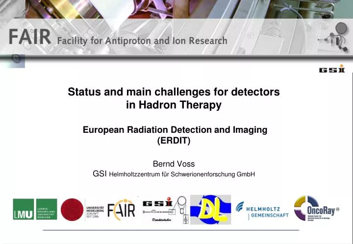
Status and main challenges for detectors in Hadron Therapy European Radiation Detection and Imaging ( ERDIT) Bernd Voss GSI Helmholtzzentrum für Schwerionenforschung GmbH. Guideline. What is Hadron Therapy about?

E N D
Status and main challenges for detectors in Hadron TherapyEuropean Radiation Detection and Imaging (ERDIT) Bernd VossGSI Helmholtzzentrum für Schwerionenforschung GmbH
Guideline What is Hadron Therapy about? What are the methods & instruments offering particles for treatment during the evolution ‘State-of-the-art’ ‘Modern’ ‘Futuristic’ accelerators? Which tasks do we have to perform and which questionsdo we have to answer in radio therapy (RT)? Which requirements & challenges for detector systems result? Are there already practical solutions? ERDIT for Horizon2020, CERN
Hadron-Therapy Light Ions vs. Photons IMRT 12C Jäkel et al, Med Phys 35 2008 • Ions… • Show inverse depth-dose profiles with a finite range and low lateral scattering at least in the plateau region • Allow superior tumor-dose conformality • Introduce increased sensibility to range uncertainties (wrong dosage)daily positioning for 30 days, intra- & inter-fractional target movement Schardt et al, Rev Med Phys 82 2010 Fragment tail ERDIT for Horizon2020, CERN
Hadron-Therapy Knowledge of base data is crucial • Depth-dose/range distributions Nuclear fragmentation cross-sections • Conversion of CT planning data (Hounsfield Units) into range of ions Existing detector equipment for the base-data collection is mature Relative ionization Rietzel et al, Rad Onc 2,14 2007 Depth in water (mm) HU: Water equivalent path length CT number ERDIT for Horizon2020, CERN
‘State-of-the-art’ Hadron Therapy facilities Beam preparation Heidelberg Ion Therapy: Beam application ‘Standard’ Accelerator structures Cyclotron, Synchrotron, Synchro-cyclotron Beam transport Mature detector equipment pick up, SEM, SCI, rest-gas monitors, IC, CG, MWPC Under investigation: GEM-TPC, Diamond, Si Basic research & Quality assurance ERDIT for Horizon2020, CERN
‘State-of-the-art’ beam delivery Minor Challenges • On-line monitoring of irradiation exhibits… • Local saturation spoiling width determination for wire based gaseous systems in high-flux areas esp. for point-like (pencil) beams with high LET radiation (12C-RT) • Potential solution Exploit robust amplification methods e.g. based on GEM technology First prototypes show feasibility ERDIT for Horizon2020, CERN
‘Modern’ Accelerators LASER driven accelerator E≥1012V/cm Relativistic e- ERDIT for Horizon2020, CERN • LASER 1PW250TW, Ti-Saphire, Neodynium-Glass, O(100m2) space req. • Pulse 25700fs, 25/w10/s conventional p-cyclotron: 100MHz • Dose Rate (2 Gy/(min l)) ~10-3Gy/pulse ~10-10 Gy/pulse • Spot point like O(10µm2), O(1021W cm-2) • Proton energy 10170MeV with exponential energy spectra (factor 2 still missing) • Targets thin foils (50nm-10µm Si,Ti,Hydro-Carbon), H2droplets Limited mass to increase proton energy at given power at decreased divergence and to obtain (quasi) mono-chromatic beams!
‘Futuristic’ Accelerators Dielectric Wall Accelerator • CT-guided rotational (200°) IMPT • Pulsed HF fields, p(200MeV) in 2m • (E,I,spot) variable pulse-to-pulse • Pulse length O(ns)@50Hz • On-line monitoring beam-application? Lawrence Livermore National Laboratory (LLNL) ERDIT for Horizon2020, CERN Y. -J. Chen et al. LLNL-CONF-414222, 2009
‘Modern’ beam delivery Major Challenges • DWA & PRIOR (???) still in a very early stage or just sketched • LASER based systems are set up; open questions: • Shielding the patient against beam contaminations (hard-X,e-,n) • Formation of irradiation-field from non-monochromatic beams • Dosimetry for exponential energy spectrum recently solved by conventional IC calibrated against FC • Measurements for (x,y,z) steering and control for ultra-fast (ns) irradiation techniques; how to do an intensity modulation & dose control particle therapy? ERDIT for Horizon2020, CERN
Intermediate Summary …ongoing detector R&D Besides ongoing attempts to optimize equipment for online monitoring of beam delivery & control (MWPC) existing detector equipment for base-data collection and state-of-the-art ion-beam application is mature R&D endeavors concentrate on: methods to reduce range uncertainties (anatomy, patient positioning, inter- and intra fractional motion of target volume) attempting to obtain 3D in-vivo on-line dosimetry & tomography using available information emerging from the target volume developing dedicated imaging detector systems ERDIT for Horizon2020, CERN
Insight into target volume Interaction & products Fragmented ions Projectile Projectile fragment Fireball radioactive nuclides Prompt g-rays Target Target fragment Nucleons & clusters 10-21 10-18 10-15 10-12 10-9 10-6 10-3 100 103 Time (s) after collision +-decay Particles Prompt -rays ERDIT for Horizon2020, CERN for 1010 protons (170 MeV, ~2Gy): (3·109) n(9·108) p(1·107) a(2·105) Aim: In-beam in-vivo single particle tomography & dosimetry Exploit information on the target volume by emerging radiation
Single particle (in-vivo) imaging ‘off-line’ Single Particle Tomography on-line / in-beam Range telescope PET ß+emitter ICT Primaries InteractionVertexImaging Light charged particles SPECT Prompt -rays in-beam in-room off-line Light ion beams Proton beams Passive collimation Slit cameras Electronic collimation Compton camera • Mostly completely new methods (except PET) • Clinical applicable technical solutions not elaborated • Appropriate detectors not commercially available Silicon Scintillator Scintillator Scintillator CdZnTe = Scatterer SCI,CZT = Absorber Single slit Multi slit ERDIT for Horizon2020, CERN
proton proton projectile 15O 16O neutron Positron Emission Tomography ‘off-line’ Single Particle Tomography on-line / in-beam Parodi et al, IEEE TNS 2005 targetfragment nucleus of tissue PET ß+emitter β+ production is a by-product of the irradiation in-beam in-room off-line 11C, 10C projectile fragment 15O, 11C, ... 12C ion projectile 12C 11C 15O 16O neutrons 15O, 11C, ... nucleus of tissue targetfragment Parodi et al, IEEE TNS 2005 Required devices: PET Camera ERDIT for Horizon2020, CERN
Positron Emission Tomography …some Hardware more… • HIMAC, Chiba • NCC, Kashiwa • HIBMC, Hyogo • MDACC, Houston • Univ. of Florida In-beam: GSI Darmstadt Off-line: MGH Boston, HIT Heidelberg • In-vivo range measurements • In-vivo dosimetry & real-time image guidance • Ongoing developments (TOF-PET, PET+CT) reduce unfavorable in-beam random coincidences/background (by 20-30%) Mature technology Courtesy W. Enghardt / OncoRay ERDIT for Horizon2020, CERN
Prompt -ray imaging Ray (IPN Lyon) Single Particle Tomography on-line / in-beam SPECT Prompt -rays Ray (IPN Lyon) Passive collimation Slit camera Electronic collimation Compton camera Silicon Scintillator Scintillator Scintillator CZT SCI,CZT Single slit Multi slit ERDIT for Horizon2020, CERN • Required devices: • Hodoscope (x,y,t) • Scatterer (x,y,E) • Absorber (x,y,z,E,t)
Prompt g-rays Nucleons and clusters Prompt -ray imaging Technique Primary ions -rays MC simulation Proton treatment plan blue FLUKA red Data 12C(75/95 AMeV) on PMMA BP position P R E L I M I N A R Y BP position Prieels et al (IBA) Dauvergneet al (IPNL Lyon) A. Ferrari and FLUKA collaboration ERDIT for Horizon2020, CERN
Prompt -ray imaging …some Hardware CZT-strip+LYSO-block Detector single slit 54x54x20 mm3 22Na multi slit 20x20x5 mm3 Scintillating-fibre Hodoscope 2x128 (1x1mm2) Timing ASIC T. Kormoll, et al., NIM A626 (2011) 114, IEEE NSS-MIC, 2011, pp. 3484 Krimmer, De Rydt IPN Lyon • Le Foulher et al. 2010 IPN Lyon t~1ns@108 s-1 ERDIT for Horizon2020, CERN
Interaction-Vertex imaging (secondary protons) Dauvergne et al 2009 Single Particle Tomography on-line / in-beam InteractionVertexImaging Light charged particles Light ion beams Proton beams AQUA Project: G4 simulations ERDIT for Horizon2020, CERN • Required devices: • Hodoscope (x,y,t) • Trackers (x,y,z,E,t) in coincidence
Nucleons (protons) Prompt g-rays Interaction-Vertex imaging Technique Single proton Primary ions Double proton Courtesy of E. Testa ERDIT for Horizon2020, CERN
Interaction-Vertex imaging …some Hardware ‘PRR30’ 2x SCI Stack (r,E) 48x3mm plastic 15cm WEPL (30-190 MeV) WLS fibres MPPC SiPM>106s-1 CMOS Hodoscope 2x2cm2 4 planes 10° PMMA GEM tracker GANIL (95 AMeV) & HIT (200-300 AMeV) 30x30cm2 2D-strips~106s-1rad.hard Courtesy of TERA ERDIT for Horizon2020, CERN
Interaction-Vertex imaging …some Results GEM-spatial 400m 6mrad Angular resolution ~0.3% (0.04 sr) Solid angle Large-angles beam diagnostics is feasible at an acquisition rate of 106 tracks/s 10 cm PRR30 1.5 m • ~5×10⁵ s-1 GEM GEM • 1010 s-1 secondary protons __ primary protons target • reconstructed vertices Courtesy of TERA ERDIT for Horizon2020, CERN
Nucleons (protons) Prompt g-rays Primary-Ion Radiography / Tomography Primary ions Single Particle Tomography on-line / in-beam Range telescope ICT Primaries • Traversing particles • Bragg peak position depends on the traversed materials • For transmission ion-imaging prior to or in-between RT ERDIT for Horizon2020, CERN • Required devices: • IC Range Telescope (r(Ei)) • (Trackers (x,y)i,e)
Primary-Ion Radiography / Tomography 3x0.6mm2 1x1mm2 12C ions Radiography X-rays 61xICs & PMMA slabs (300x300x3)mm3 Tomography Water equivalent thickness Rinaldi et al 2012 (www.ptcusa.com) Electrometer Water equivalent path length Transmission ion imaging prior to or in-between RTis feasible ERDIT for Horizon2020, CERN
12C Ion Tomography 3D ART Reconstruction P R E L I M I N A R Y P R E L I M I N A R Y Rinaldi, Gianoli et al 2012 Rinaldi, Gianoli et al 2012 ERDIT for Horizon2020, CERN
Summary • Several ‘modern’ beam production scenarios under investigation • LASER driven accelerators are not table-top likeso far Dosimetry by IC with FC calibration successfull • DWA& 4.5 GeV Proton Camera (@FAIR) are far from being reality • Detectors for Beam Control & Treatment Steering are mature • Imaging setups to gain inside in on-line dosimetry are required • Most detector systems exist on a prototype/proof-of-principle base, larger scales are needed • Several ‘new’ detector materials are under investigation (CdZnTe,LaBr,LYSO,..) • Imaging results are promising for PET, prompt gamma, secondary proton, primary-ion tomography • Serious applications as standard medical device still pending ERDIT for Horizon2020, CERN
Acknowledgement Special thanks to: Ilaria Rinaldi (Heidelberg University Hospital, Heidelberg) Katia Parodi (Ludwig-Maximilians University, Munich) Wolfgang Enghardt (OncoRay, Dresden) from whom I borrowed some of the information shown. ERDIT for Horizon2020, CERN