Download

1 / 150
1.72k likes | 2.15k Views
Neoplasia. Helpful References for the Students. Robbins textbook of pathology http://www.pathguy.com/lectures/neo-1.htm (Ed’s pathology notes) Webpath (General Pathology). Major topics of neoplasia lectures (8 hours). Introduction & Definitions Biologic behaviour of neoplasms

E N D
Helpful References for the Students • Robbins textbook of pathology • http://www.pathguy.com/lectures/neo-1.htm (Ed’s pathology notes) • Webpath (General Pathology)
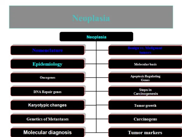
Major topics of neoplasia lectures (8 hours) • Introduction & Definitions • Biologicbehaviour of neoplasms • Histogenesis • Nomenclature • Incidence& Distribution of Cancer • Oncogenesis • Grading and Staging of Cancer • Clinicalfindingsrelatedtoneoplasms
INTRODUCTION & DEFINITIONS • Neoplasia(Latin, new growth) is an abnormality of cellular differentiation, maturation, and control of growth. • Neoplasms are commonly recognized by the formation of masses of abnormal tissue (tumors). • The term tumor can be applied to any swelling-and in that context is one of the cardinal signs of inflammation-but today it is used most commonly to denote suspected neoplasm.
1 out of about every 5 persons in the US who die this year will die of tumors (about 500,000 total). • ‘Tumor’: literally any swelling. Galen distinguished tumors that are: • ‘natural’: pregnant uterus • ‘unnatural’: pus, bony callus • ‘contrary to nature’: what we now know as neoplasms ("new growths")
Neoplasms are benignormalignant depending on several features, chiefly the ability of malignant neoplasms to spread from the site of origin. • Benign neoplasms grow but remain localized. • Cancerdenotes a malignant neoplasm (the term is thought to derive from the way in which the tumor grips the surrounding tissues with claw-like extensions, much like a crab). This feature led Hippocrates to call such tumors karkinoma after karkinos.
A neoplasm is an abnormal mass of tissue, the growth of which exceeds and is uncoordinated with that of the surrounding normal tissues and persists in the same excessive manner after cessation of the stimuli that evoked the change. Rupert Willis
"Oncology" is the study of tumors. In current usage, an oncologist is an internist or surgeon who specializes in the administration of cancer chemotherapy. • In modern usage, a tumor/neoplasm may be thought of as an attempt by the body under some stimulus to make some new sort of organ. (It develops in the wrong shape, in the wrong place, and it persists after the initiating stimulus is removed.) Tumors are like organs: • All have parenchyma and stroma. • Cells usually look similar to cells in the organ where the tumor arose. • Cells will continue to perform some of the functions of the parent organ. Tumors are different from organs: • They don't contribute to the homeostasis of the body. • They usually grow more rapidly than surrounding tissues. • Some benign and all malignant tumors never cease to grow.
BIOLOGIC BEHAVIOR OF NEOPLASMS • The biologic behavior of neoplasms constitutes a spectrum with two extremes: Benign and Malignant. • Benign: benign neoplasms grow slowly and do not invade surrounding tissues or spread to distant sites (ie, no metastasis). • Benign neoplasms are rarely life-threatening but may become so because of hormone secretion or critical location, eg, a benign neoplasm can cause death if it arises in a cranial nerve and compresses the medulla spinalis.
Malignant:Malignant neoplasmsgrow rapidly, infiltrate and destroy surrounding tissues, and metastasize throughout the body, often with lethal results. • Between Benign and Malignant; is a smaller third group of neoplasmsthat are locally invasive but have low metastatic potential. • Such neoplasms are borderline neoplasms or locally aggressive neoplasms or low-grade malignant neoplasms. An example is basal cell carcinoma of the skin and serous borderline neoplasm of the ovaries.
Benign Malignant
1. Rate of Growth: • Malignant neoplasms generally grow more rapidly than benign ones, but there is no critical rate that distinguishes malignant from benign. • Assessment of the growth rate is based upon clinical information (eg, change in size of the mass in serial examinations). • On microscopic examination, the number of mitotic figures and the metabolically active appearance of nuclei (enlarged, dispersed chromatin, large nucleoli) correlate positively with the growth rate of the neoplasm.
2. Size: • The size of a neoplasm usually has no bearing on its biologic behavior. • Many benign neoplasms become very large; conversely, highly malignant neoplasms may be lethal by virtue of extensive dissemination even though the original primary tumor is still small. • In a few neoplasms (such as endocrine neoplasms), however, size is the deciding factor in distinguishing benign from malignant growths.
3. Degree of Differentiation: • Denotes the degree to which a neoplastic cell resembles the normal mature cells of the tissue in question; this meaning is distinct from the more general use of the word to describe passage of a cell down a particular maturation pathway. • Benign neoplasms are fully (well) differentiated, ie, they closely resemble normal tissue. • Malignant neoplasms, show variable degrees of differentiation and frequently demonstrate little resemblance to normal tissue (ie, they are poorly differentiated).
In anaplasia, the neoplastic cells have no morphologic resemblance whatsoever to normal tissue. • The importance of these individual criteria varies with different neoplasms. For example, the mitotic rate is the major factor distinguishing benign from malignant smooth muscle neoplasms in the uterus; in many other neoplasms, the mitotic rate is of little relevance.
Well-differentiated SCC Anaplastic Carcinoma
4. Changes in Deoxyribonucleic Acid (DNA): • Neoplasms are associated with abnormalities in their DNA content; this abnormality increases with the degree of malignancy. • The degree of hyperchromatism (increased staining of the nucleus) provides a crude assessment of DNA content on microscopic examination; malignant cells arehyperchromatic. • When measured precisely by flow cytometry, the DNA content of malignant cells correlates well with the degree of malignancy in malignant lymphoma, bladder neoplasms, and astrocytic neoplasms. • Cytogenetic studies demonstrating aneuploidy and polyploidy also are indicative of malignancy. • Molecular techniques that demonstrate clonal deletions, translocations, or abnormalities of oncogene expression are of increasing value.
One measure of malignancy is aneuploidy by flow cytometry. The worse the neoplasm, the greater the degree of aneuploidy and the worse the prognosis.
The malignant looking tumor cell has; • Increased nuclear DNA • Increased nuclear/cytoplasmic ratio • Hyperchromatic nucleus • Coarsening of chromatin • Wrinkled nuclear edges • Multinucleation • Macronucleoli • Numerous and bizarre mitotic figures • Failure to mature along normal functional lines • Cells of widely varying sizes • Loss of orientation of cells to one another
5. Infiltration and Invasion: • Benign neoplasms are generally non-infiltrative and are surrounded by a capsule of compressed and fibrotic normal tissue. • Malignant neoplasms, have infiltrating margins. Some exceptions to this rule exist, and some benign neoplasms (eg, granular cell tumor, dermatofibroma, and carcinoid tumors) lack a capsule and have an infiltrative margin.
6. Metastasis: • The occurrence of metastasis (noncontiguous or distant growth of tumor) is absolute evidence of malignancy. • The major reason for distinguishing benign from malignant neoplasms is to be able to predict their ability to metastasize before they do so. • Gross and microscopic examination of a neoplasm usually enables a trained pathologist to classify most neoplasms as benign or malignant. • In some instances, however, this identification is difficult, and the only reliable evidence of a neoplasm's biologic behavior is the occurrence of metastasis; about 90% of pheochromocytomas are benign, but there are no reliable criteria for identifying the 10% that will metastasize.
Neoplasms can spread by seeding along body cavities, and this pattern is more typical for carcinomas than other neoplasms. • Note the multitude of small tan tumor nodules seen over the peritoneal surface of the mesentery shown here.
Both lymphatic and hematogenous spread of malignant neoplasms is possible to distant sites. • Here, a breast carcinoma has spread to a lymphatic in the lung.
CELL OR TISSUE OF ORIGIN(Histogenesis) • Neoplasms are classified and named chiefly on the basis of their presumed cell of origin. These cells have different potentials for further development into various cell types.
1. Neoplasms of Totipotent Cells: • The prototype of the totipotent cell-ie, a cell that is capable of differentiating (maturing) into any cell type in the body-is the zygote, which gives rise to the embryo, and the eventual fetus. In postnatal life, the only totipotent cells in the body are the germ cells. • These are most commonly found in the gonads but also occur in the retroperitoneum, mediastinum, and pineal region. • Germ cell neoplasms • Teratomas
The microscopic appearance of a teratoma is seen here. The three embryologic germ layers are represented by skin (ectoderm), cartilage (mesoderm) and a colonic gland (endoderm)
2. Neoplasms of Embryonic Pluripotent Cells: • Pluripotent cells can mature into several different cell types, and the corresponding neoplasms have the potential for formation of diverse structural elements; neoplasms of the renal anlage cells (nephroblastoma) commonly differentiate into structures resembling renal tubules and less often into rudiments of muscle, cartilage, and bone. These neoplasms are generally called embryomas or blastomas. • Embryonic pluripotent cells are found only in the fetal period and during the first few years of postnatal life. • The corresponding neoplasms usually occur in early childhood and only rarely in adults. • Blastomas may be completely undifferentiated -ie, are composed of small, malignant, primitive- appearing, hyperchromatic cells- or may show evidence of differentiation. Evidence of differentiation generally signifies less malignant biologic behavior.
Wilm's tumor microscopically resembles the primitive nephrogenic zone of the fetal kidney, with primitive glomeruloid structures and a cellular stroma.
3. Neoplasms of Differentiated Cells: Differentiated, adult-type cells make up most of the cells in the body in postnatal life. They show a restricted potential for differentiation, as seen when they undergo metaplasia. Most human neoplasms are derived from differentiated cells.
Nomenclature of Neoplasms of Differentiated Cells • 1. Epithelial neoplasms: • A benign epithelial neoplasm is called an adenoma if it arises within a gland (eg, thyroid adenoma, colonic adenoma) or a papilloma (Latin, papilla = nipple) when arising from an epithelial surface. • Papillomas may arise from squamous, glandular, or transitional epithelium (eg, squamous papilloma, intraductal papilloma of the breast, and transitional cell papilloma, respectively). • Not uncommonly, descriptive adjectives are incorporated in the nomenclature; eg, colonic adenomas may be villous, tubular or tubulovillous.
Malignant epithelial neoplasms are called carcinomas • Adenocarcinomas= if derived from glandular epithelia; • Squamous cell carcinoma &Transitional cell carcinoma= if originating in those kinds of epithelia. • Names may also include the organ of origin and often an adjective as well. • eg, clear cell adenocarcinoma of the kidney, Serous papillary cystadenocarcinoma of ovary, papillary adenocarcinoma of the thyroid, verrucous squamous carcinoma of the larynx.
You can add adjectives as appropriate. • papillary • well-differentiated • keratinizing • moderately well-differentiated • mucin-producing • poorly differentiated • follicular • pleomorphic • signet-ring cell • cystic (cysto-) • scirrhous • desmoplastic • medullary • comedo-