Download
1 / 46
460 likes | 475 Views
Learn about the process of cell division, which consists of nuclear division (mitosis or meiosis) and cytokinesis. Understand the phases of mitosis and how sister chromatids separate to form two genetically identical daughter cells.

E N D
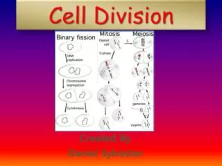
Cell division consists of two phases, nucleardivision followed by cytokinesis. • Nuclear division divides the genetic material in the nucleus, while cytokinesis divides the cytoplasm. • There are two kinds of nuclear division—mitosisand meiosis. • Mitosis divides the nucleus so that both daughter cells are genetically identical. • In contrast, meiosis is a reduction division, producing daughter cells that contain half the genetic information of the parent cell.
Sister chromatids Centromere • The first step in either mitosis or meiosis begins with the condensation of the genetic material, chromatin, into tightly coiled bodies, the chromosomes. • Each chromosome is made of two identical halves called sister chromatidsjoined at the centromere. • Each chromatid consists of a single, tightly coiled molecule of DNA, the genetic material of the cell.
In diploid cells, there are two copies of every chromosome, forming a pair, called homologous chromosomes. • In a homologous pair of chromosomes, one homologue originated from the maternal parent, the other from the paternal parent. • Humans have 46 chromosomes, 23 homologous pairs, consisting of a total of 92 chromatids.
When a cell is not dividing, the chromatin is enclosed within a clearly defined nuclear envelope. • Within the nucleus, one or more nucleoliare visible. • Outside the nucleus, two microtubule organizing centers(MTOCs, also called centrosomesin animals) lie adjacent to one another. • In animals, each MTOC contains a pair of centrioles. • These features are characteristic of interphase, the nondividing period of the cell cycle. • When cell division begins, these features change, as described below. Interphase
Mitosis There are four phases in mitosis (adjective, mitotic): prophase, metaphase, anaphase, and telophase
In prophase, three activities occur simultaneously. • First, the nucleoli disappear and the chromatin condenses into chromosomes. Prophase
Second, the nuclear envelope breaks down. • Third, the mitotic spindleis assembled. The development of the mitotic spindle begins as the MTOCs move apart to opposite ends (or poles) of the nucleus. • As they move apart, microtubules develop from each MTOC, increasing in length by the addition of tubulin units to the microtubule ends away from the MTOC. • Microtubules from each MTOC connect to a specialized region in the centromere called a kinetochore. • Microtubules tug on the kinetochore, moving the chromosomes back and forth, toward one pole, then the other. • In addition to these microtubules, the completed spindle also includes other microtubules from each MTOC that overlap at the center of the spindle and do not attach to the chromosomes.
2.Metaphasebegins when the chromosomes are distributed across the metaphase plate, a plane lying between the two poles of the spindle. Metaphase ends when the microtubules, still attached to the kinetochores, pull each chromosome apart into two chromatids. Each chromatid is connected with a centromere and a kinetochore. Once separated from its sister chromatid, each chromatid is called a chromosome. (To count the number of chromosomes at any one time, count the number of centromeres.) Metaphase
3. Anaphasebegins after the chromosomes are separated into chromatids. During anaphase, the microtubules connected to the chromatids (now chromosomes) shorten, effectively pulling the chromosomes to opposite poles. Anaphase
The microtubules shorten as tubulin units are uncoupled at their chromosome ends.
Overlapping microtubules originating from opposite MTOCs, but not attached to chromosomes, interact to push the poles farther apart. At the end of anaphase, each pole has a complete set of chromosomes, the same number of chromosomes as the original cell. (Since they consist of only one chromatid, each chromosome contains only a single copy of the DNA molecule.) Anaphase
4. Telophaseconcludes the nuclear division. During this phase, a nuclear envelope develops around each pole, forming two nuclei. The chromosomes within each of these nuclei disperse into chromatin, and the nucleoli reappear. Simultaneously, cytokinesis occurs, dividing the cytoplasm into two cells. Telophase
In animals, • microfilaments form a ring inside the plasma membrane between the two newly forming nuclei. • As the microfilaments shorten, they act like purse strings to pull the plasma membrane into the center, dividing the cell into two daughter cells. • The groove that forms as the purse strings are tightened is called a cleavage furrow.
Cell plate Wall of Daughter forming parent cell nucleus Cell wall New cell wall Vesicles Cell plate Daughter cells In plants, • vesicles originating from Golgi bodies migrate to the plane between the two newly forming nuclei. • The vesicles fuse to form a cell plate, which subsequently becomes the plasma membranes for the two daughter cells. • Cell walls develop between the membranes.
Cell cycle Once mitosis is completed and interphase begins, the cell begins a period of growth. This growth period is divided into three phases, designated G1, S, and G2 to distinguish special activities that occur. Although you can associate the labels G1 and G2 with growth and S with synthesis, it is important to recognize that growth takes place during all three phases.
In addition, S phasemarks the time during which the second DNA molecule for each chromosome is synthesized. As a result of this DNA replication, each chromosome that appears at the beginning of the next mitotic division will appear as two sister chromatids. During the G2 period of growth, materials for the next mitotic division are prepared. The time span from one cell division through G1, S, and G2 is called a cell cycle.
A cell that begins mitosis in the diploid state, that is, with two copies of every chromosome, will end mitosis with two copies of every chromosome. However, each of these chromosomes will consist of only one chromatid, or one DNA molecule. During interphase, the second DNA molecule is replicated from the first, so that when the next mitotic division begins, each chromosome will, again, consist of two chromatids.
Meiosis Meiosis (adjective, meiotic) is very similar to mitosis. Because of the similarity, however, the two processes are easily confused. The major distinction is that meiosis consists of two groups of divisions, meiosis I and meiosis II. In meiosis I, homologous chromosomes pair at the metaphase plate, and then the homologues migrate to opposite poles. In meiosis II, chromosomes spread across the metaphase plate and sister chromatids separate and migrate to opposite poles. Thus, meiosis II is analogous to mitosis. A summary of each meiotic stage follows:
Prophase Ibegins like prophase of mitosis. The nucleolus disappears, chromatin condenses into chromosomes, the nuclear envelope breaks down, and the spindle apparatus develops. Unlike mitosis, however, once the chromosomes are condensed, homologous chromosomes pair, a process called synapsis. These pairs of homologous chromosomes are variously referred to as tetrads(a group of four chromatids) or bivalents.
Tetrad Chaisma Centromere Coat-colorgenes Eye-colorgenes Tetrad(homologous pair ofchromosomes in synapsis) 1 Breakage of homologous chromatids During synapsis, corresponding regions along nonsister chromatids form close associations called chiasmata(singular, chiasma). Chiasmata are sites where genetic material is exchanged between nonsister homologous chromatids, a process called crossing over. A tetrad together with chiasmata and crossover events is referred to as a synaptonemalcomplex. 2 Joining of homologous chromatids Chiasma Separation of homologouschromosomes at anaphase I 3 Separation of chromatids atanaphase II and completion of meiosis 4 Parental type of chromosome Recombinant chromosome Recombinant chromosome Parental type of chromosome Gametes of four genetic types
2. At metaphase I, homologous pairs of chromosomes are spread across the metaphase plate. Microtubules extending from one pole are attached to the kinetochore of one member of each homologous pair. Microtubules from the other pole are connected to the second member of each homologous pair.
3. Anaphase Ibegins when homologues within tetrads uncouple as they are pulled to opposite poles.
4. In telophase I, the chromosomes have reached their respective poles, and a nuclear membrane develops around them. Note that each pole will form a new nucleus that will have half the number of chromosomes, but each chromosome will contain two chromatids. Since daughter nuclei will have half the number of chromosomes, cells that they eventually form will be haploid.
Beginning in telophase I, the cells of many species begin cytokinesis and form cleavage furrows or cell plates. In other species, cytokinesis is delayed until after meiosis II. Also, a short interphase II may begin. In any case, no replication of chromosomes occurs during this period. Instead, part II of meiosis begins in both daughter nuclei.
MEIOSIS II: Sister chromatids separate TELOPHASE IAND CYTOKINESIS TELOPHASE IIAND CYTOKINESIS PROPHASE II METAPHASE II ANAPHASE II Cleavagefurrow Sister chromatidsseparate Haploiddaughter cellsforming 5. In prophase II, the nuclear envelope disappears and the spindle develops. There are no chiasmata and no crossing over of genetic material as in prophase I.
MEIOSIS II: Sister chromatids separate TELOPHASE IAND CYTOKINESIS TELOPHASE IIAND CYTOKINESIS PROPHASE II METAPHASE II ANAPHASE II Cleavagefurrow Sister chromatidsseparate Haploiddaughter cellsforming 6. In metaphase II, the chromosomes align singly on the metaphase plate (not in tetrads as in metaphase I). Single alignment of chromosomes is exactly what happens in mitosis except that now there is only half the number of chromosomes.
MEIOSIS II: Sister chromatids separate TELOPHASE IAND CYTOKINESIS TELOPHASE IIAND CYTOKINESIS PROPHASE II METAPHASE II ANAPHASE II Cleavagefurrow Sister chromatidsseparate Haploiddaughter cellsforming 7. Anaphase IIbegins as each chromosome is pulled apart into two chromatids by the microtubules of the spindle apparatus. The chromatids (now chromosomes) migrate to their respective poles. Again, this is exactly what happens in mitosis except that now there is only half the number of chromosomes.
MEIOSIS II: Sister chromatids separate TELOPHASE IAND CYTOKINESIS TELOPHASE IIAND CYTOKINESIS PROPHASE II METAPHASE II ANAPHASE II Cleavagefurrow Sister chromatidsseparate Haploiddaughter cellsforming 8. In telophase II, the nuclear envelope reappears at each pole and cytokinesis occurs. The end result of meiosis is four haploid cells. Each cell contains half the number of chromosomes, and each chromosome consists of only one chromatid. Later in interphase, a second chromatid in each chromosome is replicated, but the cell will still have only half the number of chromosomes.
Mitosis versus Meiosis Comparing the daughter cells of mitosis and meiosis, you will find that mitosis ends with two diploid daughter cells, each with a complete set of chromosomes. True, each chromosome is composed of only one chromatid, but the second chromatid is regenerated during the S phase of interphase. Mitosis, then, merely duplicates cells, the two daughter cells essentially clones of the original cell. As such, mitosis occurs during • growth and development of multicellular organisms • and for repair (replacement) of existing cells.
In contrast, meiosis ends with four haploid daughter cells, each with half the number of chromosomes (one chromosome from every homologous pair). In order for one of these haploid cells to produce a “normal” cell with the full set of chromosomes, it must first combine with a second haploid cell to create a diploid cell. Thus, meiosis produces gametes, that is, eggs and sperm, for sexual reproduction. The fusing of an egg and a sperm, fertilization(or syngamy), gives rise to a diploid cell, the zygote. The single-celled zygote then divides by mitosis to produce a multicellular organism. Note that one copy of each chromosome in the zygote originates from one parent, and the second copy from the other parent. Thus, a pair of homologous chromosomes in the diploid zygote represents both maternal and paternal heritage.
The life cycleof a human illustrates the production of gametes by meiosis and subsequent growth by mitosis. Note that the number of chromosomes in diploid and haploid cells is indicated by 2n and n, respectively. Human cells (except gametes) contain 46 chromosomes (23 homologous pairs). Thus 2n = 46. For human gametes, n = 23. In humans, gametes are produced in the reproductive organs, the ovaries and the testes.
In other organisms, such as plants, meiosis produces spores. Spores are haploid cells that divide by mitosis to become a multicellular haploid structure, the gametophyte. Gametes are produced by the gametophyte by mitosis since the organism is already haploid. The gametes then fuse and produce a diploid cell that grows by mitosis to become the sporophyte. Specialized cells in the sporophyte divide by meiosis to produce haploid spores, which germinate to repeat the life cycle. The fern illustrates this type of reproductive cycle.
(n) (n) (2n) (n) (2n) (n) (2n) Life cycle of Ferns السَّرْخَس
Genetic Variation In mitosis, every daughter cell is exactly like the parent cell. Meiosis and sexual reproduction, however, result in a reassortment of the genetic material. This reassortment, called genetic recombination, originates from three events during the reproductive cycle:
Crossing over.During prophase I, nonsister chromatids of homologous chromosomes exchange pieces of genetic material. As a result each homologue no longer entirely represents a single parent.
2. Independent assortment of homologues.During metaphase I, tetrads of homologous chromosomes separate into chromosomes that go to opposite poles. Which chromosome goes to which pole depends upon the orientation of a tetrad at the metaphase plate. This orientation and subsequent separation is random for each tetrad. For some chromosome pairs, the chromosome that is mostly maternal may go to one pole, but for another pair, the maternal chromosome may go to the other pole.
3. Random joining of gametes.Which sperm fertilizes which egg is to a large degree a random event. In many cases, however, this event may be affected by the genetic composition of a gamete. For example, some sperm may be faster swimmers and have a better chance of fertilizing the egg.
Why Cells Divide There are two important factors that limit the size of a cell and motivate its division. The first is the relative sizeof the surface area of the plasma membrane and the volume of the cell. When a cell grows, the volume of a cell increases faster than the surface area enclosing it. This is because volume increases by the cubeof the radius (volume of a sphere = (4⁄3)πr3, where r is the radius), whereas the surface area increases by only the squareof the radius (surface area = 4πr2).
Cell Size Surface Area (length x width x 6) Volume (length x width x height) Ratio of Surface Area to Volume Ratio of Surface Area to Volume in Cells When the surface-to-volume ratio (S/V) is large, there is a large surface area relative to volume. Under these conditions, the cell can efficiently react with the outside environment. For example, adequate amounts of oxygen (for respiration) can diffuse into the cell, and waste products can be rapidly eliminated. When the S/V is small, the surface area is small compared to the volume. When this occurs, the surface area may be unable to exchange enough substances with the outside environment to service the large volume of the cell. This situation is alleviated by cell division.
A second reason for dividing is the limited capability of the nucleus. The genetic material (chromosomes) in the nucleus, collectively called its genome, “controls” the cell by producing substances which make enzymes and other biosynthetic substances. These substances, in turn, regulate cellular activities. The capacity of the genome to do this is limited by its finite amount of genetic material. As the cell grows, its volume increases, but its genome size remains constant. As the genome-to-volume ratiodecreases, the cell’s size exceeds the ability of its genome to produce sufficient amounts of materials for regulating cellular activities.
In addition to surface-to-volume and genome-to-volume ratios, other factors that are cell specific influence the onset of cell division. For example, many cells will stop dividing when the surrounding cell density reaches a certain maximum (density-dependent inhibition). Density-dependent inhibition Normal cells proliferate in culture until they reach a finite cell density, at which point they become quiescent. Tumor cells, however, continue to proliferate independent of cell density.
Other cells, such as nerve cells, will rarely divide once they have matured. When the cell cycle is interrupted and the cell stops dividing, the cell remains in an extended G1 phase (or G0 phase), never beginning the S or G2 phases until some internal or external cue initiates a resumption of the cell cycle.