Download
1 / 64
640 likes | 753 Views
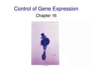
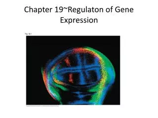
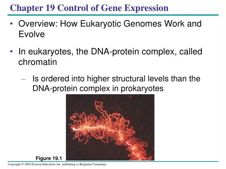
Figure 19.1. Chapter 19 Control of Gene Expression. Overview: How Eukaryotic Genomes Work and Evolve In eukaryotes, the DNA-protein complex, called chromatin Is ordered into higher structural levels than the DNA-protein complex in prokaryotes.

E N D
Figure 19.1 Chapter 19 Control of Gene Expression • Overview: How Eukaryotic Genomes Work and Evolve • In eukaryotes, the DNA-protein complex, called chromatin • Is ordered into higher structural levels than the DNA-protein complex in prokaryotes
Concept 19.1: Chromatin structure is based on successive levels of DNA packing • Eukaryotic DNA • Is precisely combined with a large amount of protein • Eukaryotic chromosomes • Contain an enormous amount of DNA relative to their condensed length
Nucleosomes, or “Beads on a String” • Proteins called histones • Are responsible for the first level of DNA packing in chromatin • Bind tightly to DNA • The association of DNA and histones • Seems to remain intact throughout the cell cycle
2 nm DNA double helix Histone tails His- tones 10 nm Histone H1 Linker DNA (“string”) Nucleosome (“bead”) (a) Nucleosomes (10-nm fiber) • In electron micrographs • Unfolded chromatin has the appearance of beads on a string • Each “bead” is a nucleosome • The basic unit of DNA packing Figure 19.2 a
30 nm Nucleosome (b) 30-nm fiber Higher Levels of DNA Packing • The next level of packing • Forms the 30-nm chromatin fiber Figure 19.2 b
Protein scaffold Loops Scaffold 300 nm (c) Looped domains (300-nm fiber) • The 30-nm fiber, in turn • Forms looped domains, making up a 300-nm fiber Figure 19.2 c
700 nm 1,400 nm (d) Metaphase chromosome • In a mitotic chromosome • The looped domains themselves coil and fold forming the characteristic metaphase chromosome Figure 19.2 d
19.2 Regulation of Gene Expression • All organisms • Must regulate which genes are expressed at any given time • During development of a multicellular organism • Its cells undergo a process of specialization in form and function called cell differentiation
Differential Gene Expression • Each cell of a multicellular eukaryote • Expresses only a fraction of its genes • In each type of differentiated cell • A unique subset of genes is expressed
Signal NUCLEUS Chromatin Chromatin modification: DNA unpacking involving histone acetylation and DNA demethlation DNA Gene available for transcription Gene Transcription Exon RNA Primary transcript Intron RNA processing Tail mRNA in nucleus Cap Transport to cytoplasm CYTOPLASM mRNA in cytoplasm Degradation of mRNA Translation Polypetide Cleavage Chemical modification Transport to cellular destination Active protein Degradation of protein Degraded protein Figure 19.3 • Many key stages of gene expression • Can be regulated in eukaryotic cells
Regulation of Chromatin Structure • Genes within highly packed chromatin • Are usually not expressed
Chromatin changes Transcription RNA processing Translation mRNA degradation Protein processing and degradation Histone tails DNA double helix Amino acids available for chemical modification Histone Modification • Chemical modification of histone tails • Can affect the configuration of chromatin and thus gene expression Figure 19.4a (a) Histone tails protrude outward from a nucleosome
Acetylated histones Unacetylated histones (b) Acetylation of histone tails promotes loose chromatin structure that permits transcription Figure 19.4 b • Histone acetylation • Seems to loosen chromatin structure and thereby enhance transcription
DNA Methylation • Addition of methyl groups to certain bases in DNA • Is associated with reduced transcription in some species
Regulation of Transcription Initiation • Chromatin-modifying enzymes provide initial control of gene expression • By making a region of DNA either more or less able to bind the transcription machinery
Poly-A signal sequence Termination region Proximal control elements Enhancer (distal control elements) Exon Intron Intron Exon Exon DNA Downstream Upstream Promoter Transcription Poly-A signal Exon Exon Intron Intron Exon Cleared 3 end of primary transport Primary RNA transcript (pre-mRNA) 5 Chromatin changes RNA processing: Cap and tail added; introns excised and exons spliced together Transcription Intron RNA RNA processing Coding segment mRNA degradation Translation mRNA P Protein processing and degradation G P P Start codon Poly-A tail Stop codon 3 UTR (untranslated region) 5 Cap 5 UTR (untranslated region) Organization of a Typical Eukaryotic Gene • Associated with most eukaryotic genes are multiple control elements • Segments of noncoding DNA that help regulate transcription by binding certain proteins Figure 19.5
The Roles of Transcription Factors • To initiate transcription • Eukaryotic RNA polymerase requires the assistance of proteins called transcription factors Dsads
Enhancers and Specific Transcription Factors • Proximal control elements • Are located close to the promoter • Distal control elements, groups of which are called enhancers • May be far away from a gene or even in an intron Jjj Lll Gene sequence
Distal control element Promoter Activators Gene Enhancer TATA box General transcription factors Activator proteins bind to distal control elements grouped as an enhancer in the DNA. This enhancer has three binding sites. 1 DNA-bending protein Group of Mediator proteins A DNA-bending protein brings the bound activators closer to the promoter. Other transcription factors, mediator proteins, and RNA polymerase are nearby. 2 RNA Polymerase II Chromatin changes The activators bind to certain general transcription factors and mediator proteins, helping them form an active transcription initiation complex on the promoter. 3 Transcription RNA processing RNA Polymerase II mRNA degradation Translation Protein processing and degradation Transcription Initiation complex RNA synthesis • An activator • Is a protein that binds to an enhancer and stimulates transcription of a gene Figure 19.6
Some specific transcription factors function as repressors • To inhibit expression of a particular gene • Some activators and repressors • Act indirectly by influencing chromatin structure
Coordinately Controlled Genes • Unlike the genes of a prokaryotic operon • Coordinately controlled eukaryotic genes each have a promoter and control elements • The same regulatory sequences • Are common to all the genes of a group, enabling recognition by the same specific transcription factors
Mechanisms of Post-Transcriptional Regulation • An increasing number of examples • Are being found of regulatory mechanisms that operate at various stages after transcription
Chromatin changes Transcription RNA processing mRNA degradation Translation Protein processing and degradation Exons DNA Primary RNA transcript RNA splicing or mRNA RNA Processing • In alternative RNA splicing • Different mRNA molecules are produced from the same primary transcript, depending on which RNA segments are treated as exons and which as introns Figure 19.8
mRNA Degradation • The life span of mRNA molecules in the cytoplasm • Is an important factor in determining the protein synthesis in a cell • Is determined in part by sequences in the leader and trailer regions
The micro- RNA (miRNA) precursor folds back on itself, held together by hydrogen bonds. The bound miRNA can base-pair with any target mRNA that contains the complementary sequence. One strand of each short double- stranded RNA is degraded; the other strand (miRNA) then associates with a complex of proteins. 3 The miRNA-protein complex prevents gene expression either by degrading the target mRNA or by blocking its translation. An enzyme called Dicer moves along the double- stranded RNA, cutting it into shorter segments. 5 4 2 1 2 Chromatin changes Transcription RNA processing mRNA degradation Translation Protein complex Protein processing and degradation Dicer Degradation of mRNA OR miRNA Target mRNA Blockage of translation Hydrogen bond • RNA interference by single-stranded microRNAs (miRNAs) • Can lead to degradation of an mRNA or block its translation 5 Figure 19.9
Initiation of Translation • The initiation of translation of selected mRNAs • Can be blocked by regulatory proteins that bind to specific sequences or structures of the mRNA • Alternatively, translation of all the mRNAs in a cell • May be regulated simultaneously
Protein Processing and Degradation • After translation • Various types of protein processing, including cleavage and the addition of chemical groups, are subject to control
Enzymatic components of the proteasome cut the protein into small peptides, which can be further degraded by other enzymes in the cytosol. 3 The ubiquitin-tagged protein is recognized by a proteasome, which unfolds the protein and sequesters it within a central cavity. 2 1 Multiple ubiquitin mol- ecules are attached to a protein by enzymes in the cytosol. Chromatin changes Transcription RNA processing Proteasome and ubiquitin to be recycled Ubiquitin Translation mRNA degradation Proteasome Protein processing and degradation Protein fragments (peptides) Protein to be degraded Ubiquinated protein Protein entering a proteasome • Proteasomes • Are giant protein complexes that bind protein molecules and degrade them Figure 19.10
19.3 • Cancer results from genetic changes that affect cell cycle control • The gene regulation systems that go wrong during cancer • Turn out to be the very same systems that play important roles in embryonic development
Types of Genes Associated with Cancer • The genes that normally regulate cell growth and division during the cell cycle • Include genes for growth factors, their receptors, and the intracellular molecules of signaling pathways
Oncogenes and Proto-Oncogenes • Oncogenes • Are cancer-causing genes • Proto-oncogenes • Are normal cellular genes that code for proteins that stimulate normal cell growth and division
Proto-oncogene DNA Translocation or transposition: gene moved to new locus, under new controls Point mutation within a control element Point mutation within the gene Gene amplification: multiple copies of the gene Oncogene Oncogene New promoter Normal growth-stimulating protein in excess Hyperactive or degradation- resistant protein Normal growth-stimulating protein in excess Normal growth-stimulating protein in excess • A DNA change that makes a proto-oncogene excessively active • Converts it to an oncogene, which may promote excessive cell division and cancer Figure 19.11
Tumor-Suppressor Genes • Tumor-suppressor genes • Encode proteins that inhibit abnormal cell division
Interference with Normal Cell-Signaling Pathways • Many proto-oncogenes and tumor suppressor genes • Encode components of growth-stimulating and growth-inhibiting pathways, respectively
1 Growth factor MUTATION Hyperactive Ras protein (product of oncogene) issues signals on its own Ras GTP G protein (a) Cell cycle–stimulating pathway. This pathway is triggered by a growth factor that binds to its receptor in the plasma membrane. The signal is relayed to a G protein called Ras. Like all G proteins, Ras is active when GTP is bound to it. Ras passes the signal to a series of protein kinases. The last kinase activates a transcription activator that turns on one or more genes for proteins that stimulate the cell cycle. If a mutation makes Ras or any other pathway component abnormally active, excessive cell division and cancer may result. Ras 1 P P 2 GTP P P 3 P P Protein kinases (phosphorylation cascade) Receptor 4 5 NUCLEUS Transcription factor (activator) 3 5 4 2 DNA Gene expression Protein that stimulates the cell cycle Figure 19.12a • The Ras protein, encoded by the ras gene • Is a G protein that relays a signal from a growth factor receptor on the plasma membrane to a cascade of protein kinases
(b) Cell cycle–inhibiting pathway. In this pathway, DNA damage is an intracellular signal that is passed via protein kinases and leads to activation of p53. Activated p53 promotes transcription of the gene for a protein that inhibits the cell cycle. The resulting suppression of cell division ensures that the damaged DNA is not replicated. Mutations causing deficiencies in any pathway component can contribute to the development of cancer. 1 2 Protein kinases 2 MUTATION 3 Defective or missing transcription factor, such as p53, cannot activate transcription UV light Active form of p53 3 DNA damage in genome 1 DNA Protein that inhibits the cell cycle • The p53 gene encodes a tumor-suppressor protein • That is a specific transcription factor that promotes the synthesis of cell cycle–inhibiting proteins Figure 19.12b
Mutations that knock out the p53 gene • Can lead to excessive cell growth and cancer (c) Effects of mutations. Increased cell division, possibly leading to cancer, can result if the cell cycle is overstimulated, as in (a), or not inhibited when it normally would be, as in (b). EFFECTS OF MUTATIONS Protein overexpressed Protein absent Cell cycle not inhibited Cell cycle overstimulated Increased cell division Figure 19.12c
The Multistep Model of Cancer Development • Normal cells are converted to cancer cells • By the accumulation of multiple mutations affecting proto-oncogenes and tumor-suppressor genes
Colon 1 Loss of tumor- suppressor gene APC (or other) 4 Loss of tumor-suppressor gene p53 2 Activation of ras oncogene Colon wall 3 Loss of tumor- suppressor gene DCC 5 Additional mutations Normal colon epithelial cells Larger benign growth (adenoma) Small benign growth (polyp) Malignant tumor (carcinoma) • A multistep model for the development of colorectal cancer Figure 19.13
Certain viruses • Promote cancer by integration of viral DNA into a cell’s genome
Inherited Predisposition to Cancer • Individuals who inherit a mutant oncogene or tumor-suppressor allele • Have an increased risk of developing certain types of cancer
19.4 • Eukaryotic genomes can have many noncoding DNA sequences in addition to genes • The bulk of most eukaryotic genomes • Consists of noncoding DNA sequences, often described in the past as “junk DNA” • However, much evidence is accumulating • That noncoding DNA plays important roles in the cell
The Relationship Between Genomic Composition and Organismal Complexity • Compared with prokaryotic genomes, the genomes of eukaryotes • Generally are larger • Have longer genes • Contain a much greater amount of noncoding DNA both associated with genes and between genes
Exons (regions of genes coding for protein, rRNA, tRNA) (1.5%) Repetitive DNA that includes transposable elements and related sequences (44%) Introns and regulatory sequences (24%) Unique noncoding DNA (15%) Repetitive DNA unrelated to transposable elements (about 15%) Alu elements (10%) Simple sequence DNA (3%) Large-segment duplications (5-6%) • Now that the complete sequence of the human genome is available • We know what makes up most of the 98.5% that does not code for proteins, rRNAs, or tRNAs Figure 19.14
Transposable Elements and Related Sequences • The first evidence for wandering DNA segments • Came from geneticist Barbara McClintock’s breeding experiments with Indian corn Figure 19.15
New copy of transposon Transposon DNA of genome Transposon is copied Insertion Mobile transposon (a) Transposon movement (“copy-and-paste” mechanism) New copy of retrotransposon Retrotransposon DNA of genome RNA Insertion Reverse transcriptase (b) Retrotransposon movement Movement of Transposons and Retrotransposons • Eukaryotic transposable elements are of two types • Transposons, which move within a genome by means of a DNA intermediate • Retrotransposons, which move by means of an RNA intermediate Figure 19.16a, b
Sequences Related to Transposable Elements • Multiple copies of transposable elements and sequences related to them • Are scattered throughout the eukaryotic genome • In humans and other primates • A large portion of transposable element–related DNA consists of a family of similar sequences called Alu elements