Download
1 / 26
270 likes | 534 Views
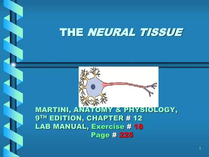
THE NEURAL TISSUE MARTINI, ANATOMY & PHYSIOLOGY, 9 TH EDITION, CHAPTER # 12 LAB MANUAL, Exercise # 18 Page # 225. NOTE:. THIS IS A STUDY GUIDE , NOT AN ALL INCLUSIVE REVIEW. THERE MIGHT BE THINGS NOT COVERED BY THIS STUDY GUIDE THAT MIGHT BE ASKED IN YOUR PRACTICUMS /

E N D
THE NEURAL TISSUEMARTINI, ANATOMY & PHYSIOLOGY,9TH EDITION, CHAPTER # 12LAB MANUAL, Exercise#18Page# 225
NOTE: • THIS IS A STUDY GUIDE, NOT AN ALL INCLUSIVE REVIEW. • THERE MIGHT BE THINGS NOT COVERED BY THIS STUDY GUIDE THAT MIGHT BEASKED IN YOUR PRACTICUMS / QUIZZES. • STUDENTS ARE RESPONSIBLE FOR READING THEIR TEXBOOK (S) AND FOR ALL THE MATERIAL COVERED DURING THE LABORATORY PERIOD, AS PER THE COURSE SYLLABUS
Competency 6: The Nervous System • Upon successful completion of this laboratory, the students will be able to demonstrate an understanding of the structural and functional features of the nervous system including the special sense organs by: • Identifying the parts of a neuron. • Explaining the structural and functional classification of the neurons.
Competency 6: The Nervous System • Describing the structure and functions of the supporting cells of the nervous system. • Distinguishing between neuron, nerve and track. • Explaining the structure and functions of the central nervous system and the cranial nerves.
NEURAL TISSUE NEURON STRUCTURE CLASSIFICATION OF NEURONS BY STRUCTURE CLASSIFICATION OF NEURONS BY FUNCTION SCHWANN CELLS GREY MATTER WHITE MATTER
NEURON STRUCTURE • CELL BODY (SOMA) • NEUROFIBRILS(Black or brown lines in model) • F- intracellular transport • NISSIL BODIES- Blue in one model & blue with red dots in another model • F- protein synthesis • DENDRITES- They are short citoplasmatic • projections from cell bodies • F- to receive the nerve impulse or signal • AXON- It is a citoplasmaticprojection from the • cell body • F-to carry away the nerve impulse or signal
AXON HILLOCK- The place where the • the neurofibrils converge • F- trigger zone for nerve impulse • AXON TERMINALS- End part of the neuron • that connects with another neuron • F- to release the neurotransmiter
Dendrites Perikaryon Cell body Dendritic spines Dendrite Nucleus Nissl bodies (RER and free ribosomes) Axon Telodendria Mitochondrion (a) Regions of a neuron Axon hillock Initial segment of axon Axolemma Axon Telodendria Golgi apparatus Neurofilament Synaptic terminals See Figure 12-2 Nucleolus Nucleus PRESYNAPTIC CELL POSTSYNAPTIC CELL (b) Structural components of a neuron
NEURONS BY STRUCTURE • ANAXONIC • BIPOLAR • UNIPOLAR • MULTIPOLAR
Bipolar neuron Multipolar neuron Anaxonic neuron Unipolar neuron Dendrites Dendrites Initial segment Dendrites Axon Axon Axon Synaptic terminals Axon Synaptic terminals Synaptic terminals Figure 12-3: A Structural Classification of Neurons
SCHWANN CELLS- They surround the axon • f- to produce the myelin sheath • MYELIN SHEATH- Layers of cell membrane that • surround the axon (yellow in the models • f- to isolate the axon • Neurilemma- outermost layer with nucleus of the Schwann cell • NODES OF Ranvier- area between two Schwann cells • f- to produce saltatory conduction • (jumping conduction), to increase the • speed of nerve impulse transmission
NEURONS BY FUNCTIONS • SENSORY (Afferent)- it carries iformation • towards the cns (central nervous system) • MOTOR (EFFERENT)-It carries information • away from cns • ASSOCIATION NEURONES OR INTERNEURONS- • They carry impulses between • sensory & motor neurons located at cns
Anatomy of a Spinal Nerve ALFONSO A. PINO. MD.
GREY MATTER- Non myelinated parts of the • Neurons & glia (cell bodies) • It forms nuclei in the cns • WHITE MATTER- Myelinated fibers • It forms tracs in the cns • It forms nerves in pns (peripheral • nerve system)
GRAY MATTER & WHITE MATTER (CNS) ALFONSO A. PINO. MD.
WHITEMATTER (CNS) ALFONSO A. PINO. MD.
REMEMBER!GO TO THE TUTORING ROOM AND PRACTICE WITH MODELS.ROOM 3326.