Download
1 / 75
1.15k likes | 2.94k Views
Glomerulonephritis in Children. Dr. Hala Wannous pediatric nephrologist damascus university faculty of medicine children ’ s hospital. 1. Glomerulonephritis. Glomeulonephritis is an immune-mediated renal disease induced by two mechanisms: 1- Antibody-mediated immunity:

E N D
Glomerulonephritis in Children • Dr. HalaWannous • pediatric nephrologist • damascus university • faculty of medicine • children’s hospital 1
Glomerulonephritis Glomeulonephritis is an immune-mediated renal disease induced by two mechanisms: 1- Antibody-mediated immunity: • In situ immune complexes:Antibodies (autoantibodies) that bind to planted (exogenous) antigens, or interact with intrinsic glomerular cell surface antigens, as a result of changes to the cell surface ( anti-GBM antibody nephritis, membranous glomerulonephritis). • Circulating immune complex:Pre-formed immune complexes from the circulation that trapped by glomerulous. Regardless of whether they are endogenous (lupus nephritis), or exogenous (acute post streptococcal GN), the antigens are not glomerular in origin. 2
Glomerulonephritis 2- Cell-mediated immunity : Caused by sensitized T-cells, cytotoxic action of T cells which may damage the intrinsic glomerular cells or alter glomerular filtration barrier (e. In pauci-immune glomerular diseases, such as ANCA-related crescentic glomerulonephritis). Then, Activation of classical or alternative complement pathway, followed by secretionof secondary inflammatory mediators, including cytokines and chemical mediators, derived from activated leukocytes. Following the initiation of immune-mediated glomerular injury, various cytokines, chemokines, and growth factors are significantly involved in the promotion of glomerular injury as second messengers. 3
Terminology • Glomerulonephritis : inflammation of the glomeruli • Glomerulopathy : disease of the glomeruli • All glomeruli are involved: Diffuse (generalized) • Some glomeruli are involved: Focal In one glomerulus: • if the whole glomerulus is involved : Global • if only a part of the glomerulus is involved: Segmental Other terminologies in common use: • Proliferative: increase in the number of cells • Sclerosing: Hardening of the tissue
Glomerular lesion The lesion Consists of: • Infilteration of leucocytes. • Proliferation of endothelial, mesangial and epithelial cell. • Formation of deposits. • Also immunoglobulins and complements form deposits (granular deposits). The formed deposits lie at three sites: • In the mesangium (mesangial deposits ). • Between the endothelial cells and glomerular basement membrane (GBM ) (subendothelial deposits ). • Between the outside of GBM and podocytes (subepithelial deposits).
Glomerular injury • Glomerulonephritis arises from the responses of intrinsic glomerular cells to inflammatory reactions. Glomerular deposition, hypercellularity (intrinsic and inflammatory cells), and capillary destruction are typical features of glomerular injury. • Extensive inflammatory damage to glomeruli → Impairment of selective filtering properties of the kidney leading to a decreased GFR and eventually produce uremic symptoms with salt and water retention, leading to edema and hypertension. • Molecules normally not filtered such as constituents of the blood, pass into the urine and are excreted. 8
Light micrograph of a normal glomerulus. There are only 1 or 2 cells per capillary tuft, the capillary lumens are open, the thickness of the glomerular capillary wall (long arrow) is similar to that of the tubular basement membranes (short arrow), and the mesangial cells and mesangial matrix are located in the central or stalk regions of the tuft (arrows). Courtesy of Helmut G Rennke. 14
Electron microscopy • Electron micrograph of a normal glomerular capillary loop showing the fenestrated endothelial cell (Endo), the glomerular basement membrane (GBM), and the epithelial cells with its interdigitating foot processes (arrow). The GBM is thin and no electron dense deposits are present. Two normal platelets are seen in the capillary lumen. Courtesy of Helmut Rennke, MD. 15
Possible Clinical Manifestations: • Proteinuria: asymptomatic • Haematuria: asymptomatic (especially dysmorphic red cells, red cell casts) • Hypertension • Nephrotic syndrome • Nephritic syndrome • Acute renal failure • Rapidly progressive renal failure • End stage renal failure 16
Acute Nephritic Syndrome: Syndrome characterised in typical cases by: • haematuria • oliguria • oedema • hypertension • reduced GFR • proteinuria • fluid overload • Phase contrast microscopy showing dysmorphic red cells in a patient with glomerular bleeding. Acanthocytes can be recognized as ring forms with vesicle-shaped protrusions (arrows). Courtesy of Hans Köhler, MD. 19
Clinical Features of the Acute Nephritic Syndrome: • haematuria is usually macroscopic with smoky brownurine (like coca cola) • oliguria may be overlooked or absent in milder cases • oedema is usually mild and is often just peri-orbital, weight gain may be detected • hypertension common and associated with raised urea and creatinine • proteinuria is variable but usually less than in the nephrotic syndrome 20
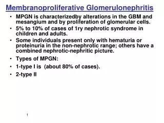
Etiology of the Nephritic Syndrome • Most common cause is Acute PoststreptococcalGlomerulonephritis • Subacute Bacterial Endocarditis • Lupus Nephritis (SLE) • Anti-glomerular Basement Membrane Disease • IgA Nephropathy • ANCA-related glomerulonephritis(Wegener's granulomatosis) • Henoch-Schonleinpurpura • MembranoproliferativeGlomerulonephritis • Crescentic glomerulonephritis 21
Management issues in the nephritic syndrome • Appropriate investigations: skin and throat swabs,strep serology, complement, urea, creatinine, electrolytes, urinalysis and CXR • BP, urine output and daily weight • Fluid and diet management • Treat hypertension and fluid overload • Treat infection 22
Complications of the Nephritic Syndrome • Hypertensive encephalopathy (seizures, coma) • Heart Failure (pulmonary oedema) • Uraemia requiring dialysis 23
Prognosis in the Nephritic Syndrome • More than 95% of children make a complete recovery. • Chronic renal impairment in the longer term is uncommon in children. • Bad prognostic features include severe renal impairment at presentation and continuing heavy proteinuria and hypertension. • Adults more likely to have long term sequelae than children. 24
Acute Post-Streptococcal Glomerulonephritis(APSGN) Post-streptococcal glomerulonephritis is an immune- mediated disease involving: - Streptococcal antigens (endostreptosin) - Circulating (and in situ) immune complexes - Activation of complement,and glomerular infiltration of lymphocytes and macrophages in association with cell-mediated immunity . Poststreptococcal glomerulonephritis is prototypical for acute endocapillary proliferative glomerulonephritis. 25
APSGN: Pathogenesis It is caused by an exogenous antigen (endostreptosin) following infection of the pharynx or skin with strain of group A β-hemolytic streptococcus. Specific M proteins in group A streptococci were initially considered “nephritogenic,” Host susceptibility factors (HLA-DR) 26
APSGN: Pathogenesis • Studies in epidemics revealed that APSGN was associated with pyodermitis due to Group A streptococci of M types 47, 49, 55, 57 and 60, and with upper respiratory infections due to streptococci of M types 1, 2, 4, 12, 18 and 25. • Immune complexes formed against nephritogenic streptococcal antigen(s) may be formed in circulation, and deposited in the glomeruli, or formed in situ against antigenic fractions planted in the glomeruli. 27
APSGN: Clinical Characteristics • Children from 4 to 14 years are more frequently • affected by APSGN, It is rare below the age of 2 • and above the age of 20. • Twice more frequent in males than in females. • The latent period between infection and nephritis • is longer after skin infections (3–5 weeks) than • after upper respiratory infections (1–2 weeks). 28
APSGN: Clinical Characteristics • The classic presentation is an acute nephritic syndrome with hematuria (usually gross and the urine looks smoky brown), pyuria, red blood cell casts, edema, hypertension, and oliguric renal failure, which may, exceptionally, be severe enough to appear as RPGN (crescentic GN). and more rarely with heavy proteinuria (nephrotic). • Systemic symptoms ofslight fever, headache, nausea, malaise, anorexia, and flank pain (due to swelling of the renal capsule) are reported in as many as 50% of cases. • Subclinical disease is characterized by a reduction of serum complement, microscopic hematuria and normal or increased blood pressure in asymptomatic patients. 29
APSGN: Serological Findings • The most constant serological finding is the reduction in serum complement levels that occurs in more than 90% of the cases. The activation of the complement system is usually via the alternative complement pathway and reduced C3 with normal C1 and C4. • IgG and IgM serum levels are elevated in 80–90% of the patients with APSGN. • Rising antistreptococcal antibody titers are the usual clinical indication of a preceding streptococcal infection since positive cultures are obtained in only 20–25% of the cases. 30
APSGN: Serological Findings • Antistreptolysin O titers and anti-DNAse B titers are the most frequently elevated antibody titers after streptococcal throat infections and after streptococcal skin infection, respectively. • Throat cultures are usually negative by the time patient presents with GN, but ASLO, anti-DNAse B should be high along with low complements. 31
APSGN: Pathology Renal biopsy is usually not done in patients with APSGN. If the clinical presentation is atypical or resolution of the disease delayed, then renal biopsy is mandatory: 1- The serum complement is normal 2- The serum complement remains low after 8 weeks 3- Proteinuria in the nephrotic range 4- Depressed GFR > 3 – 4 weeks 5- Gross hematuria > 2 - 3 weeks. 6-Persistent proteinuria > 3 - 6 months 7- Hypertension > 2 – 3 weeks 8- Severe anuria and rapid progressive course 32
APSGN: Pathology • Biopsy findings in APSGN are those of endocapillary proliferative glomerulonephritis. glomeuli enlarged due to swelling and hypercellularity ( mesangial and endothelial cell proliferation). Glomerular basement membrane is normal. Macrophages and lymphocytes can be found in increased numbers in the glomeruli and in tubulointerstitial areas. • the more remarkable histologic characteristics are found in the glomeruli that present a diffuse increment in cellularity. • The immunofluorescent appearance: Glomerular granularsubendothelial immune deposits C3, IgG and IgM are usually present and they may be demonstrated in the mesangium and in the glomerular basement membrane. 33
APSGN: Pathology The hallmark of APSGN is the electron microscopy findings of large subepithelial deposits ( humps ), believed to be immune complexes formed in situ against antigenic fractions planted in the glomeruli. Follow-up studies performed several years after the initial episode of acute poststreptococcal glomerulonephritis,have revealed immune deposits and a variable degree of mesangial sclerosis and obliteration, even in the absence of clinical manifestations of renal disease. 34
APSGN: Diagnosis • The clinical presentation is that of a child of 4–15 years who suddenly develops dark and scanty urine and swelling of the face and legs. • History and physical examination determines a high blood pressure and the absence of a systemic illness. • A history of a precedent upper respiratory infection and/or skin infections suggests a post-streptococcal etiology that maybe confirmed by a positive culture or rising anti- streptococcal antibody titers. • The serum complement C3 is low. 37
APSGN: Differential Diagnosis The complement is a first line diagnostic test that permits consideration of acute glomerulonephritis that present low (C3): • APSGN • Lupus nephritis • shunt nephritis • endocarditis • cryoglobulinemia • membranoproliferative glomerulonephritis The reduction of C1, C4 and C3 levels, indicating activation of the classic pathway of complement, is characteristic of lupus erythematosus. 38
APSGN: Treatment Antibiotic treatment for streptococcus:Even without infection (penicillin or, in allergic individuals, erythromycin) should be given to all patients and their cohabitants. Antibiotic treatment after the onset of APSGN does not alter the course of the disease. Antibiotic prophylaxis given to family members may reduce the risk of spread of APSGN. Treatment of the Acute Nephritic Syndrome: Supportive • Restrictions of fluid and sodium intake are the cornerstones of the treatment. • Cases that present significant edema, hypertension and circulatory congestion benefit from the administration of loop diuretics. 39
APSGN: Treatment • Antihypertensive treatment in cases of severe hypertension: Nifedipine, Parenteral hydralazine may be required with close observation.Angiotensin converting enzyme inhibitors carry the risk of hyperkalemia. • Hemodialysis or peritoneal dialysis may be required occasionally in children for the treatment of hyperkalemia, uremia or severe circulatory congestion. • Steroids are not generally indicated but are used in patients who have renal failure or rapidly progressive GN, which is usually associated with crescentic APSGN. There are reports of beneficial effects of pulse methyprednisolone therapy. 40
APSGN: Prognosis Majority of children with APSGN are believed to make a full recovery without significant long-term consequences. Recurrences of APSGN are rare. Progression to chronic kidney disease (CKD) and end-stage renal disease (ESRD) may occur in exceptionally severe cases of APSGN. Complete resolution of the hematuria and proteinuria in children occurs within 3–6 weeks of the onset of nephritis. 41
APSGN: Follow-up Follow-up of APSGN is important to document full recovery. • Proteinuria usually resolves by 6–8 weeks. Prolonged proteinuria for more than 3 months may indicate irreversible renal injury. • Gross hematuria usually improves within 1–3 weeks. • Microscopic hematuria can persist for up to 1 year and is not an indicator of poor prognosis. • Recovery of serum complement C3 to normal usually takes 6–8 weeks. Persistent hypocomplementemia beyond 8 weeks requires a careful search for other diagnoses, such as MPGN and SLE nephritis. 42
Other acute post-infectious GN Acute GN follows other infections: • Bacterial sepsis • Acute or subacute bacterial endocarditis • Visceral abscess • Infected ventriculoperitoneal shunt • Osteomyelitis Treatment is aimed at eradicating the primary disease. 43
IgA Nephropathy (Berger’s Disease) IgA nephropathy (IgA N) is the most frequent type of chronic glomerulonephritis in children. Pathogenesis: IgA N is generally considered to be an immune complex- mediated or aggregated IgA - mediated glomerulonephritis. Genetic factors have been suggested as important in the development of IgA Nephropathy. Because of the frequent association between upper respiratory tract or gastrointestinal infection and the onset of macroscopic hematuria, It has been suggested that certain viral or bacterial infections may lead to IgA nephropathy. 44
IgA Nephropathy: Pathogenesis Many antigens, including herpes simplex virus, cytomegalovirus, Epstein-Barr virus, adenovirus and milk antigen, have been Identified. Circulating IgA immune complexes have been detected,often associated with IgG immune complex. Serum levels of IgA are increased in 50–70% of patients with IgA nephropathy, IgA production is T cell-dependent, and the increased production in IgA nephropathy may indicate altered T cell function. 45
IgA Nephropathy: Pathology Light microscopic findings: are quite variable, often mild to moderate mesangial cell proliferation. The most characteristic abnormality is mesangial enlargement, caused by various combinations of hypercellularity and increase in matrix. Progression of IgA nephropathy leads to gradual resolution of mesangial hypercellularity and an increase of matrix associated with The development of sclerosis. 47
IgA Nephropathy: Pathology Immunofluorescence (IF): - The diagnostic immunopathological pattern of IgAN is the presence of IgA in the glomerular mesangium as the sole or predominant Ig (IgA1 deposits). - There are also deposits of IgG and/or IgM with the same staining pattern as IgA but with lesser intensity and frequency. - C3 deposits were observed in a similar distribution pattern in 64% of cases. 48
IgANephropathy: Clinical Features IgA N occurs at all ages but is most common during the second and third decades of life. Male > Females 2 to 1 The clinical presentation varies: • Some patients have asymptomatic persistent microscopic hematuria with or without proteinuria. • Another classic presentation is recurrent episodes of macroscopic haematuria typically occur during a URI (less frequently, in association with other infections) and clearing after a period of 2–4 days. The interval between the precipitating infection and the appearance of hematuria ranges from 1 to 2 days compared with 1 or 2 weeks in acute postinfectious glomerulonephritis. 49
IgANephropathy: Clinical Features • The blood pressure and renal function at onset are normal. • Hypertension is infrequent and usually mild to moderate. • Some patients present with acute nephritic syndrome or nephrotic syndrome. • Renal function is usually normal but occasionally a patient will present with acute renal failure due to acute tubular necrosis secondary to the gross haematuria. • Less than 5% have chronic renal insufficiency (CRI) at diagnosis • Some patients develop extensive crescents and arapidly progressive course. 50
IgA Nephropathy: Diagnosis • There is no specific serologic marker for IgA Nephropathy. • Diagnosis depends on IF examination: The presence of IgA as the sole or predominant Ig in the glomerular mesangium. • Serum concentrations of C3 and C4 are normal. • Although serum IgA level is usually above the mean and often significantly elevated, the sensitivity and specificity are too low for its use as a diagnostic test. 51
IgA Nephropathy: Management • There is no clear guidelines for the treatment of pediatric IgA N. • Aggressive control of blood pressure and proteinuria with ACEI’s or AR2B’s. • Combination therapy with ACEI and AR2B has an additive dose-dependent antiproteinuric effect compared to monotherapies. • Corticosteroid therapy is indicated for pediatric patients with IgA N who have the nephrotic syndrome. • Rapidly progressive or severe crescentic IgA N are usually treated with high-dose intravenous methylprednisolone pulses followed by oral corticosteroids and sometimes other immunosuppressive agents such as cyclophosphamide. 52
IgA Nephropathy: Prognosis • The outcome for pediatric patients with IgA N ranges from complete resolution of all clinical signs of disease to ESRD. • Slowly progressive. • By 20 years, 50% have end stage kidney disease. • Clinical markers at diagnosis that associate with worse prognosis and progression to CRI and/or ESRD are: 1- proteinuria >1g/day 2- severe histologic features such as the presence of crescents and/or segmental sclerosis, glomerular fibrosis. 3- hypertension. 4- increased creatinine. 53