Download
1 / 1
10 likes | 98 Views
A hybrid approach to the Skull Stripping problem in MRI F.Ségonne 1,2 A.M.Dale 2 E.Busa 2 M.Glessner 2 D.Salat 2 H.K.Hahn 3 B.Fischl 2 1 MIT-AI Lab, 2 MGH-NMR Center, 3 MeVis Bremen. INTRODUCTION

E N D
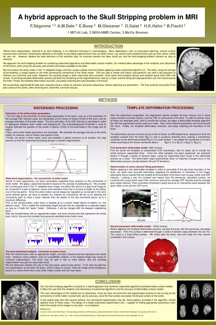
A hybrid approach to the Skull Stripping problem in MRI F.Ségonne1,2A.M.Dale2E.Busa2M.Glessner2D.Salat2H.K.Hahn3 B.Fischl2 1 MIT-AI Lab, 2 MGH-NMR Center, 3 MeVis Bremen INTRODUCTION Whole brain segmentation, referred to as skull stripping, is an important technique in neuroimaging. Many applications, such as presurgical planning, cortical surface reconstruction and brain morphometry, depend on the ability to accurately segment brain from non-brain tissue, e.g. remove extra-cerebral tissue such as skull, sclera, orbital fat, skin, etc. However, despite the clear definition of this essential step, no universal solution has been found yet, and the skull-stripping problem remains an area of research. We approach the skull-stripping problem by combining watershed algorithms and deformable surface models. Our method takes advantage of the simplicity and robustness of the former, while using the accuracy and surface information available to the latter. We first localize the white matter in the T1-weighted image, and then create a global minimum before applying a basic watershed algorithm [1]. The latter, using the concept of pre-flooding, is simply based on the three dimensional connectivity of the white matter. This first step is simple and robust, and performs very well in the presence of intensity non-uniformity and noise. However, the resulting image is often inaccurate and unsmooth, since some extra-cerebral tissues and cerebral spinal fluid (CSF) still remain. A shrinking template deformation process [2] does the smoothing of the segmented brain used as a mask, and the fitting last-step parameters are then found around the limits. Finally, the template deformation resumes, accurately matching the outer boundary of the brain. We successfully segmented 60 data sets, acquired using a variety of scanners and pulse sequences, without adjusting any parameters. The final surfaces accurately fit the pial surface of the cortex, often removing thin, sheet-like, non-brain tissues. METHODS • WATERSHED PROCESSING • Extraction of the white matter parameters: • The first step is the extraction of some basic parameters of the brain, such as a first estimation of the average CSF intensity value, the coordinates of the Center of Gravity (COG) of the brain, and an average estimation of the brain radius. The first estimation of the CSF intensity is calculated in order to distinguish between brain matter and background. The brain/background threshold is used to roughly estimate the position of the center of gravity (COG) of the brain/head in the image and its radius. • Next, some white matter parameters are estimated. We calculate the average intensity of the white matter, its variance, and its inferior and superior limits. • Finally, we locate a white matter voxel and establish a global minimum at its location; this step assures us that the watershed processing will give a relevant segmentation. • Watershed segmentation: the connectivity of white matter • Similar to other approaches, our basic assumption regarding brain anatomy is the connectivity of white matter. We apply a watershed transform based on the concept of preflooding [1]. We consider the inverted gray level in the T1-weighted brain image: we define two points of a gray level image to be connectedif a path of adjacent voxels exists between them that is at least as bright as the darker one of the two points. Since the white matter cannot always be regarded as connected in the strict sense defined above, we have to weaken our criterion for connectivity. We do so by allowing the connectivity path to show a lower intensity than the darker of the two connected points up to a maximum difference. • Prior to the transformation, each basin is flooded up to a certain height above its bottom (i.e. the darkest voxel). This is called the preflooding heightand will only be regarded as a separate region as long as it holds the “water” inside. Otherwise, it will be merged with the deepest neighboring basin. • After the transformation with an appropriate height, one basin should exist that represents the whole brain, that is, the one that includes the previously identified white matter voxel. • The post-watershed analysis: correction of some errors • After the transformation with an appropriate height, one basin should exist that represents the whole brain. However, some artifacts, such as susceptibility artifacts, in the original image may cause an irrelevant segmentation. The brain may be split in two or more basins, and the resulting segmentation may give an inaccurate result. • We first determine whether the size of the main basin seems to be correct. If not, then we add this first segmentation to the latter the basin, which is likely to correct. Then we merge all the ambiguous basins (i.e. those which have many white matter voxels) with the main basin. TEMPLATE DEFORMATION PROCESSING After the watershed computation, the segmented volume contains all brain tissues, but in some cases contains non-brain tissues, such as CSF, or some parts of the skull. In order to remove many of these non desirable parts, and to smooth the surface, we apply a deformable template algorithm [2], with the segmented volume used as a mask. Next, some relevant parameters are found around the limits. Finally, the template deformation resumes, accurately matching the boundary of the brain. The deformation process is driven by two kinds of forces:an MRI-based force, designed to drive the template outward from the brain FM( t ), and a curvature reducing force, placing a smoothness constraint on the deformed template FS( t ). On each iteration t, we update the coordinate xk of each vertex according to the forces mentioned above :Xk( t + 1 ) = Xk ( t ) + FS( t ) + FM ( t ). First parametric deformable model:2562 vertices First we center the template at recalculated COG coordinates, with its radius set to include the whole previous segmented brain. Once the initial template has been positioned, we gradually deform it through a series of iterative steps, using the segmented brain found in the watershed process as a mask. The deformation stops automatically when no important changes occur in the deformation process (usually between 50 and 70 iterations). Determination of some relevant fitting parameters: Before applying a last deformable template in order to accurately fit the pial outer boundary of the brain, we need more accurate information regarding the distribution of intensities in the image, particularly tissue classes that are located at the boundary of the brain such as gray matter and CSF intensity. Looking a few mm outward and inward from the previously calculated surface, we construct two curves which reach their maximum around the average CSF intensity and the average gray matter intensity. Then a transition threshold is easily found as an intersection point between the gray matter curve and the CSF curve. Second parametric deformable model:10242 vertices Before applying the template deformation process, we peel the brain with the previously calculatedtesselation. Then this surface is deformed through a series of iterative steps (between 50 and 70). The result is a brain-fitting surface. We finally peel the brain, and only keep the final volume contained in the surface. CONCLUSION Our new skull-stripping algorithm is based on a hybrid approach that combines watershed algorithms and deformable surface models. It offers the user both the simplicity and robustness of watershed algorithms and the accuracy of deformable surface models. The main advantages of this method are its robustness, since we have not found this fast segmentationprocess to fail as long as the connectivity of white matter is preserved; and its accuracy, since the final surface accurately fits the pial surface of the cortex. In low quality data sets with several artifacts, this normalized segmentation may fail. Some options, provided in the algorithm, should address most of these cases. The design of a totally automated segmentation tool – capable of finding appropriate parameters even when the quality of the images is very poor – is in progress. References [1] Horst K. Hahn, Heinz-Otto Peitgen: The Skull Stripping Problem in MRI Solved by a Single 3D Watershed Transform. MICCAI 2000, Pittsburgh, Pennsylvania, USA 134-143. [2] Dale, A.M., Fischl, Bruce, Sereno, M.I., (1999). Cortical Surface-Based Analysis I: Segmentation and Surface Reconstruction. NeuroImage 9(2):179-194.