Download
1 / 111
1.44k likes | 2.08k Views
Urology. Hematuria Stones Tumours. Outline. Hematuria DDx General Work up Renal Colic Stones Malignancy Renal Bladder Scrotal masses. Hematuria. Objectives 1. Taking a Hx . 2. Lab & Radiologic Invx’s . 3. Which pt’s to refer to Urologist. Hematuria.

E N D
Urology Hematuria Stones Tumours
Outline • Hematuria • DDx • General Work up • Renal Colic • Stones • Malignancy • Renal • Bladder • Scrotal masses
Hematuria • Objectives • 1. Taking a Hx. • 2. Lab & Radiologic Invx’s. • 3. Which pt’s to refer to Urologist.
Hematuria General Approach • Stones • Infection • Trauma • Tumors • GU Endometriosis • Glomerulonephritities • AV Fistulas • Vascular Malformations • Infection • Tumor • Myo/hemoglobinuria • Coagulation disorders • Pseudohematuria • (beets, dyes, laxatives)
Hematuria • Etiology by Age
Hematuria General Approach • Stones • Infection • Trauma • Tumors • GU Endometriosis • Glomerulonephritities • AV Fistulas • Vascular Malformations • Infection • Tumor • Myo/hemoglobinuria • Coagulation disorders • Pseudohematuria • (beets, dyes, laxatives)
HematuriaDDx • Stones • Infections • Tumours • Trauma
Hematuria HPI • Stones: • Flank/Abdo pain, dysuria, PHx Stones. • Infection: • Suprapubic pain, dysuria, frequency, fever/chills +/- flank pain. • Malignancy • Wt loss, night sweats, flank pain, voiding changes, Occupational Hx (petroleum exposure), smoking Hx, FMHx of Cancer • Trauma • Recent encounters with Chuck Norris.
Gross HematuriaInvx • Laboratory Work up: • 1. CBC • Hgb - severity of blood loss. • WBC – infection. • Platelet loss/coagulopathy. • 2. Cr • Renal impairment. • 3. INR/PTT • Coagulopathy. • 4. U/A • Leukocytes, Nitrites – Infection. • R&M – if dysmorphicRBC’s +/- Protein = Glomerular cause, crystals stones. • C&S – Infection.
HematuriaInvx Radiology Investigations • Painless Gross Hematuria • Triphasic CT: arterial/venous/ureteric phases • Microscopic Hematuria • Start with Renal U/S. • Flank Pain • Plain film KUB, CT KUB (non con). • Signs of infection • Start with U/S, if findings may consider CT with contrast
Hematuria Referral • When to refer to Urologist? • Gross hematuria NEED cystoscopy +/- Retrograde Pyelogram!! • Pt’s with GU Malignancies, stones, trauma. • What should be done prior to referral? • Hx, PE, Lab Invx’s, Imaging • Initial management and stabilization of pt.
Hematuria Acute Rx • ABC’s. • Stabilize Pt, Blood products if needed. • Invx to determine cause • Treat underlying cause. • Continuous Bladder Irrigation • Call Urology. • Manually irrigate all clots out of bladder first! • Surgical management • Cystoscopy + Fulgaration. • Hyperbaric Oxygen • IR embolization. • Cystectomy and Urinary diversion.
Hematuria Summary 1. Painless Gross Hematuria • Malignancy until proven otherwise. 2. Stones, infections & trauma • Rarely asymptomatic Hx.. Hx.. Hx.. 3. Workup • Hx, PE • Lab: CBC, Cr, U/A C&S, INR/PTT • Imaging: CT or U/S • Referral to Urologist: cystoscopy +/- Retrograde pyelogram 4. Management • Stabilize Pt, +/- CBI, +/- Surgical intervention
Hematuria Cases • GeeyuMalignansey, a 67 yo female with 114 pk/yr smoking hx presents with Gross Hematuria. • Wazun Mi, 23 yo male minding his own business gets stabbed to the flank, while voiding and notices he urine becomes red… • 24 yo Engineering student comes in with dysuria after holding her urine for 14 hours playing ‘Call of duty’, she has leuks, nitrites and RBC’s on U/A…
Outline • Hematuria • DDx • General Work up • Renal Colic • Stones • Malignancy • Renal • Bladder • Scrotal masses
Renal Colic Objectives; • Give a differential diagnosis for acute flank pain including two life-threatening conditions • Describe the laboratory and radiologic evaluation of a patient with renal colic • Know 4 different kinds of kidney stones and the risk factors for stone formation • Know 3 indications for emergency drainage of an obstructed kidney
Renal Colic DDx • Gyne • Pelvic inflammatory Disease • Ovarian Torsion/Rupture • Endometriosis • GU • Renal/Ureteric Calculi • Renal Abscess • Pyelonephritis • Renal Vein Thrombosis • Acute Glomerulonephritis • Other • Acute lumber disc herniation • Herpes Zoster • Fitz-Hugh-Curtis Syndrome • Life Threatening: • Abdominal Aortic Dissection • Abdominal Aortic Aneurysm Rupture • Appendicits • Ectopic Pregnancy • Septic Stone • GI • Cholecystitis • Biliary Colic • Acute Pancreatitis • Diverticulitis • Duodenal Ulcer • Inflammatory Bowel Disease • Viral gastritis • Splenic Infarct
Renal Colic Invx • Rocky, a 32 yo Male comes to ED with microscopic hematuria and is writhing with Lt Flank pain. • What Laboratory Invx’s do you order? • What initial imaging do you order?
Stones – Acute Lab Invx’s • CBC • WBC – increased indicates inflammation or infection. • Creatinine • Assess for impaired renal function (obstruction). • Urine Microscopy • Bacteriuria, pyuria, pH
Renal Colic – 1st Imaging Test • Plain Film KUB! • ~85% of stones are Radio-opaque on plain film. • No info on degree of obstruction though.
Renal Colic – Other imaging options IVP • Intravenous Pyelogram • Visualizes most stones (radiolucent stones will appear as filling defects) • Excellent Functional Study • Requires IV contrast, thus risk of allergic reactions and nephrotoxicity
Renal Colic – Radiologic Evaluation • CT Scan, hold the contrast. • Aka CT-KUB. • Fast Inexpensive • Imaging choice in most emergency rooms. • Degree of obstruction inferred by presence of hydronephrosis.
Stones - Factoids • They are common! • Lifetime risk in North American Male is 1 in 8. • M:F ratio is 3:1 • Presenting complaint • Renal colic due to acute obstruction of ureter by stone. • Initial Evaluation • Focuses on excluding other potential causes of abdominal or flank pain. • Non-obstructing stones • Should not cause pain unless they are associated with Urinary tract infection.
Ureteric Stone • 3 Common sites of Obstruction
Ureteric Stones • Spontaneous passage? • Pharmacologic aid in spontaneous passage? • Alpha blockers! Flomax
Renal and Ureteric Stones • So you have established that there is a stone. • When is ‘immediate’ referral to a Urologist Necessary?
Immediate Referral to Urology • Obstructed ureter + Fevers/chills, bacteriuria or elevated WBC • Risk of Urosepsis - emergency • Obstructed Ureter + Insulin dependent DM • Risk of papillary necrosis or emphysematous pyelonephritis • Solitary Kidney • Significant co-morbid conditions • Eg. CHF, pregnancy etc.
Calcium Oxalate • Most common type. • Risk Factors: • Dietary Hyperoxaluria: chocolate, nuts, tea, strawberries, peanut butter, cabbage or excessive restriction of dietary calcium. • Hypercalciuria • Inherited increased absorption, or incr PTH • Dietary Hypercalciuria
Calcium Phosphate • Second most common stone type. • Often seen in pt’s with Metabolic Abnormalities: • Primary Hyperparathyroidism. • Distal Renal tubular acidosis. • Hypercalcemia due to Malignancy or Sarcoid.
Uric Acid • Radiolucent on Plain X-Rays, but is visualized on CT scan • Risk Factors: • Persistent Acidic urine: iel • Low urine volumes • Chronic diarrhea • Excessive sweating • Inadequate fluid intake • Gout (Hyperuricemia) • Excess dietary purine (Meataholics) • Chemotherapy for lymphoma, leukemia
Struvite (Infection Stones) • Composed of MAP • Magnesium + Ammonium Phosphate & Calcium • Can only form if urine pH >8.0! • Thus: usually only in presence of urease +ve bacteria • Proteus, Klebsiella, Providentia, Pseudomonas, Staph Aureus • Note: E Coli does NOT produce urease • Tend to form Staghorn stones
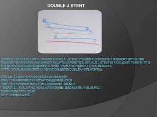
Ureteric Stents • “Double J Stents” • Stay in place b/c of curled ends • Can place these Antegrade or Retrograde • Typically requires General Anesthetic. • Low risk of bleeding.
PercutaneousNephrostomy Tubes • “Neph Tubes” • Placed under local anesthetic by Interventional Radiology • Increased Risk of Bleeding.
Treating/Removing Stones • Ways to Treat stones. • Conservative passage + Alpha Blocker (Flomax) + Hydration + NSAID (if Normal GFR) • Extracorporeal Shockwave Lithotripsy (ESWL) • Ureteroscopy + Basket or Laser • PercutaneousNephrolithotomy
Treating Stones • Conservative passage + Alpha Blocker (Flomax) + Hydration + NSAID (if Normal GFR) • Indications • Pain can be controlled with Ketorolac + Narcotic • No renal impairment • No Intractable Vomiting (aka pt not hypovolemic) • No sign of infection. • No previous failed trials of conservative passage.
Treating Stones • Extracorporeal Shockwave lithotripsy • Indication: • <~1.5cm renal or ureteric stone. • Stone is localized by X-Ray. • ~3000 Shocks targeted to gradually fragment stone. • Fragments passed in urine.
Treating Stones • Ureteroscopy • + Basket • If stone is small enough to adequately remove by basket. • + Holmium Laser • If stone is ‘impacted’ or cannot simply be basketed out.
Treating Stones • PercutaneousNephrolithotomy • Indications • Large Proximal ureteric or Renal Calculi >~1-1.5cm • Treatment of Staghorn Calculi • Risks: • Bleeding • Renal Perforation or Avulsion