Download
1 / 19
230 likes | 565 Views
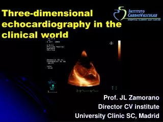
3 Dimensional TEE Echocardiography. Roberto M Lang, MD. University of Chicago. An internist heard a murmur …. 47 year old physics professor Bicycles over 100 miles a week Noted a mild decrease in exercise tolerance.

E N D
3 Dimensional TEE Echocardiography Roberto M Lang, MD University of Chicago
An internist heard a murmur … • 47 year old physics professor • Bicycles over 100 miles a week • Noted a mild decrease in exercise tolerance
When we look at biplane long axis views, we see that the patient has a flail P2 segment with jet of mitral regurgitation which is moving and which is going in an inferior direction. Qualitatively the severity of the mitral regurgitation is severe.
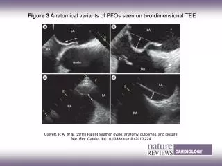
These are zoomed views of the mitral valve showing the presence of prolapse in the P2 segment. If you look carefully, particularly in the image on the right in which we are visualizing the left atrium from the left atrial perspective, we see two flail leaflets
P2 Flail We see long axis views in which we see 3D color Doppler in the formation of a 3D Pisa.
LV Function Pre and Post MV Repair EDV = 141 ml ESV = 72 ml LV EF = 49% EDV = 93 ml ESV = 33 ml LV EF = 64%
Model Valve P2 and P3
Interatrial Septum 90 % IAS FO MV LV LA View Sugeng L, Lang RM; J Am Coll Cardiol
Evaluation Device Position LA disc RA disc
Conclusions • RT3D TEE is feasible and provides an unique views of the MV, LAA, PV and IAS • RT3D TEE imaging of prosthetic valves yields unique perspectives • RT3D TEE will be the method of choice to monitor different percutaneous procedures