Download
1 / 81
840 likes | 1.49k Views
PREVENTION OF DIABETIC FOOT ULCERS AND LOWER EXTREMITY AMPUTATION. Barry Stults, M.D. Scott Clark, D.P.M Thomas Miller, M.D. University of Utah Medical Center. ©2006. American College of Physicians. All Rights Reserved. CASE: Mr. M.C. 64 yr-old obese white male, not seen x 12 mo

E N D
PREVENTION OF DIABETIC FOOT ULCERS AND LOWER EXTREMITY AMPUTATION Barry Stults, M.D. Scott Clark, D.P.M Thomas Miller, M.D. University of Utah Medical Center ©2006. American College of Physicians. All Rights Reserved.
CASE: Mr. M.C. • 64 yr-old obese white male, not seen x 12 mo • Type 2 DM (15 yrs) BP (18 yrs) Dyslipidemia (18 yrs) CABG (10 yrs ago) Claudication (today; 25 yds) • Insulin/Metformin/Statin/ARB/Hctz/CCB/ASA • “Sore on my left foot, Doc” ©2006. American College of Physicians. All Rights Reserved.
CASE: Mr. M.C. • Clinical evaluation of heel ulcer: • Probe reached bone • Extensive subcutaneous abscess • MRI: extensive osteomyelitis • ABI: 0.2 • Angiography: severe infrapopliteal, suprapopliteal obstruction • Not amenable to revascularization • Uncontrolled infection despite antibiotics/drainage ©2006. American College of Physicians. All Rights Reserved.
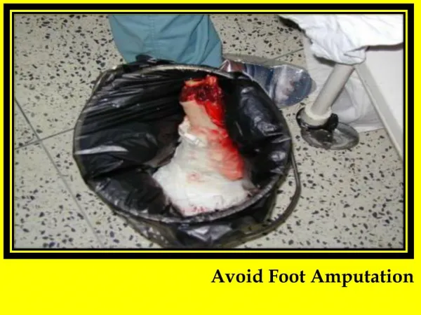
AMPUTATIONS IN DIABETES Common: • Worldwide – amputation 2 to diabetes q 30 sec. • U.S.A. – 80,000 amputations/y (2002) • Higher rates in men, racial/ethnic minorities Costly: • $60,000/amputation • $2 billion/y total costs Lancet 2005; 366:1719 DiabetesCare 2004; 27:1598 DiabetesCare 2003; 26:495 ©2006. American College of Physicians. All Rights Reserved.
AMPUTATIONS IN DIABETES Tragic: “Rule of 50” • 50% of amputations transfemoral/transtibial level • 50% of patients 2nd amputation in 5y • 50% of patients Die in 5y ClinicalCareoftheDiabeticFoot, 2005 ©2006. American College of Physicians. All Rights Reserved.
FOOT ULCERS IN DIABETES Precipitate 85% of amputations: “Rule of 15” • 15% of diabetes patients Foot ulcer in lifetime • 15% of foot ulcers Osteomyelitis • 15% of foot ulcers Amputation ClinicalCareoftheDiabeticFoot, 2005 ©2006. American College of Physicians. All Rights Reserved.
FOOT ULCERS IN DIABETES Costly: • $30,000/ulcer • $9 billion/y total costs Tragic: • Quality of life: ulcer patient amputation patient • Burden of non-weight-bearing as ulcer heals • Lifetime behavioral adaptations to prevent recurrence • Fear of recurrent ulcer/amputation • 70% ulcer recurrence in 3y FootAnkleInt 2005; 26:32, 128 ClinInfectDis 2004; 39(Suppl 2):S129 ©2006. American College of Physicians. All Rights Reserved.
TEAM CARE REDUCES ULCERS/AMPUTATIONS Five clinical trials: • Format: integrated, risk-stratified interventions • ID high-risk patients with exam: • Frequent follow-up to detect early problems • Educate/motivate self-care behaviors • Prophylactic nail/skin care by podiatry • Therapeutic footwear, if needed • Prompt, multidisciplinary Rx of ulcers Lancet 2005; 366:1676 ©2006. American College of Physicians. All Rights Reserved.
TEAM CARE REDUCES ULCERS/AMPUTATIONS Efficacy of team care: • 50-80% reductions in ulcers/amputations • Economic modeling studies of team care: • Cost-effective if 25-40% reduction in ulcer rate • Cost-saving if > 40% reduction in ulcer rate Applicable only to high-risk patients Lancet 2005; 366:1719 DiabetesCare 2004; 27:901 ©2006. American College of Physicians. All Rights Reserved.
PATHOGENESIS OF DIABETIC FOOT ULCER AND AMPUTATION Sensory Joint Motor Autonomic PAD Neuropathy Mobility Neuropathy Neuropathy Protective Muscle atrophy and Sweating Ischemia sensation 2° foot deformities 2° dry skin Foot pressure Foot pressure Fissure Healing Minor trauma esp. over recognition bony prominences Callus Pre-ulcer ULCER Infection AMPUTATION Minor Trauma: Interdigital Maceration Mechanical (Moisture, Fungus) Chemical Thermal ©2006. American College of Physicians. All Rights Reserved.
OTHER RISKS FOR ULCER/AMPUTATION Failure to adequately care for the feet: • Inadequate patient education • Inadequate patient motivation • Depression, anxiety, anger more common in diabetes • Physical disability • Cannot see feet 2 to retinopathy • Cannot reach feet 2 to obesity, age (?50% of patients) • Limited access to podiatry services AgeAgeing 1992; 21:333 DiabetesCare 2003; 29:495 DiabMetabResRev 2004; 20(Suppl 1):S13 ©2006. American College of Physicians. All Rights Reserved.
CAUSAL PATHWAYS FOR FOOT ULCERS % Causal Pathways NEUROPATHY Neuropathy: 78% Minor trauma: 79% DEFORMITY Deformity: 63% Behavioral issues ? MINOR TRAUMA - Mechanical (shoes) POOR SELF- - Thermal FOOT CARE - Chemical ULCER DiabetesCare 1999; 22:157 ©2006. American College of Physicians. All Rights Reserved.
DETECTING FEET-AT-RISK • History: • Prior amputation • Prior foot ulcer • PAD: known or claudication at < 1 block • Exam: • Insensate to 5.07/10g monofilament • Major foot deformities • PAD • Absent DP and PT pulses • Prolonged venous filling time • Reduced Ankle-Brachial Index (ABI) • Pre-ulcerative cutaneous pathology ArchInternMed 1998; 158:157 ©2006. American College of Physicians. All Rights Reserved.
RISK STRATIFY FOR FOOT ULCERATION Foot Ulcer, % Office Patients Risk Level%/yr(diabetes clinics) 3: prior amputation 28.1% 7% prior ulcer 18.6% 2: insensate 6.3% 10% and foot deformity or absent pedal pulses 1: insensate 4.8% 17 - 30% 0: all normal 1.7% 66% DiabetesCare 2001; 24:1442 DiabetesMetab 2003; 29:261 ©2006. American College of Physicians. All Rights Reserved.
ANNUAL DIABETIC FOOT EXAMS2000 Behavioral Risk Factor Surveillance System, CDC ©2006. American College of Physicians. All Rights Reserved.
PHYSICAL EXAMINATION OF THE FEET IN PERSONS WITH DIABETES ©2006. American College of Physicians. All Rights Reserved.
SENSORY NEUROPATHY IN DIABETES • Loss of protective sensation in feet • Sensory loss sufficient to allow painless skin injury • Major risk factor for foot ulcer in diabetes • Detect with 5.07/10g Semmes-Weinstein monofilament • Prevalence of insensate feet to 10g monofilament: • Age > 40y: 30% of diabetic patients • Age > 60y: 50% of diabetic patients • Up to 50% have no neuropathic symptoms DiabetesCare 2006; 29(Suppl 1):S24 DiabetesCare 2004; 27:1591 ©2006. American College of Physicians. All Rights Reserved.
UTILITY OF MONOFILAMENT TESTING Predicts ulcer/amputation in 5 prospective studies: • NPV (normal sensing) = 90-98% PPV (fail to sense) = 18-36% • Prospective 32 mo observational study: • 80% of ulcers/100% of amputations in insensate feet • Superior predictive value to other tests: • Pin prick, cotton wisp, symptoms • ? 128 Hz tuning fork? • ADA recommendation, 2006: also test vibration DiabetesCare 2006; 29(Suppl 1):S25 JFamPract 2000; 49:S30 DiabetesCare 1992; 15:1386 ©2006. American College of Physicians. All Rights Reserved.
USING THE 5.07/10gm MF (Tool-Kit) • Demonstrate sensation on the forearm or hand • Place monofilament perpendicular to test site • Bow into C-shape for one second • Test four sites/foot: Predicts 95% of ulcer formers vs. 8 sites • Heel testing does not discriminate ulcer formers • Avoid calluses, scars, and ulcers ©2006. American College of Physicians. All Rights Reserved.
USING THE 5.07/10g MF (Tool-Kit) • Minimize bias: • Test sites in random sequences • Test each site X3, sham test as 1 of 3 • Do you feel it? Yes or No? • Retest site if patient fails (misses 2/3 responses) • Insensate at 1 site = insensate feet • Falsely insensate with edema, cold feet • Test annually when sensation normal • Use < 100x/d; replace if bent; replace q 3 mo. • Purchase calibrated MF (See Tool-Kit) ©2006. American College of Physicians. All Rights Reserved.
PAD IN DIABETES • Prevalence (ABI < 0.9): 20-30% • 10-20% in type 2 diabetes at Dx • 30% in diabetics age 50y • 40-60% in diabetics with foot ulcer • Complications: • Claudication and functional disability • Increases risk for concurrent CAD and CVD • Delays ulcer healing • Increases amputation risk • Not increase foot ulcer risk JACC 2006; 47:921 DiabetMed 2005; 22:1310 DiabetesCare 2003; 26:3333 ©2006. American College of Physicians. All Rights Reserved.
HX TO DETECT PAD IN DIABETES • Claudication at < 1 block suggests severe ischemia Vascular LevelSite of Pain Aorto-iliac Buttocks/Thigh Femoral Calf Tibioperoneal Foot/Ankle • Rest pain indicates critical ischemia • Toes and forefoot • Difficult to distinguish from neuropathic pain ©2006. American College of Physicians. All Rights Reserved.
Ischemic Rest Pain Unilateral (usually) Continuous; hs With dependency Absent DP/PT pulses Neuropathic Pain Bilateral (usually) Wax/wane No change with dependency Variable DP/PT pulses HX TO DETECT PAD IN DIABETES (After Pompogelli and Campbell, 2002) ©2006. American College of Physicians. All Rights Reserved.
HX TO DETECT PAD IN DIABETES • Asymptomatic, severe PAD common in diabetes • Tibio-peroneal disease predominance: • Unrecognized ankle/foot claudication • No claudication • Sensory neuropathy blunts/eliminates pain sensation of claudication and rest pain DiabetesCare 2003; 26:3333 ©2006. American College of Physicians. All Rights Reserved.
EXAM TO DETECT PAD IN DIABETES • Pedal pulse exam: • Absent DP and PT: LR = 3.0-3.8 for severe PAD • Absent DP or PT not predict PAD • Non-palpable DP (8%) or PT (3%) in normals • Present DP and PT not R/O PAD! • 30% with PAD have one palpable pulse (collaterals) • High PAD suspicion vascular testing • Claudication, foot ulcer JAMA 2006; 295:536 ArchInternMed 1998; 158:1357 DiabetesCare 2003; 26:3333 ©2006. American College of Physicians. All Rights Reserved.
EXAM TO DETECT PAD IN DIABETES • Venous filling time • Technique: • Sitting: ID pedal vein bulging above skin • Supine: Elevate leg to 45° for 1 min • Sitting: time to pedal vein bulging above skin JClinEpidemiol 1997; 50:659 ArchInternMed 1998; 158:1357 ©2006. American College of Physicians. All Rights Reserved.
EXAM TO DETECT PAD IN DIABETES • Venous filling time • Filling time > 20 sec predicts ABI < 0.5 • Sensitivity = 22%; Specificity = 94%; LR = 3.9 JClinEpidemiol 1997; 50:659 ArchInternMed 1998; 158:1357 ©2006. American College of Physicians. All Rights Reserved.
OTHER EXAM FINDINGS FOR PAD • Helpful: • Femoral bruit (LR = 4.7–5.7) • Unilateral cool extremity • Not predictive of PAD: • Atrophic skin • Hair loss • Capillary refill > 5 sec DiabetesMed 2005; 22:1310 ArchInternMed 1998; 158:1357 ©2006. American College of Physicians. All Rights Reserved.
VASCULAR LAB TO DETECT PAD • Ankle/Brachial BP Index or ABI Testing • Screening: 2004 ADA recommendation • “Consider” at age 50 and q 5 yr • Screen earlier if multiple CVD risks • Diagnosis: • Claudication, absent DP/PT pulses, foot ulcer • Limitations: • Underestimate severity if medial artery Ca++ • Consider pulse volume recording, systolic toe BP, vascular consultation if uncertain about PAD DiabetesCare 2005; 28:2206 DiabetesCare 2004; 27(Suppl 1): S15-S35 ©2006. American College of Physicians. All Rights Reserved.
INTERPRETATION OF THE ABI ABI Normal 0.91-1.30 Mild obstruction 0.71-0.90 *Moderate obstruction 0.41-0.70 *Severe obstruction 0.40 **Poorly compressible >1.30 2° to medial Ca++ *Poor ulcer healing with ABI 0.50 **Further vascular evaluation needed ©2006. American College of Physicians. All Rights Reserved.
MOTOR NEUROPATHY AND FOOT DEFORMITIES • Hammer toes • Claw toes • Prominent metatarsal heads • Hallux valgus • Collapsed plantar arch ©2006. American College of Physicians. All Rights Reserved.
Hammer Toes • Claw Toes From Levin and Pfeifer, TheUncomplicatedGuidetoDiabetesComplications, 2002 ©2006. American College of Physicians. All Rights Reserved.
Hallux Valgus From Levin and Pfeifer, TheUncomplicatedGuidetoDiabetesComplications, 2002 ©2006. American College of Physicians. All Rights Reserved.
From Boulton, et al DiabeticMedicine 1998, 15:508 ©2006. American College of Physicians. All Rights Reserved.
PRE-ULCER CUTANEOUS PATHOLOGY Neuropathy inappropriate footwear: • Persistent erythema after shoe removal • Callus • Callus with subcutaneous hemorrhage: “pre-ulcer” Autonomic neuropathy and secondary dry skin: • Fissure ulceration • Augment callus formation Poor self-care of the feet: • Interdigital maceration with fungal infection • Nail pathology ©2006. American College of Physicians. All Rights Reserved.