Download
1 / 38
380 likes | 383 Views
This section discusses the mechanisms and processes involved in synaptic transmission in the nervous system, including the generation and propagation of action potentials and the anatomy of synapses. It also explores the distribution of pumps and channels in neurons and the basic steps of synaptic transmission.

E N D
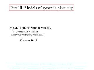
Reading: Anatomy &Physiology: An Integrative approach, McKinley et al., Chapter 12, Nervous System:Nervous Tissue, Section 12.8 ANPS 19 Neurophysiology part 3: Synaptic Transmission Dr. Tompkins Department of Neurological Sciences Given D408 john.tompkins@uvm.edu
+ + Now to make the model look more like a neuron. will represent K+ ions, will represent Na+ ions, and will represent Cl- ions. Also, most of the anion charge inside cells is fixed, to proteins or larger molecules, and cannot move out. This is shown as . - - + + - - - + + - + - + + - Na+/K+-ATPase pump + + + + - + - - - There are more of these channels in the membrane and they contributes the most to the resting membrane potential (RMP). - + - K+ leak channel + + + - + - - - + + + + - + - Na+ leak channel There are few of these channels. + + - + - - - + + in out In this scenario, the potential difference in charge between inside and outside, or the resting membrane potential, is set by the separation of charge due to a diffusion of potassium out of the cell (down its chemical gradient) and a small leak of sodium into the cell (down its chemical and electrical gradients). On the next slide we’ll discuss what the actual potential is in mV. - + chemical gradients V + voltmeter - electrical gradients
Generation of an Action Potential The unstimulated axon has a resting membrane potential of –70 mV. 1 1 2 3 4 5 +30 Graded potentials reach axon hillock and are added together. 2 +10 Depolarization phase of action potential occurs when the threshold (–55 mV) is reached; voltage-gated Na+ channels open and Na+ enters rapidly, reversing the polarity from negative to positive (–55 mV +30 mV). 3 0 –10 –30 mV Repolarization phase of the action potential occurs due to closure of voltage-gated Na+ channels (inactivation state) and opening of voltage-gated K+ channels. K+ moves out of the cell into the interstitial fluid and polarity is reversed from positive to negative (+30 mV –70 mV). 4 Threshold –50 –70 Resting membrane potential Hyperpolarization phase of the action potential occurs when voltage-gated K+ channels stay open longer than the time needed to reach the resting membrane potential; during this time the membrane potential is less than the resting membrane potential of –70 mV. 5 –90 0 1 2 3 Time (msec)
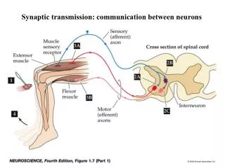
Propagation of the Action Potential 1) action potential first generated in axon hillock, then 2) action potential propagated down axon 1 2
The neuronal membrane can be broken into 4 functional segments. The function of each segment is dictated by the type of ion channels expressed in those regions. Fig. 12.14 axon (definition of hillock – a small hill) Neurotransmitter axon hillock Propagation of action potential Initial segment Conductive segment Transmissive segment Summation of graded potentials; initiation of action potential Propagation of action potential Action potential causes release of neurotransmitter Receptive segment Binding of neurotransmitter released from presynaptic neuron; production of graded potentials Now we will discuss these two segments.
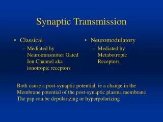
Anatomy of the Synapse • two type of synapses : chemical and electrical • this is a chemical synapse microtubules neurofilaments synaptic knob/bouton/terminal presynaptic membrane mitochondria synaptic cleft postsynaptic membrane (i.e. muscle, nerve or gland)
Electrical vs Chemical Synapse electrical synapse chemical synapse • uncommon • found in limited regions of brain and eyes • most common • in CNS and PNS
Distribution of Pumps and Channels in the Plasma Membrane of a Neuron(Figure 12.11) Receptive segment Chemically gated cation channel Chemically gated Cl– channel Chemically gated K+ channel (b) Cell body Initial segment Dendrites Voltage-gated K+ channel Voltage-gated Na+ channel Axon hillock Na+/K+ pump Na+ leak channel K+ leak channel (c) Conductive segment Entire neuron Voltage-gated Na+ channel Voltage-gated K+ channel Axon Transmissive segment (d) Voltage-gated Ca2+ channel Ca2+ pump we’re here (a) (e) Synaptic bulb
Voltage-gated Ca2+-channel opened by depolarization allows calcium into cell Na+ (145 mM) Na+ (10 mM) Cl- (110 mM) Cl- (20 mM) Concentration gradient maintained by calcium-pumps and intracellular buffers Ca++ (0.0001 mM) Ca++ (2 mM) K+ (140 mM) K+ (5 mM) inside outside
Basic steps of synaptic transmission Fig. 12.21a Synaptic cleft Voltage-gated Ca2+channels open and Ca2+ enters the synaptic terminal and binds with proteins of synaptic vesicles. Receptor Ca2+ Ca2+ Action potential reaches synaptic terminal. + + + + + Synaptic vesicles merge with synaptic terminal plasma membrane and neurotransmitter is released by exocytosis. + + + Neurotransmitter + + + + + + + + Voltage-gated Ca2+ channels Neurotransmitter crosses synaptic cleft and attaches to receptors on post-synaptic membrane. Synaptic vesicle (contains neurotransmitter) Synaptic terminal
Anatomy of a Synaptic Vesicle Synaptic cleft Synaptic vesicles (40 nM dia.) Muscle cell Synaptic knob Proteins associated with a synaptic vesicle Release of synaptic vesicles can be modulated by the proteins associated with the vesicle
Basic steps of synaptic transmission Fig. 12.21a Synaptic cleft Voltage-gated Ca2+channels open and Ca2+ enters the synaptic terminal and binds with proteins of synaptic vesicles. Receptor Ca2+ Ca2+ Action potential reaches synaptic terminal. + + + + + Synaptic vesicles merge with synaptic terminal plasma membrane and neurotransmitter is released by exocytosis. + + + Neurotransmitter + + + + + + + + Voltage-gated Ca2+ channels Neurotransmitter crosses synaptic cleft and attaches to receptors on post-synaptic membrane. Synaptic vesicle (contains neurotransmitter) Synaptic terminal
Removal of neurotransmitters from the synaptic cleft • Temporary association between neurotransmitter and receptor • Necessary to eliminate molecule after stimulation • Removal occurs by two major mechanisms • Occurs by degradation • neurotransmitter chemically inactivated in synaptic cleft • e.g., breakdown of ACh by acetylcholinesterase • Occurs by reuptake • neurotransmitter reabsorbed by transport protein in presynaptic neuron • “recycled” into another synaptic vesicle for reuse
Basic Steps in Synaptic Transmission • Action potential propagated down axon to synaptic terminal • Action potential opens voltage-gated Ca2+ channels allowing calcium ions to move into the synaptic terminal and bind to synaptic vesicle proteins • Vesicles fuse with the with the neuronal plasma membrane and neurotransmitter is released into the synaptic cleft by exocytosis • Neurotransmitter binds to specific post-synaptic receptors (chemically-gated ion channels)
Neurotransmitters • Classes of neurotransmitters • Neurotransmitters, various small organic compounds • Released at synaptic cleft • Approximately 100 known • Classified into major groups • Can be either excitatory (causing depolarization of the membrane) or inhibitory (causing hyperpolarization of the membrane) • Neurons typically release one type of transmitter and are named accordingly (i.e. cholinergic)
Neurotransmitters and Neuromodulation • Classes of neurotransmitters (continued) • Acetylcholine (cholinergic neuron) • excitatory or inhibitory neurotransmitter • released in both CNS and PNS • molecule released from motor neuron at neuromuscular junction • Amino acids • building blocks of proteins • some also neurotransmitters • e.g., glutamate, glycine, GABA
Neurotransmitters and Neuromodulation • Classes of neurotransmitters (continued) • Monoamines • derived from certain amino acids • carboxyl group removed and functional group added • subgroup added determines type • includes subgroup, catecholamines (norepinephrine, epinephrine, dopamine) • Neuropeptides • chains of amino acids • include enkephalins and somatostatin See Table 12.4: Neurotransmitters
Basic steps of synaptic transmission Fig. 12.21a Synaptic cleft Voltage-gated Ca2+channels open and Ca2+ enters the synaptic terminal and binds with proteins of synaptic vesicles. Receptor Ca2+ Ca2+ Action potential reaches synaptic terminal. + + + + + Synaptic vesicles merge with synaptic terminal plasma membrane and neurotransmitter is released by exocytosis. + + + Neurotransmitter + + + + + + + + Voltage-gated Ca2+ channels Neurotransmitter crosses synaptic cleft and attaches to receptors on post-synaptic membrane. Synaptic vesicle (contains neurotransmitter) Synaptic terminal
Acetylcholine and other Neurotransmitters can produce graded potentials on neurons! Example of an ionotropic receptor
Distribution of Pumps and Channels in the Plasma Membrane of a Neuron(Figure 12.11) Receptive segment now we’re here Chemically gated cation channel Chemically gated Cl– channel Chemically gated K+ channel (b) Cell body Initial segment Dendrites Voltage-gated K+ channel Voltage-gated Na+ channel Axon hillock Na+/K+ pump Na+ leak channel K+ leak channel (c) Conductive segment Entire neuron Voltage-gated Na+ channel Voltage-gated K+ channel Axon Transmissive segment (d) Voltage-gated Ca2+ channel Ca2+ pump (a) (e) Synaptic bulb
Chemically-gated Ion Channels • normally closed • opened by very specific molecule (i.e. ligand/ neurotransmitter) think “lock & key” • allows specific ions to diffuse when open • chemical energy transduced into physical energy Closed Open Neurotransmitter binds and opens gate K+ • (Figure 12.10c) (c) Chemically gated channels
Introduction to Neuron Physiology: Changing the Membrane Potential When the post-synaptic receptors are activated a graded potential is produced at the post-synaptic membrane. A graded potential is a small change in membrane potential that can be either hyperpolarizing or depolarizing.
Introduction to Neuron Physiology: Changing the Membrane Potential Graded potentials versus action potentials • Graded potentials • May result in depolarization or hyperpolarization • depends on channel that opens • Degree dependent on stimulus magnitude – not all or none • larger stimulus opening more chemically gated channels • flow of more ions • Decreases in intensity with distance along the membrane • Short-lived • lasts until local ion current ceases
Release of Excitatory Neurotransmitter and Generation of EPSP (Figure 12.15a) Axons of presynaptic neuron Postsynaptic neuron Release of excitatory neurotransmitter and generation of EPSP Excitatory neurotransmitter released from presynaptic neuron binds to receptors, which are chemically gated cation channels, causing them to open. 1 Axons of presynaptic neuron Excitatory neurotransmitter Postsynaptic neuron 0 Chemically gated cation channel Na+ flows into neuron. 2 –20 Synaptic knob Na+ Inside of neuron becomes more positive (less negative); called EPSP (e.g., –68 mV). 3 –40 Voltage (mV) Threshold EPSP Synaptic vesicles containing excitatory neurotransmitter –60 Stimulus EPSP propagates toward axon hillock. 4 –70 Resting membrane potential –80 Time (msec) Synaptic cleft (a)
Physiologic Events in the Neuron Segments: Receptive Segment Generation of EPSPs • Sequence of events • Excitatory neurotransmitter crosses synaptic cleft. • binds to receptor • opens a chemically gated cation channel • More Na+ moves into neuron than K+ moves out. 3) Inside becomes slightly more positive (depolarized). • less negative state called excitatory postsynaptic potential (EPSP) • Local current of Na+ becomes weaker • decreases in intensity with distance traveled
Physiologic Events in the Neuron Segments: Receptive Segment • Generation of EPSPs (continued) • Degree of change in RMP • dependent on amount of neurotransmitter bound per unit time • More excitatory neurotransmitter released • more cation channels open • greater change in the positive direction
Release of Inhibitory Neurotransmitter and Generation of IPSP (Figure 12.15b) Release of inhibitory neurotransmitter and generation of IPSP Inhibitory neurotransmitter binds to either chemically gated K+ channels or chemically gated Cl– channels, causing them to open. 1 Axons of presynaptic neuron Inhibitory neurotransmitter Postsynaptic neuron 0 Chemically gated K+ channel Chemically gated Cl– channel –20 2 Either K+ flows out of, or Cl– flows into, the neuron, depending on the type of channel stimulated. Synaptic vesicles containing inhibitory neurotransmitter K+ –40 Cl– Voltage (mV) Inside of neuron becomes more negative; called IPSP (e.g., –72 mV). 3 Threshold –60 Cl– Stimulus –70 IPSP propagates toward axon hillock. 4 Resting membrane potential IPSP –80 Time (msec) (b)
Physiologic Events in the Neuron Segments: Receptive Segment Generation of IPSPs • Sequence of events • Inhibitory neurotransmitter crosses synaptic cleft. • binds to chemically gated K+ channel or Cl- channel • depends on neurotransmitter and channels present • If neurotransmitter gates K+ channel, K+ moves out of neuron. If neurotransmitter binds Cl-channel, Cl- flows into neuron. 3) Inside of the cell becomes slightly more negative • more negative state termed inhibitory postsynaptic potential (IPSP) 4) Local current of ions becomes weaker. • decreases in intensity with distance traveled toward initial segment
Physiologic Events in the Neuron Segments: Receptive Segment • Simultaneous release • Excitatory and inhibitory neurotransmitters • may be simultaneously released from different neurons • Varied frequency of releasing neurotransmitter • Result: many EPSPs, many IPSPs, or both
Several Presynaptic Neurons with a Postsynaptic Neuron(Figure 12.16) IPSP EPSP Postsynaptic neuron Synaptic knob Presynaptic axons Axons of presynaptic neuron Dendrites Cell body of postsynaptic neuron Myelin sheath Axon SEM 80,000x Axons of presynaptic neuron
Physiologic Events in the Neuron Segments: Initial Segment • Summation • Addition of graded postsynaptic potentials (IPSPs and EPSPs) • Occurs at the initial segment • Determines if threshold membrane potential is reached • -55 mV, +15 mV from RMP • If threshold reached • voltage-gated channels open • action potential generated that travels along axon
Physiologic Events in the Neuron Segments: Initial Segment • Summation (continued) • Spatial summation • release of neurotransmitter from multiple presynaptic neurons • action potential initiated if enough EPSPs generated • Temporal summation • repeated release of excitatory neurotransmitter at same location • effects added if occur within small timeframe • action potential initiated if threshold reached
Spatial Summation at the Axon Hillock(Figure 12.7a) Spatial summation +30 Initial segment Action potential 0 Membrane potential (mV) P5 Axon hillock –55 Threshold P4 P3 P2 P1 –70 Time (m sec) Dendrites Cell body of postsynaptic neuron P1 P2 Myelin sheath P3 EPSPs P4 P5 Axon Axons of presynaptic neurons (P1–P5) (a) Spatial summation
Temporal Summation at the Axon Hillock(Figure 12.7b) Temporal summation +30 Action potential 0 Membrane potential (mV) P2 Threshold –55 –70 Time (m sec) Axon of presynaptic neuron (P2) Postsynaptic neuron P2 Axon EPSPs (b) Temporal summation