Download
1 / 31
310 likes | 316 Views
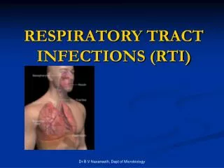
Mycobacteria & Fungal Respiratory Tract Pathogens. Prof. Dr. Asem Shehabi Faculty of Medicine University of Jordan. Global prevalence of TB.

E N D
Mycobacteria & Fungal Respiratory Tract Pathogens Prof. Dr. Asem Shehabi Faculty of Medicine University of Jordan
Global prevalence of TB • The World Health Organization (WHO) estimates that approximately one-third of the global population is infected with M. tuberculosis (TB). • Around 9 to 10 million new cases of TB are being reported each year, 2-3 million deathsoccur each year worldwide.. 95% in developing countries • After emerging (HIV)/AIDS, TB is the secondmost common cause of death in AIDS patients due to an infectious disease.
Mycobacterium Tuberculosis Tubercle Bacilli.. Acid-Fast Bacilli.. Widely distributed in Human, Animals, Birds, Environment.. TB bacilli grow slowly, Resistant to Dryness, low Acidity.. survive years in nature.. But Susceptible to UV-light, Heat. M. tuberculosis.. Causes 95% of human TB cases.. mostly pulmonary .. Respiratory infection ..Few cells.. Lung positive person may infect hundred of susceptible person.. All ages.. mostly children.. with malnutrition . Optimal conditions for transmission include: overcrowding, Large cities, poor conditions & Low standard public hygiene..
Pulmonary Infection • Primary Tuberculosis: 90% Pulmonary TB Children, Asymptomatic, TB Bacilli infect Alveolar macrophages.. Develop small lung lesions..Fibrosis, Calcification, Hypersensitivity.. Infected person becomes Positive for Tuberculin Skin test. • Few cases.. TB bacilli may spread from primary Lesion by direct extension to lymphatic system, bronchi, blood, Kidneys Gastrointestinal, Meningitis (children).. rarely developing Military tuberculosis.
Post-Pulmonary Infection-2 • Post primary tuberculosis:Reactivation old lesions.. Common in young adults & elderly persons.. Developing Large Lung lesions.. Cavities , Less lymphatic involvement , intensive Granuloma & Caseation.. May spread rapidly to other body part, CNS, Gastrointestinal/Urinary Tract .. positive TB urine culture • Clinical Features:Productive Cough..bloody sputum, Low continuous Fever, Night-time sweating , Loss weight & Appetite.. General weakness, Lesions/ Cavities can be detected easily by chest x-ray..Sputum culture +ve
Tuberculin Test • Symptomatic/ asymptomatic infected persons..develop positiveTuberculin skin test.. Reaction to Cell WallMycolicacids+lipoproteins • Mantoux/ Tuberculin skin test.. is produced from boiling culture of M. tuberculosis. • 1,1o, 100 TU .. intra-cutaneously injected in the forearm. The test is read after 48-72 hours. • Positive tuberculin: Indurations, Edema & Erythematic skin> 1 cm, Interpretation: +/-ve. • Vaccination with BCG ( Bacilli- Celements-Guerin).. Attenuated M. bovis ..Protection 30-78%..result in positive Tuberculin test.
Other Human Pathogenic Mycobacteria species • M. bovis:common in domestic animal.. rare human.. Infection.. source: milk, dairy products, meat.. begins mostly intestinal infection.. may spread to other parts.. Slow grower • Atypical mycobacteria: Widely distributed in nature.. water, soil, birds, animals and man including: • M. kansasii: Photochromogenic.. Rapid grower, produce yellow/orange color during incubation in light.. Mostly Lung tuberculosis.. immuno-suppressed persons, AIDS. • M. marinumMostly localized Skin ulcers-soft tissues Swimming pool, Granuloma.. Lymph nodes.. (water, low temperature)..Rapid grower • M.ulcernas. Skin lesions, necrosis, More Resistant to anti-tuberculosis drugs.. Slow grower • M. avium complex (animals, water).. Skin Lesions, rarely Pulmonary disease.. Slow grower
Diagnosis & Treatment-1 • Rapidly growing Mycobacteria species:Rarely cause non-pulmonary diseases, mostly non-pathogens.. M. smegmatis.. Found in on extra genital tract.. May contaminate urine culture. • Diagnosis & treatment: Tuberculosis is confirmed by positive Direct AF Smear/ Culture, Real-time PCR X-ray, Positive tuberculin Test. • Clinical specimens: Sputum, Urine, CSF, Tissues, Culture Loewenstein-Jensen Agar.. 4-8 Weeks.. No Blood Serological test. • Treatment Multiple Antibiotics: 6-24 Months.. Rifampicin, Isonaized, Pyrazinamid, Ethambutol, development of Multidrug resistant MB tuberculosis.. At present 1-10% worldwide..Completing treatment is essential for cure
Nocardiosis • Nocardia asteroids/ N. barsiliensis. Aerobic G+ve.. Pleomorphic Bacilli-Branched Filaments.. Slightly Acid Fast.. Common as Environmental Saprophytes. • Human Exogenous Infection.. Mostly Pulmonary Localized Abscesses.. Necrosis.. small Cavities.. spread to Brain, Kidneys.. Common in Immuno-suppressed, Lung malignancy • Chronic suppuration.. Abscess.. Granulomas, Draining sinuses containing granules.. Muscles, Bones, Feet, Hands and other body parts. • Diagnosis & Treatment: Sputum/biopsies culture on blood 3-30 days at 45 C.. Co-trimoxazole, Rifampicin, Amikacin.. 4-6 Weeks.
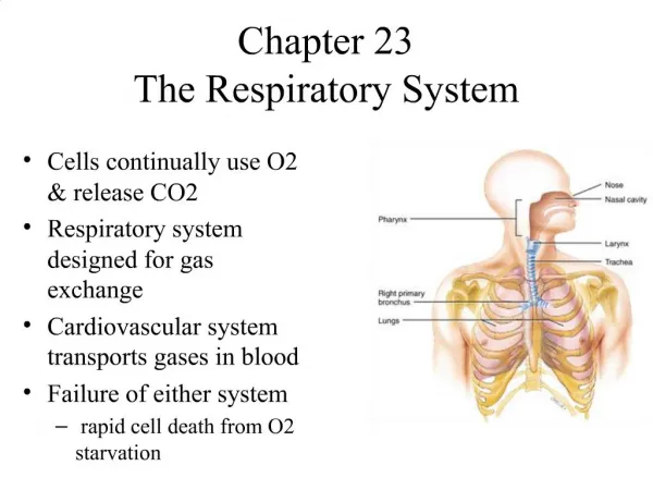
RespiratoryFungal Agents • Fungal respiratory diseases can be divided into: • Generally healthy individuals • Immuno-suppressed patients. • Fungal agents ..Widely distributed in Environment.. Cause mostly infection in immuno-compromised individuals.. receiving immunosuppressive therapy.. undergoing bone marrow transplantation or solid-organ transplant .. HIV infection. • Clinical presentations of fungal respiratory infections are non-specific and often overlap with other infectious and non-infectious processes. The causative agents can be opportunistic Yeast or exogenous filmentous Fungi /Molds
Yeast Form:Oral Candidiasis • Part of oral /intestinal/vaginal flora.. causes characteristic mucosa patches of a creamy-white to grey pseudomembrane composed of Blastoconidiaand Pseudohyphae ..C. albicans often after long antibiotics treatment . • Oral candidiasis may spread.. Esophagus, bronchi, lungs, gastro-intestinal tract, or become systemic .. Candidiaemia, endocarditis, meningitis. • Systemic candidiasis is common in patients with cell-mediated immune deficiency, receiving aggressive cancer, immunosuppression, transplantation therapy. • C. albicans, C.glabrata,C. tropicalis, C. krusei,
Predisposing Factors for the Development of Candidiasis • Impaired Epithelial Barrier: Burns, Wounds / abrasions, Hydration/maceration, Indwelling catheters, Foreign bodies (dentures, etc), Increased gastric pH, Cytotoxic/ Antibiotics agents.. Irradiation • Systemic Disorders: Diabetes mellitus, Pregnancy/oral contraceptives, Malnutrition, Malabsorption, Iron deficiency. • Malignancy / Haematologic Disorders: Neutropnea / MacrophageLeukemia, Lymphoma, advanced cancer, immunodeficiency.. AIDS • Sytemic treatment: fluconazole , amphotericin B, Voriconazole, Itraconazole, Caspofungin.. • Local Ointment: Nystatin, micronazole, clotrimazole
Yeast: Cryptococcosis • EncapsulatedC. neoformans..cause a chronic, subacute to, acute pulmonary.. systemic or meningitic disease.. Often isolated from pigeon, Birds excreta. • Primary pulmonary infections ..Inhalation.. have no specific diagnostic symptoms.. usually subclinical. Dissemination may include central nervous system, skin, bones and other visceral organs. • C. neoformanshas a world-wide distribution.. now one of the most significant opportunistic pathogens in humans.. immunodifficient ..AIDS patients..
Molds: Aspergillosis-1 • Aspergillus speciesare common in nature: A. fumigatus, A. flavus, A. niger. • Human Aspergillosis include: 1) Mycotoxicosis due to ingestion of contaminated foods with fungal toxin .. • A. flavus .. Produce Aflatoxins.. Liver cirrhosis..Death 2) Allergic BronchopulmonaryAspergillosis: Presence of conidia or transient growth of the organism in body Respiratory tract.. Sinuses.. often associated with Allergic reaction ..Eosinophilia.. Asthma.. 3) Colonization without extension in preformed cavities and debilitated tissues.. Common in Tuberculosis & Lung carcinoma..
Aspergillosis-2 4) Pulmonary Aspergilloma( Fungus Ball).. Invasive Aspergillosis.. Pre-existing lung cavity, inflammatory, granulomatous, necrotizing disease of lungs, May spread.. other organs.. Causing Thrombosis.. Rhino-cerebral lesions rarely systemic and fatal disseminated disease. • Treatment:Fluconazole, Itraconazole, CaspofunginAmphotericin B .. • Less common Respiratory fungi are Zygomycetes (Zygmycosis / Mucormycosis).. Mucor, Rhizopusspp, Absidia spp. Fusarium spp. Others.. Common Rhino-cerebral mucormycosis
Dimorphic Fungus: Histoplasmosis-1 • Histoplasmacapsulatum.. Dimorphic fungus with conidia and yeast forms at body temperature and hyphae & marcoconidia in vitro culture.. Common in soil enriched with excreta of birds. Endemic in southern U.S.A, Australia.. Less other countries. • The primary site of infection is usually pulmonary.. inhalation dust with microconidia.. Phagocytosed by macrophages, obligate intracellular parasites.. Causing slight inflammatory reaction.. Most cases of histoplasmosis are asymptomatic /subclinical, benign.. Flu-like syndrome. • Few may develop chronic progressive lung disease.. Granuloma & fibrosis, chronic cutaneous or systemic disease involve any internal organ.. Fatal systemic disease. • All infected persons become positive by histoplasmin skin test.
Coccidioidomycosis &Blastomycosis-2 • Coccidioidesimmitis & Blastomycesdermatitidis.. soil inhabiting Dimorphic Fungus.. Endemic in south-western U.S.A., northern Mexico and various parts South America. • Respiratory infection, resulting from the inhalation of microconidia, often resolves rapidly leaving the patient with a strong specific immunity to re-infection. • Some individuals the disease may progress to a chronic pulmonary condition or a systemic disease involving the meninges, bones, joints, subcutaneous, cutaneous tissues.. Antigen Skin test positive.. Not significant in diagnosis.
Laboratory Diagnosis • Direct microscopy and culture should be performed on all specimens (sputum, bronchial washings, CSF, pleural fluid tissue biopsies from various visceral organs ). • wet mounts in 10% KOH with india ink.. Ovoid-budding yeast cells (b) Gram-stain smear.. • Cultures on Sabouraud dextrose agar should be maintained for one month at 25C.... fungal growths & Wet Mount.. Identification ..produces hyphae-like conidio-phores & Spores.. Color of fungal growth • Serological tests are of limited value.. not significant • Detection of Histoplasm antigen in blood & urine is significant
Pneumocystis (carinii, Rats type) P. jiroveci (Human type) • PC is a fungus ..Yeast like Cells ..has suspected infection reservoir (Rats).. Associated with contaminated dust. • Pneumocystisinfection occurs by inhalation .. It is widely common found in the lungs of healthy individuals. • Asymptomatic Infection mostly started in children & increased in Adults .. Worldwide. • Clinical Disease occurs only when defects exist in both cellular immunity and humoral immunity, suppressed immunity Once inhaled, the trophic formof the organism attaches to the lung alveoli.. Encyst & multiple in host tissues.
Pneumocystis-2 • PC clinical disease .. Pneumonia.. Organism is usually found in the interstitial fluid in the lungs, Lung tissue.. of immunocompromised patients.. AIDS ..may disseminate to other internal body organs.. Associated with high mortality. • Sputum /lung biopsy specimens are usually used for PC detection. • Silver – Giemsa-, Stain.. Immunofluorescent Antigen (IFA)..Treatment: Cotrimoxazole alone or with intravenous Pentamidine in sever cases.