Download
1 / 76
770 likes | 942 Views
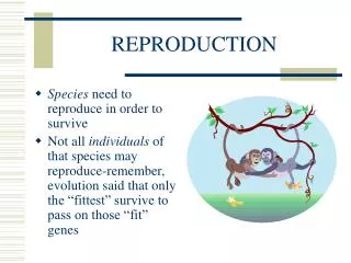
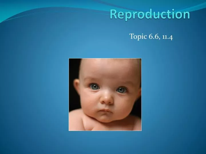
Reproduction. Topic 6.6, 11.4. Sexual reproduction. A population transcends the limit of finite life spans only by reproduction, the creation of new individuals from existing ones.

E N D
Reproduction Topic 6.6, 11.4
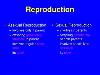
Sexual reproduction • A population transcends the limit of finite life spans only by reproduction, the creation of new individuals from existing ones. • Sexual reproduction is the creation of offspring by the fusion of two haploid (n) sex cells, or gametes, to form a diploid (2n) zygote (fertilized egg). • The male gamete, the sperm, is a relatively small cell that moves by means of a flagellum. • The female gamete, the unfertilized egg, or ovum (plural, ova) is a much larger cell that is not self-propelled. • The zygote—and the new individual it develops into—contains a unique combination of genes carried from the parents via the egg and sperm.
Sexual Reproduction • Sexual reproduction increases genetic variability among offspring. • Meiosis and random fertilization can generate enormous genetic variation. • The variability produced by the reshuffling of genes in sexual reproduction may provide greater adaptability to changing environments. • In humans, internal fertilization occurs by which sperm are deposited in to the female reproductive tract, and gametes unite within the tract. • Requires copulation, or sexual intercourse. • Also requires complex reproductive systems, including organs for gamete storage and transport and organs that facilitate intercourse. • Both females and males have gonads (ovaries or testes) where the gametes are produced, a system of ducts that house and conduct the gametes, and structures that facilitate copulation.
Reproductive anatomy of the human female • A woman’s ovaries are each about an inch long, with a bumpy surface. • The bumps are follicles, each consisting of a single developing egg cell surrounded by one or more layers of follicle cells that nourish and protect the developing egg cell. • In addition to producing egg cells, the ovaries produce hormones. • Specifically, the follicle cells produce the female sex hormones estrogen. • Most or all of the 400,000 follicles a woman will ever have are though to be formed before her birth, but only several hundred will release egg cells during her reproductive years. • Starting at puberty and continuing until menopause, one follicle (or rarely tow are more ) matures and releases its egg cells about every 28 days. • An egg cell is ejected from the follicle in a process called ovulation.
Reproductive anatomy of the human female • After ovulation, the remaining follicular tissue grows within the ovary to form a solid mass called the corpus luteum (Latin for “yellow body”): • Secretes the hormone progesterone, which helps maintain the uterine lining during pregnancy, and additional estrogen. • If the egg is not fertilized, the corpus luteum degenerates, and a new follicle matures during the next cycle. • **we will discuss ovulation and female hormonal cycles in more detail later on…**
Reproductive anatomy of the human female • Each ovary lies next to the opening of an oviduct, also called a fallopian tube. • The oviduct opening resembles a funnel fringed with fingerlike projections. • The projections touch the surface of the ovary, but the ovary is actually separated from the opening of the oviduct by a tiny space. • When ovulation occurs, the egg cell passes across the space and the oviduct, where cilia sweep it toward the uterus. • Fertilization usually occurs in the upper third of the oviduct. • The resulting zygote starts to divide, thus becoming an embyro, as it moves along within the oviduct.
Reproductive anatomy of the human female • The uterus, also known as the womb, is the actual site of pregnancy. • The uterus is only about 3 inches long in a woman who has never been pregnant, but during pregnancy it expands considerably to accommodate a baby. • The uterus has a thick muscular wall, and its inner lining, the endometrium, is richly supplied with blood vessels. • The embryo implants (digests a place for itself) in the endometrium, and development is completed there. • The term embryo is used for the stage of development from the first division of the zygote until body structures begin to appear, about the ninth week in humans. • From the ninth week until birth, a developing human is called a fetus.
Reproductive anatomy of the human female • The uterus is the normal site of pregnancy. • However, in about 1% of pregnancies, the embryo implants somwhere else, resulting in an ectopic pregnancy. • Most ectopic pregnancies occur in the oviduct and are called tubal pregnancies. • Ectopic pregnancies require surgical removal; otherwise, they can rupture surrounding tissues, causing severe bleeding and even death of the mother.
Reproductive anatomy of the human female • The narrow neck of the uterus is the cervix, which opens into the vagina. • The vagina is a thin-walled, but strong, muscular chamber that serves as the birth canal through which the baby is born. • It is also the repository for sperm during copulation. • The vagina opens to the outside just behind the opening of the urethra, the tube through which urine is excreted. • A pair of slender skin folds, the labia minora, border the openings, and a pair of thick, fatty ridges, the labia majora, protect the vaginal opening. • Until sexual intercourse or vigorous physical activity ruptures it, a thin piece of tissue called the hymen partly covers the vaginal opening. • Bartholin’s glands, near the vaginal opening, secrete mucus during sexual arousal, lubricating the vagina and facilitation intercourse. • The vagina, labia minora, and a structure called the clitoris all engorge with blood and enlarge during sexual activity. • The sole function of the clitoris is sexual arousal
Reproductive anatomy of the human female Refer to p. 538 Figure 27.2A
Reproductive anatomy of the human female Refer to p. 539 Figure 27.2C
Reproductive anatomy of the human male • The male gonads, or testes, are each housed outside the abdominal cavity in a sac called the scrotum. • Sperm cannot develop at human core body temperature, but the scrotum keeps the sperm-forming cells cool enough to function normally.
Reproductive anatomy of the human male • Path of sperm from one of the testes out of the male’s body: • From each testis, sperm pass into a coiled tube called the epididymis, which stores the sperm while they continue to develop. • Sperm leaves the epididymis during ejaculation, the expulsion of sperm-containing fluid from the penis. • At that time, muscular contractions propel the sperm from the epididymis through another duct called the vas deferens. • The vas deferens passes upward into the abdomen and loops around the urinary bladder. • Next to the bladder, the vas deferens joins a short duct from a gland, the seminal vesicle. • The two ducts unit to form a short ejaculatory duct, which joins its counterpart conveying sperm from the other testis. • The union of the two ejaculatory ducts forms the urethra, which conveys both urine and sperm out through the penis, although not at the same time. • Thus, unlike the female, the male has a connection between the reproductive and excretory systems.
Reproductive anatomy of the human male • In addition to the testes and ducts, the male reproductive systems contains three sets of glands: the seminal vesicles, the prostate gland, and the bulbourethral glands.: • The two seminal vesicles secrete a thick fluid that contains fructose, which provides most of the energy used by the sperm. • The prostate gland secretes a thin fluid that further nourishes the sperm. • The two bulbourethral glands secrete a clear, alkaline mucus that balances the acidity of any traces of urine in the urethra.
Reproductive anatomy of the human male • The sperm and the glandular secretions make up semen, the fluid discharged (ejaculated) from the penis during orgasm. • About 2-5 mL (1 teaspoonful) of semen are discharged during a typical ejaculation. • About 95% of the fluid consists of glandular secretions. • The other 5% is made up of 50-130 million sperm, only one of which may eventually fertilize a single egg. • The alkalinity of the semen helps neutralize the acidic environment of the vagina, protecting the sperm and increasing their motility.
Reproductive anatomy of the human male • The human penis consists mainly of tissue that can fill with blood to cause an erection during sexual arousal. • Erection is essential for insertion of the penis into the vagina. • Like the clitoris, the penis consists of a shaft that supports the glans, or head. • The glans is richly supplied with nerve endings and is highly sensitive to stimulation. • As in the female, a fold of skin called the prepuce, or foreskin, covers the glans. • Circumcision, the surgical removal of the prepuce, arose from religious traditions. • Scientific studies have not proved that circumcision has an overally positive or negative impact on a man’s health or hygiene.
Reproductive anatomy of the human male • Ejaculation occurs in two stages: • 1.At the peak of sexual arousal, muscles in the epididymis, seminal vesicles, prostate gland, and vas deferens contract. • These contractions force secretions from the glands into the vas deferens and propel sperm from the epididymis. • At the same time, a sphincter muscle at the base of the bladder contracts, preventing urine from leaking into the urethra from the bladder. • Another sphincter also contracts, closing off the entrance of the urethra into the penis. • The section of the urethra between the two sphincters fills with semen and expands. • 2.In the second stage, the expulsion stage, the sphincter at the base of the penis relaxes, admitting semen into the penis. • Simultaneously, a series of strong muscle contractions around the base of the penis and along the urethra expels the semen from the body.
Reproductive anatomy of the human male • Hormones control sperm production by the testes: • Influence by signals from other parts of the brain, the hypothalamus secretes a releasing hormone that regulates release of follicle-stimulating hormone (FSH) and luteinizing hormone (LH) by the anterior pituitary. • FSH increase sperm production by the testes, while LH promotes the secretion of androgens, mainly testosterone. • Androgens stimulate sperm production. • In addition, androgens carried in the blood help maintain homeostasis by a negative-feedback mechanism, inhibiting secretion of both the releasing hormone and LH. • Under the control of this chemical regulatory system, the testes produce hundreds of millions of sperm every day, from puberty well into old age.
Gametogenesis • Spermatogenesis • Formation of sperm cells, takes about 65-75 days in the human male.
Gametogenesis • Spermatogenesis • Sperm develop in the testes in coiled tubes called the seminiferous tubules. • Diploid cells that begin the process are located near the outer wall of the tubules. • These cells multiply constantly by mitosis, and each day about 3 million of them differentiate into primary spermatocytes, the cells that undergo meiosis. • Meiosis I or a primary spermatocyte produces two secondary spermatocytes, each with the haploid number of chromosomes (n=23) • The chromosomes are still in their duplicated state, each consisting of two identical chromatids. • Meiosis II then forms four cells, each with the haploid number of single-chromatid chromosomes. • A sperm cell develops by differentiation of each of these haploid cells and is gradually pushed toward the center of the seminiferous tubule. • From there it passes in to the epididymis, where it matures, becomes motile, and is stored until ejaculation.
Gametogenesis • Oogenesis • The development of mature ova (egg cells)
Gametogenesis • Oogenesis • Most of the process occurs in the ovary. • Oogenesis actually begins prior to birth, when a diploid cell in each developing follicle begins meiosis. • At birth, each follicle contains a dormant primary oocyte, a diploid cell that is resting in prophase of meiosis I. • A primary oocyte can be hormonally triggered to develop further. • After puberty, about overy 28 days, FSH (follicle-stimulating hormone) from the pituitary stimulates one of the dormant follicles to develop. • The follicle enlarges, and the primary oocyte completes meiosis I and begins meiosis II. Meiosis then halts again at metaphase II. • In the female, the division of the cytoplasm in meiosis I is unequal, with a single secondary oocyte receiving almost all of it. • The smaller of the two duaghter cells, called the first polar body, receives almost no cytoplasm.
Gametogenesis • Oogenesis (continued)… • The secondary oocyte is the stage released by the ovary during ovulation. • It enters the oviduct, and if a sperm cell penetrates it, the secondary oocyte completes meiosis II. Meiosis II yields a second polar body and the actual ovum. • The haploid nucleus of the ovum can then fuse with the haploid nucleus of the sperm cell, producing a zygote. • Polar body formation leaves the ovum with nearly all the cytoplasm and thus the bulk of the nutrients contained in the original diploid cell.
Gametogenesis • Oogenesis (continued…) • Development of an ovarian follicle • An actual ovary would have thousands of dormant follicles, each containing a primary oocyte. • Usually, only one follicle has a dividing oocyte at any one time, and as it develops, that follicle stays in one place in the ovary. • Meiosis I occurs as the follicle matures • About the time the secondary oocyte forms, the pituitary hormone LH (luteinizing hormone) triggers ovulation, the rupture of the follicle and expulsion of the secondary oocyte. • The ruptured follicle then develops into a corpus luteum. • Unless fertilization occurs, the corpus luteum degenerates before another follicle starts to develop.
Gametogenesis Development of an ovarian follicle
Gametogenesis • Key differences between spermatogenesis and oogenesis: • 1. only one ovum results from each diploid cell that undergoes meiosis. The other products of oogenesis, the polar bodies, degenerate. • In spermatogenesis, all four products of meiosis develop into mature gametes • 2. although the cells from which sperm develop continue to divide by mitosis throughout the male’s life, this is not the case for the comparable cells in the human female • 3. oogenesis has long “resting” periods, whereas spermatogenesis produces mature sperm in an uninterrupted sequence.
Ovarian and Menstrual cycle • The reproductive cycle is actually one integrated cycle involving cycles in tow different reproductive organs: the ovaries and the uterus. • in discussing oogenesis, we were locating at the ovarian cycle, cyclic events that occur about every 28 days in the human ovary . • Hormonal messages synchronize the ovarian cycle with related events in the uterus called the menstrual cycle. • The hormones are complex and involve intricate feedback mechanisms. Refer to Table 27.5 on p. 544
Ovarian and Menstrual cycle • The ovarian cycle is divided into two phases separated by ovulation: • the pre-ovulatory phase, when a follicle is growing and a secondary oocyte is developing, • and the post-ovulatory phase, after the follicle has become a corpus luteum. • Events in the menstrual cycle are synchronized with the ovarian cycle. • By convention, the first day of a woman’s “period” is designated day 1 of the menstrual cycle. • Uterine bleeding, called menstruation, usually persists for 3-5 days. This corresponds to the pre-ovulatory phase of the ovarian cycle. • During menstruation, the endometrium (inner lining of the uterus) breaks down and leaves the body through the vagina. • The menstrual discharge consists of blood, small clusters of endometrial cells, and mucus. • After menstruation, the endometrium regrows. It continues to thicken through the time of ovulation, reaching a maximum at about 20-25 days. • If an embryo has not implanted in the uterine lining by this time, menstruation begins again, marking the start of the next ovarian and menstrual cycles.
Ovarian and Menstrual Cycle • Hormones that regulate ovarian and menstrual cycles: • The five hormones listed in Table 27.5 synchronize events in the ovarian cycle (the growth of the follicle and ovulation) with events in the menstrual cycle (preparation of the uterine lining for possible implantation of an embryo). • A releasing hormone from the hypothalamus in the brain regulates secretion of the two pituitary hormones FSH and LH. • The blood levels of FSH, LH, and two other hormones—estrogen and progesterone—coincide with specific events in the ovarian and menstrual cycles.
Ovarian and Menstrual Cycle • Hormonal events before ovulation: • 1.The releasing hormone from the hypothalamus stimulates the anterior pituitary to increase its output of FSH and LH. • 2. True to its name, FSH stimulates the growth of an ovarian follicle, in effect starting the ovarian cycle. In turn, the follicle secretes estrogen. Early in the pre-ovulatory phase, the follicle is small and secretes relatively little estrogen. • 3. As the follicle grows, it secretes more and more estrogen, and the rising but still relatively low level of estrogen exerts negative feedback on the pituitary. This keeps the blood levels of FSH and LH low for most of the pre-ovulatory phase. As the time of ovulation approaches, hormone levels change drastically, with estrogen reaching a critical peak just before ovulation. This high level of estrogen exerts positive feedback on the hypothalamus which then • 4. makes the pituitary secrete bursts of FSH and LH. • 5. Then, ovulation occurs.
Ovarian and Menstrual Cycle • Hormonal events at ovulation and after: • LH: • stimulates the completion of meiosis, transforming the primary oocyte in the follicle into a secondary oocyte. • it also signals enzymes to rupture the follicle, allowing ovulation to occur, and triggers the development of the corpus luteum from the ruptured follicle. • it also promotes the secretion of progesterone and estrogen by the corpus luteum. • Estrogen and progesterone • High levels of these hormones in the blood following ovulation have a strong influence on both ovary and uterus. • The combination of both hormones exerts negative feedback on the hypothalamus and pituitary, producing • 6. falling FSH and LH levels. This drop prevents follicles from developing and ovulation from occurring during the post-ovulatory phase. Also, the LH drop is followed by gradual degeneration of the corpus luteum. Near the end of the post-ovulatory phase, unless an embryo has implanted in the uterus, the corpus luteum stops secreting estrogen and progesterone.
Ovarian and Menstrual Cycle • Hormonal events at ovulation and after • 7.When the levels of estrogen and progesterone drop, the endometrium begins to slough off. Menstrual bleeding begins soon thereafter, on day 1 of a new cycle. • 8. As blood levels of FSH and LH drop, they hypothalamus once gain can stimulate the pituitary to secrete more FSH and LH, and a new cycle begins.
Ovarian and Menstrual Cycle • Control of the menstrual cycle: • Hormonal control of the menstrual cycle is simpler than that of the ovarian cycle. • The menstrual cycle is directly controlled by estrogen and progesterone alone. • Starting around day 5 of the cycle, the endometrium thickens in response to the rising levels of estrogen and later progesterone. • When the levels of these hormones drop, the endometrium begins to slough off. • Menstrual bleeding begins soon thereafter, on day 1 of a new cycle.
Fertilization • Embryonic development begins with fertilization, the union of a sperm and an egg to form a diploid zygote. • Fertilization combines haploid sets of chromosomes from two individuals and also activates the egg by triggering metabolic changes that start embryonic development.
Fertilization • The Properties of Sperm Cells • Of all of the millions of sperm that surround a human egg cell, only one will enter and fertilize the egg. • All the other sperm will die; the one sperm that penetrates the egg adds its unique set of genes to those of the egg and contributes to the next generation. • In a mature human sperm, we can see how form fits function: • The sperm’s streamlined shape is adaptation for swimming through fluids in the vagina, uterus, and oviduct of the female. • The sperm cell’s thick head contains a haploid nucleus and is tipped with a vesicle, the acrosome, which lies just inside the plasma membrane. It contains enzymes that help the sperm penetrate the egg. • The neck and middle piece of the sperm contain a long, spiral mitochondrion. • The sperm absorbs high-energy nutrients, especially the sugar fructose, from the semen. • Thus fueled, its mitochondrion provides ATP from movement of the tail, which is actually a flagellum. • By the time a sperm has reached the egg, it has consumed much of the energy available to it. But a successful sperm will have enough energy left to penetrate the egg and deposit its nucleus in the egg’s cytoplasm.
Fertilization • The Process of Fertilization: • To reach the egg nucleus, the sperm nucleus must pass through three barriers: the egg’s jelly coat (yellow), a middle region of glycoproteins called the vitelline layer (pink), and the egg cell’s plasma membrane.
Fertilization • The Process of Fertilization: • 1. As a sperm approaches and then • 2. contracts the jelly coat of the egg, the acrosome in the sperm head releases a cloud of enzyme molecules that digest a cavity into the jelly. • 3. When the sperm head reaches the vitelline layer, species-specific protein molecules on its surface bind with specific receptor proteins on the vitelline layer. • The binding between these proteins ensures that sperm of other species cannot fertlize the egg. • 4. After the specific binding occurs, the sperm proceeds through the vitelline layer and the sperm’s plasma membrane fuses with that of the egg. • 5. Fusion of the two membranes makes it possible for the sperm nucleus to enter the egg. • Fusion of the sperm and egg plasma membranes triggers a number of important changes in the egg. • Two such changes prevent other sperm from entering the egg. About 1 second after the membranes fuse, the entire egg plasma membrane becomes impenetrable to other sperm cells
Fertilization • The Process of Fertilization (continued): • 6. Shortly thereafter, the vitelline layer hardens and separates from the plasma membrane. The space quickly fills with water, an the vitelline layer becomes the so-called fertilization envelope, another barrier impenetrable to sperm. • If these events did not occur an egg were fertilized with more than one sperm, the resulting zygote nucleus would contain too many chromosomes, and the zygote could not develop normally. • Membrane fusion also triggers a burst of metabolic activity in the egg. In preparation for the enormous growth and development that will follow fertilization, the egg gears up from near dormancy, increasing cellular respiration and protein synthesis. • 7. Next, the egg and sperm nuclei fuse, producing the diploid nucleus of the zygote.
Fertilization- Animation • http://www.uchsc.edu/ltc/Fertilization.html • http://www.dnatube.com/video/1127/Human-Reproduction-Fertilization-and-Fetal-Development
Pregnancy • Pregancy, or gestation, is the carrying of developing young within the female reproductive tract. • It begins at conception, the fertilization of the egg by a sperm and continues until birth. • Duration of pregnancy varies considerably among animal species, in humans, it averages 266 days (38 weeks) from conception, or 40 weeks from the start of the last menstrual cycle. • Conception occurs in the oviduct. • Cleavage starts about 24 h ours after fertilization and continues as the embryo moves down the oviduct toward the uterus. • By the sixth or seventh day after fertilization the embryo has reached the uterus, and cleavage has produced about 100 cells.
Embryonic Development- Cleavage • The transformation from a zygote to the multicellular state is truly phenomenal.
Embryonic Development- Cleavage • Cleavage is a rapid succession of cell divisions that produces a ball of cells—a multicellular embryo—from the zygote. • DNA replication, mitosis, and cytokinesis occur rapidly, but gene transcriptin virtually shuts down, and few new proteins are synthesized. • As a result, the embryo of most animals does not grow larger during cleavage. Nutrients stored in the egg nourish the dividing cells, and the cell divisions partition the zygote into many smaller cells. • The first cleavage is completed after 36 hours, and each succeeding division takes less time. • After 3 days, successive cleavages have produced a solid mass of cells called the morula, which is still about the same size as the original zygote.