Download
1 / 65
690 likes | 995 Views
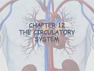
Chapter 12 Circulatory System. Workbook Assignment True and False pg 147 Key Term Assessment pg 148 Evaluation of Learning pg 140 #1-76 Crossword Workbook and Terminology Book due on the day of the test. Circulatory System. Heart – pump

E N D
Chapter 12 Circulatory System Workbook Assignment True and False pg 147 Key Term Assessment pg 148 Evaluation of Learning pg 140 #1-76 Crossword Workbook and Terminology Book due on the day of the test
Circulatory System • Heart – pump • Blood Vessels – form the pathway for which blood can flow throughout the body • Blood – transport medium for nutrients, oxygen, blood cells, etc
FACTS • Perfusion: blood flowing throughout the body to supply the tissues and organs with oxygen and nutrients • Decreased blood flow results in organ shut down, loss of brain cells and cardiac muscle cells. • Then you die……
Heart • Muscular Pump • Strong enough to pump blood to all the tissues of the body (5 L of blood per minute) • Tissues need constant supply of oxygen and nutrients • Waste products must be removed regularly or they will build up and cause damage
HEART - Size and Location • Located in the thoracic cavity between the lungs, behind the sternum(posterior) and in front of the spine (anterior) • 2/3 of the heart lie to the left of the sternum Apex – pointed end (bottom) Base – wider end (top) Size – varies with each pt (fist)
Pericardium • Pericardium – double layered sac that encloses the heart, loose fitting
Coverings continued • Fibrous pericardium: outer layer of the pericardium • Parietal pericardium: membrane that lines the fibrous pericardium • Visceral pericardium: membrane that faces the heart aka: epicardium Pericardial Cavity: space between the parietal and visceral layers, contains fluid that reduces friction
Layers of the heart wall 1. Epicardium: outer layer • Thin protective layer • contains blood vessels that nourish the heart wall 2. Myocardium: middle muscular layer • bulk of the heart wall • contraction of this muscle forces the blood out of the heart and into the blood vessels 3. Endocardium - smooth inner lining heart wall • Blood moves easily thru the heart • forms the valves of the heart
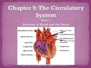
Heart Chambers Atria : top 2 chambers of the heart -thin walled receiving chambers Ventricles: 2 lower chambers • larger and thicker walled - pumping chambers
Right Atrium • Receiving chamber – deoxygenated blood from the body • Superior Vena Cava: Returns blood to the heart from the head, neck, & upper extremities. • Inferior Vena Cava: Returns blood to the heart from the thorax, abdomen, pelvis & lower extremities.
Left Atrium • Receiving chamber – oxygenated blood from the lungs through the 4 pulmonary veins
Interatrial Septum - Thin partition that separates the R and L atria Within the interatrial septum is a fossa ovalis: thinner part of the interatrial septum. • In the fetal heart there is an opening called a foramen ovale. Closes after birth on most. Can remain open through adulthood. Referred to as a Patent Foramen Ovale
Ventricles • R Ventricle: Receives blood from the R atrium and pumps it to the lungs where it receives oxygen • L Ventricle: Receives blood from the L atrium and pumps it to the body tissue • Interventricular Septum: thick muscular partition between the ventricles
4 Valves of the Heart • Keep the fluid moving in one direction • Heart has 2 types: Atrioventricular & Semilunar Valves Mitral Valve
Atrioventricular Valves • Permit the blood to flow through the atria into the corresponding ventricle • Prevent backflow of blood from ventricles into atria • Tricuspid valve (3) cusps – R atrium/R ventricle • Bicuspid valve – (2) cusps – L atrium/L ventricle
Semilunar Valves - 2 • Located at the base of the arteries that carry blood from the ventricles • Pulmonary SL valve: exit of the right ventricle in the base of the pulmonary trunk • Aortic SL valve: exits of the left ventricle and base of the aorta
Pathway of blood through the heart • Both atria contract at the same time followed by both ventricles • Right side of the heart: Pulmonary Circulation pumps blood to the lungs • Left side of the heart: Systemic Circulation Pumps blood to the body (organs and tissues)
Step by Step - Handout • 1 superior and inferior vena cava, • 2 to the right atrium • 4 through the tricuspid valve • 4 to the right ventricle • 5 through the pulmonary semilunar valve • 6 to the pulmonary artery (2) • to the lungs - not pictured • The blood picks up oxygen in the lungs, flows back to the heart thru • 7 pulmonary veins (4) • 8 into the left atrium • 9 through the mitral valve (bicuspid Valve) • 10 to the left ventricle • 11 through the aortic semilunar valve • 12 to the aorta • to the body drops off O2 and nutrients and returns with waste through the vena cavas
Video Pathway of blood through the heart • http://www.youtube.com/watch?v=JA0Wb3gc4mE
Blood supply to the myocardium • Myocardium – muscle layer of the heart • needs a constant supply of oxygen and nutrients • Two arteries that branch off the aorta supply heart muscle with blood • Right and left coronary arteries • Coronary Arteries branch off many times and supply the whole heart with blood
Angiogram • http://www.bing.com/videos/search?q=cardiac+angiogram+you+tube&view=detail&mid=D83E420B64E73B87ABFAD83E420B64E73B87ABFA&first=0&qpvt=cardiac+angiogram+you+tube
Function of the Heart - Pump • Pump blood to the lungs: pulmonary circulation • pick up O2 and nutrients • Pump blood to the body: systemic circulation • to deliver O2 and pick up CO2 • Blood is pushed around the body by contractions of the heart muscle (myocardium) • The conduction system of the heart stimulates the heart to contract and relax at a regular and efficient rate.
Components of the Conduction System - 5 • Sinoatrial Node (SA Node) • Located in the right atrium • Initiates impulses without neural stimulation • Called the hearts natural pacemaker • Impulses travel through both atria at the same time causing the atria to contract
Conduction System • AtrioventricularNode - (AV Node) • Located in the floor of the right atrium • Receives impulse from the SA node and briefly delays it - gives the atria time to finish contracting before the ventricles begin 3. Bundle of HIS 4. Right and Left bundle branches 5. Conduction Myofibers(purkinje’s fibers)
Cardiac Cycle • Consists of one complete heartbeat • Both atria contract at the same time • Atria rest while the ventricles contract at the same time • Systole: contraction phase of the chambers • Diastole: relaxation phase of the chambers
Systole and Diastole • Atrial Systole – contraction of the atria • Ventricles are in diastole (relaxed and filling) • Ventricular Systole – contraction of the ventricles • Atria are in diastole (relaxed and filling) • While the Aortic and Pulmonary Vales are closing the physician can hear “heart sounds” or “Lubb-Dubb”
BLOOD • Transport medium (vehicle) for nutrients and oxygen to support the tissues of the body. • Removing waste and carbon dioxide • Composed of cells and cell fragments suspended in plasma • The average adult has (blood volume) • Female 4-5 Liters • Males 5-6 Liters
Blood Functions: Transportation, Regulation and Protection Transportation: (circulating blood) transports oxygen & nutrients to cells transports carbon dioxide to the lungs transports waste to the kidneys transports hormones Regulation: regulates body temperature by removing heat from active areas (skeletal muscle) and transporting it to other areas (skin) regulates fluid and electrolyte balance
Function continued • Protection: • Clotting mechanism prevent blood loss - platelets • Phagocytic white blood cells engulf bacteria • Antibodies help protect against disease Cells move around the circulatory system in PLASMA (liquid portion of the blood)
Composition of Blood Blood sample centrifuged in the lab separates into 1. cells aka formed elements 2. liquid Plasma is the liquid – 55% Buffy Coat - cells Red blood Cells – 45% Plasma: 90% water and 10% solutes
10% dissolved solutes • Plasma Proteins • Albumins • Globulins- (lipids and antibodies) • Fibrinogen • Electrolytes • Sodium, Potassium, Calcium, Chloride, Phosphate, Bicarbonate • Nutrients • Amino acids (protein digestion) • Glucose (carbohydrates digestion) • Fatty Acids (lipid digestion) • Respiratory Gases: (transported as dissolved gasses) • Oxygen • Carbon dioxide
10% solutes con’t • Fibrinogen • Fibrinogen is a soluble plasma protein. When the body is injured, enzymes are released which cause a chemical reaction that changes the fibrinogen into fibrin. Fibrin is insoluble (does not dissolve in water) and forms the foundation of a blood clot.
Formed Elements or cells • Erythrocytes – Red Blood Cells – RBC • Leukocytes –White blood cells – WBC • Thrombocytes – platelets
Hematopoiesis • Production of the blood cells - hemocytoblasts • Before birth: in the Liver and the Spleen • After birth: red bone marrow of specific bones • Hemocytoblast: • Stem cell which can develop into 7 different blood cells
Erythrocytes (RBC) • Most numerous of the formed elements • Females-4.5 – 5.5 million • Males – 4.5 – 6.2 million • Shape: Biconcave shaped disc • Thin in the middle and thicker around the outside. Shape provides flexibility for moving through capillaries and a greater surface area for diffusion of gases
Erythrocytes - RBC • Reticulocyte – immature RBC with a nucleus. Move from the bone marrow while still immature to the blood • RBC matures it loses it’s nucleus to create more room on the cell for the hemoglobin. Function of the erythrocyte: transport oxygen and carbon dioxide
Erythrocyte • Hemoglobin –takes up 1/3 of each erythrocyte • Two parts: Heme and globin • Heme: formed from a pigment that contains iron • Globin: protein Hemoglobin combines with the oxygen in the blood it forms oxyhemoglobin – bright red After the oxygen is released into the tissues it becomes deoxyhemoglobin – darker red
Production of Erythrocytes • Erythropoietin is a hormone manufactured by the liver and secreted into the blood in an inactive state. • When blood oxygen level gets low the kidneys produce REF renal erythropoietic factor which activates the erythropoietin already circulating in the blood, and it stimulates the red bone marrow to increase the production of RBCs. • Once 02 levels stabilize, the kidneys decrease the REF and RBC production decreases
Production of Erythrocytes • In order for new RBC to be produced, the body must have Iron, Folic Acid and Vitamin B12. The stomach produces Intrinsic factor – a protein, so Vitamin B12 can be absorbed. • Pernicious Anemia: low RBC count due to Vitamin B12 deficiency (alcoholism, celiac, Charon's)
Destruction of Erythrocytes Lifespan of RBC – 120 days As the cell ages, the membrane becomes fragile and the cell breaks down Macrophages (phagocytic cells in the spleen and liver) remove them from circulation and new ones are created a the same time. Hemoglobin separates into Heme and globin Heme: bilirubin, yellow bile pigment globin: protein broken down into an amino acid