Download
1 / 65
650 likes | 763 Views
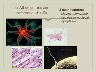
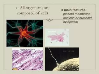
Why Are Organisms Made of Cells. Chapter 4.

E N D
Why Are Organisms Made of Cells Chapter 4
Antonie van Leeuwenhoek, a Dutch merchant made glass lenses by polishing bits of glass and mounting lenses between gold plates to examine and magnify things. He was first to see red blood cells, little animalcules, and that insects hatched from eggs.
He sent these reports to the Royal Society of London (over 375 reports). In 1683 he was the first person to see bacteria. Robert Hooke was curator of instruments for the Royal Society. In 1665 he published a book, Micrographia. He also coined the term cellular from looking at cork. Cells resembled the little rooms in a monastery.
Neither man’s work was taken seriously because they were commoners. Also there was a great deal of bias left from the Middle Ages where science was left to magic and superstition.
Leeuwenhoek’s lenses magnified up to 300x, sophisticated them but primitive now. In the 1820’s better microscopes led to the discovery of nuclei and that “juice” described by Hooke was protoplasm.
Lenses of electron microscopes are electromagnets that bend the path of electrons. Two kinds of them the TEM (transmission electron microscope) and SEM (scanning electron microscope) can produce an image on a screen. In the TEM light passes through the specimen revealing internal structure. The SEM shows surface detail.
In 1838 Schleiden theorized that all plants were made of cells. In 1839 Schwann said the same about animals. Noncellular organisms do not exist. They proposed cells crystallized out of shapeless material but eventually in 1858 Virchow, a physician who saw cells divide, formalized the phrase”all cells from cells”.
This concluded that cells could not come from non-living matter and that diseases are caused by changes in cells. Their work makes up the “cell theory”. 1.All organisms come from cells. 2.Cells are units of structure and function of organisms. 3.Cells come only from other cells.
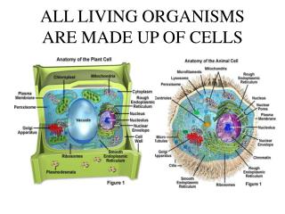
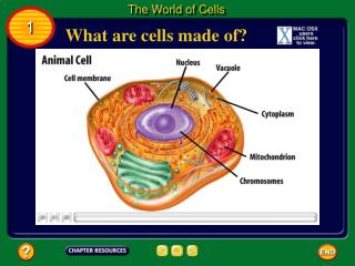
Every cell consists of a boundary, cell body, and set of genes. The plasma membrane is the boundary, highly organized and responsive –defines the limits and regulates the internal environment.
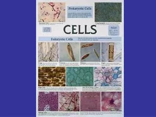
The genes are DNA, in eukaryotes contained in a nucleus. Prokaryotes are the eubacteria and archaebacteria. DNA is in nucleid with no membrane. Organelles do specialized tasks. Cytoplasm not contained in organelles is cytosol. Most of the cell’s biochemical work takes place there.
Protein fibers run through it forming a cytoskeleton, giving the cell shape and helps in cell movement. A cell is alive because it is made of organized parts, performs chemical reactions, responds to the environment, changes over time, reproduces, and shares evolutionary history.
Organelles work together to maintain homeostasis. Most capture energy from glucose, oxidize it to CO2 and H2O which takes place in organelles or in the cytosol.
Cells change over time chemically and mechanically: muscle cells shorten, some change size and shape. To do this eukaryotes have a cytoskeleton. Cells can copy genes for reproduction ( read and duplicate DNA).
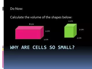
Different cells make different proteins. Wastes, CO2, and ammonia are excreted. Most cells are in a similar size range 10-100 micrometers. (eukaryotes) Prokaryotes are 0.4-0.5micrometers. Ostrich eggs are huge. Plant fibers can be meter- long cells, and over meter long cells in a giraffe’s leg.
Size is limited by the cell’s need to regulate its internal environment. Cells need to maintain homeostasis, same internal pH, concentration of salts, take in useful molecules, and get rid of wastes. Size is limited by the plasma membrane’s ability to do this. Protists are able to do these functions (Didinium and Paramecium).
Cells are limited by the surface – to – volume ratio. Larger cells have smaller surface to volume ratios to regulate the internal environment. Large cells have a hard time getting nutrients, getting rid of wastes, and regulating internal concentrations of ions and molecules.
How can eukaryotic cells be larger than prokaryotic? Eukaryotes have special adaptations to increase their surface areas, convoluted membranes and elaborate internal membrane systems. RBC’s carry O2, muscle cells contract, cells of plants absorb nutrients.
Cells live and die independently of the whole organism. Skin, blood, and intestines replace themselves. The life of a multicellular organism can extend beyond the life of a cell. Many different organelles can be compared to a walled city- there are power stations, a library, warehouses to package proteins.
The development of the TEM in the 1940’s-50’s surprised scientists with the complexity of internal membranes, and number of compartments. Vesicles (empty sacs can be 100 nm., and can be 95% of a plant cell.
The Nucleus is 5-19% of the cell. Inside are chromosomes – complexes of DNA and protein.The nucleus is the library and contains instructions for forming new cells.The boundary is a double – membrane, a nuclear envelope with nuclear pores. The pores are channels between the inside and the cytoplasm to control movement of materials in and out of the nucleus.
The DNA is the code for building all polypeptides the body will ever need. The cytosol is about ½ of the cell volume. It can be separated from the cell by breaking open cells and spinning the solution in a centrifuge at 100,000x gravity (2,000 – 80,000 r.p.m.’s)
Heavier larger cell fragments concentrate at the bottom. There are thousands of enzymes in the cytosol that produce building blocks, degrade small molecules, and synthesize proteins.
Cytosol is aqueous but about 20% protein giving it a viscosity like jello. Granules of energy rich droplets of fat called ribosomes (15-30 nanometers) and smaller proteosomes contain RNA and are where proteins are put together with peptide bonds. They are bound to thr rough E.R. or float freely. Proteosomes brek up old proteins and recycle amino acids.
Peroxisomes contain enzymes that transfer hydrogen from substrates to toxygen forming H2O2. They can break down fatty acids and detoxify substances. Glyoxysomes are found in fat storing seeds of plants.
The endoplasmic reticulum makes proteins and lipids. It forms a convoluted network throughout the cell. The ER membrane encloses a network of cavities and channels called the lumen that make up 15% of the cell’s volume. The E.R. consists of the rough area dotted with ribosomes on the cytosol side to make proteins to be exported from the cell.
The smooth E.R. has no ribosomes; it synthesizes lipids and breaks down toxins. Both rough and smooth E.R. are in eukaryote cells. Specialized cells may have more of one type than the other.
The pancreas has a lot of rough E.R. (makes digestive enzymes and insulin). Cells that produce lipid and steroid hormones (adrenal glands and liver) have a lot of smooth E.R.
The Golgi Complex is a packaging center. In cell reproduction and maintenance it forms structures that stay in the cell like lysosomes and prepares materials for export. It is made of sets of flattened discs (in 6’s) with small vesicles at the ends. Cells that make glycoproteins have more.
Glycoproteins are proteins with attached sugars. (albumin in egg white for ex.). Palade and Farquhar labeled new glycoproteins with radioactive tracers. Proteins to be exported appeared first in the rough E.R., then the Golgi complex. The Golgi complex modifies the glycoproteins and packages them in secretory vesicles.
Vesicles fuse with the plasma membrane and discharge their contents. The Golgi complex manages the flow of proteins to different destinations by modifying the carbohydrates on glycoproteins- labels them with tags that direct them to specific locations, lysosomes, or outside the cell.
Lysosomes are in all eukaryotic cells. They contain enzymes that break down proteins, nucleic acids, sugars, and lipids. Vacuoles of plant cells are like large lysosomes. Lysosomes are numerous in phagocytic cells that consume and digest food. (amebas, other protists, and our white blood cells).
The membrane of a lysosome keeps enzymes from digesting the cell’s cytosol. If the membrane breaks down the cell digests itself. Lysosomes are formed by the Golgi complex. Its enzymes are made by ribosomes of the rough E.R.
Mitochondria obtain energy from nutrients. They make most of the ATP for the chemical reactions of the cell. Mitochondria convert sugar to ATP. Under the TEM they are the most numerous organelles of a eukaryotic cell.
Different kinds of cells have different numbers of them. Liver and heart cells may contain thousands (1/4 the cell’s volume. A TEM shows 2 membranes. The double membrane and having their own DNA suggests mitochondria evolved from eukaryotes that captured bacteria which evolved into mitochondria.
Plastids also have a double membrane. Chloroplasts make and store sugar for food. Most organisms depend on them for food. They are generally large, round, and green.
Chloroplasts have internal folded membranes, thylakoids, piled in stacks of 10 called grana. They have DNA and make protein so are thought to have evolved as free-living organisms captured by early eukaryotic cells that became chloroplasts.
Chromoplasts contain yellow, orange, or red pigments and form from chloroplasts that reshape the membrane and break down chlorophyll. (tomatoes ripen) Amyloplasts store starches in roots of potatoes and seeds like wheat and rice.
The cytoskeleton is a network of protein filaments that are visible with the TEM. 3 types of filaments are: microtublues, actin, and intermediate filaments. The cytoskleton gives support and force for cell movement, changes in shape and transport of materials through the cell.
Some proteins are specilaized for muscle movement (actin). Microtubules are cylinders functioning in cell division and dividing materials to daughter cells. Microtubules originate from microtubule organizng centers (MTOC’s) near the nucleus in a zone called the centrosome that contains a centriole.
Microtubules consist of 2 globular protein molecules called tubulins. 50 proteins can form microtubules. Actin are finer, anchored to the cell surface in muscle fibers, necessary for contraction.
Intermediate filaments, are fibrous like keratin that forms hair. They are in parts of cells that are subject to stress. Cilia (short and numerous) and flagella (longer for propulsion) have the same arrangement in cross section. Around the periphery are 9 pairs and 2 in the center.
Membranes limit cell size because the membrane increases in size more slowly than the volume. It must supply nutrients and O2 and remove wastes. Its functions are: 1.Forms a boundary 2.Regulates contents. 3.Place for chemical reactions and secretion of enzymes. 4.Participates in energy conversion.
Membranes of cells have lipid bilayers. RBC’s have been studied. Biochemists break them open, extract the hemoglobin, leaving RBC “ghosts”. The most important lipids are phospholipids which are amphipathic (hydrophilic heads and hydrophobic tails).
Proteins occupy the 2 layers of the plasma membrane (peripheral) and also the space between (transmembrane). Membrane proteins are amphipathic. Enzymes can break down lipids in the outer or inner layer but not the other. Some carbohydrates appear only on the outer surface attached only to proteins (glycoproteins) or to lipids (glycolipids)
The inner and outer layers differ in how they interact with membrane proteins . Proteins that span the entire thickness orient themselves in a certain direction. The orientation of the protein is crucial to allowing molecules to pass through.
Diffusion and Osmosis • Water and small molecules can diffuse through the membrane. Other molecules must be selectively pumped. Some lipids move freely through the membrane. About half of the proteins move freely within each layer, half remain tightly bound.
Lipids and proteins move easily because they are in layers of fluid. In 1972 Singer and Garth proposed it was a fluid mosaic model and a lipid bilayer. Proteins are peripheral or transmembrane. The membrane is a hydrophobic barrier. Some membrane proteins transport molecules across the membrane.
Membranes are selectively permeable. Proteins and ions cannot pass through. Homeostasis is dependent on the membrane molecules that actively pull small molecules and ions through..
Water diffuses. If you add solute like salt to water its concentration is highest where it was added but it diffuses until it is distributed evenly. Still the salt particles continue to move randomly. A difference in concentration between 2 areas is a concentration gradient..
Diffusion and mixing eliminate the gradient. Rate of diffusion is affected by temperature, size of molecules, steepness of the gradient (greater with greater difference in concentrations). Osmosis is diffusion of water toward where concentration of water molecules is less. Water will move into a cell with a high solute concentration.