Download
1 / 29
290 likes | 427 Views
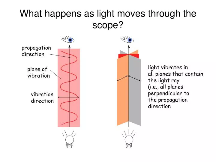
propagation direction. light vibrates in all planes that contain the light ray (i.e., all planes perpendicular to the propagation direction. plane of vibration. vibration direction. What happens as light moves through the scope?. Light and colors reach eye!.

E N D
propagation direction light vibrates in all planes that contain the light ray (i.e., all planes perpendicular to the propagation direction plane of vibration vibration direction What happens as light moves through the scope?
Light and colors reach eye! Light vibrating in many planes and with many wavelengths 3) Now insert a thin section of a rock west (left) Unpolarized light east (right) Light vibrating E-W How does this work??
Some generalizations and vocabulary • All isometric minerals (e.g., garnet) are isotropic – they cannot reorient light. Light does not get rotated or split; propagates with same velocity in all directions • These minerals are always black in crossed polars. • All other minerals are anisotropic–they are all capable of reorienting light (transmit light under cross polars). • All anisotropic minerals contain one or two special directions that do not reorient light. • Minerals with one special direction are called uniaxial • Minerals with two special directions are called biaxial
Isotropic minerals: light does not get rotated or split; propagates with same velocity in all directions • Anisotropic minerals: • Uniaxial - light entering in all but one special direction is resolved into 2 plane polarized components that vibrate perpendicular to one another and travel with different speeds • Biaxial - light entering in all but two special directions is resolved into 2 plane polarized components… • Along the special directions (“optic axes”), the mineral thinks that it is isotropic - i.e., no splitting occurs • Uniaxial and biaxial minerals can be further subdivided into optically positive and optically negative, depending on orientation of fast and slow rays relative to xtl axes
Isometric • All crystallographic axes are equal • Hexagonal, tetragonal • All axes c are equal but c is unique Orthorhombic, monoclinic, triclinic • All axes are unequal How light behaves depends on crystal structure Isotropic Uniaxial Biaxial
Double images: Ray 2 rays with different propagation and vibration directions Each is polarized ( ^ each other) O E Anisotropic crystals Calcite experiment anddouble refraction O-ray (Ordinary) ω Obeys Snell's Law and goes straight Vibrates ^ plane containing ray and c-axis (“optic axis”) E-ray (Extraordinary) ε deflected Vibrates in plane containing ray and c-axis ..also doesn't vibrate ^ propagation, but we'll ignore this Fig 6-7 Bloss, Optical Crystallography, MSA
O E • IMPORTANT: A given ray of incoming light is restricted to only 2 (mutually perpendicular) vibration directions once it enters an anisotropic crystal • Called privileged directions • Each ray has a different n • w = no • e = nE • in the case of calcite w < e • …which makes the O-ray dot appear above E-ray dot • If each ray has a different velocity, then each has a different wavelength because velocity=l Both rays vibrate parallel to the incident surface for normal incident light, so the interface x-section of the indicatrix is still valid, even for the E-ray Thus our simplification of vibration ^ propagation works well enough From now on we'll treat these two rays as collinear, but not interacting, because it's the vibration direction that counts Fig 6-7 Bloss, Optical Crystallography, MSA
If I slow down 1 ray and then recombine it with another ray that is still going faster, what happens??
‘Splitting’ of light what does it mean? • For some exceptionally clear minerals where we can see this is hand sample this is double refraction calcite displays this • Light is split into 2 rays, one traveling at a different speed, and this difference is a function of thickness and orientation of the crystal Norden Bombsight patented in 1941 utilized calcite in the lenses to gauge bomb delivery based on speed, altitude of plane vs target • ALL anisotropic minerals have this property, and we can ‘see’ that in thin sections with polarized light!
Difference between our 2 rays • Apparent birefringence – d – difference in refractive index (speed) between the 2 rays • Retardation – D distance separating the 2 rays • Retardation therefore is a function of the apparent birefringence and the thickness of the crystal ideally all thin sections are 0.3 mm, but mistakes do happen…
w e polarizer Polarized light going into the crystal splits into two rays, going at different velocities and therefore at different wavelengths (colors) one is O-ray with n = w other is E-ray with n = e When the rays exit the crystal they recombine When rays of different wavelength combine what things happen?
Estimating birefringence 1) Find the crystal of interest showing the highest colors (D depends on orientation) 2) Go to color chart thickness = 30 microns use 30 micron line + color, follow radial line through intersection to margin & read birefringence Suppose you have a mineral with second-order green What about third order yellow?
w 1.544 1.553 e Example: Quartz w = 1.544 e = 1.553 Data from Deer et al Rock Forming Minerals John Wiley & Sons
Sign?? (+) because e > w e - w = 0.009 called the birefringence (d) = maximum interference color (when seen?) What color is this?? Use your chart. Example: Quartz w = 1.544 e = 1.553
Colors one observes when polars are crossed (XPL) Color can be quantified numerically: d = nhigh - nlow
Rotation of crystal? • Retardation also affected by mineral orientation! • As you rotate a crystal, observed birefringence colors change • Find maximum interference color for each in practice
Extinction • When you rotate the stage extinction relative to the cleavage or principle direction of elongation is extinction angle • Parallel, inclined, symmetric extinction • Divided into 2 signs of elongation based on the use of an accessory plate made of gypsum or quartz (which has a retardation of 550 nm) which changes the color for a grain at 45º from extinction look for yellow (fast) or blue (slow)
Twinning and Extinction Angle • Twinning is characteristic in thin section for several common minerals – especially feldspars • The twins will go from light to dark over some angle • This is characteristic of the composition • Stage of the petrographic microscope is graduated in degrees with a vernier scale to measure the angle of extinction precisely
Vernier scale 1.23
Appearance of crystals in microscope • Crystal shape – how well defined the crystal shape is • Euhedral – sharp edges, well- defined crystal shape • Anhedral – rounded edges, poorly defined shape • Subhedral – in between anhedral and euhedral • Cleavage – just as in hand samples! • Physical character – often note evidence of strain, breaking, etching on crystals – you will notice some crystals show those features better than others…
xl has particular interference color = f(biref, t, orientation) • Points of equal thickness will have the same color • isochromes = lines connecting points of equal interference color • At thinner spots and toward edges will show a lower color • Count isochromes (inward from thin edge) to determine order So far, all of this has been orthoscopic (the normal way) All light rays are ~ parallel and vertical as they pass through the crystal Orthoscopic viewing Fig 7-11 Bloss, Optical Crystallography, MSA