Download
1 / 21
220 likes | 376 Views
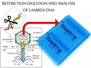
Restriction Analysis and Digestion of Lambda DNA. DNA is tightly p ackaged into c hromosomes which r eside in the nucleus. Model of DNA DNA is Comprised of Four Base Pairs. DNA Restriction Enzymes. • Used by bacteria to protect against viral DNA infection

E N D
DNA is tightly packaged into chromosomes which reside in the nucleus
DNA Restriction Enzymes • Used by bacteria to protect against viral DNA infection • Endonucleases = cleave within DNA strands • Over 3,000 known enzymes
Enzyme Site Recognition Restriction site Palindrome • Each enzyme digests (cuts) DNA at a specific sequence = restriction site • Enzymes recognize 4- or 6- base pair, palindromic sequences (eg GAATTC) Fragment 2 Fragment 1
Common Restriction Enzymes EcoRI – Eschericha coli – 5 prime overhang Pstl – Providencia stuartii – 3 prime overhang
Your tasks: • Cut lambda DNA into a series of fragments using restriction enzymes • To separate and sort a large group of DNA molecules according to theirsize
Important note: First add DNA, then restriction buffer, and then the enzymes to the tubes. Use a fresh pipette tip for restriction buffer and each enzyme.
The DNA Digestion Reaction Restriction Buffer provides optimal conditions • NaCI provides the correct ionic strength • Tris-HCI provides the proper pH • Mg2+ is an enzyme co-factor
Place the sample tubes in a 37°C water bath or oven for approximately 30 minutes • While you are waiting, this a good time to cast your agarose gel
DNA Digestion Temperature Why incubate at 37°C? • Body temperature is optimal for these and most other enzymes What happens if the temperature is too hot or cool? • Too hot = enzyme may be denatured (killed) • Too cool = enzyme activity lowered, requiring longer digestion time
Part 1. Prepare Your Samples for Electrophoresis • Add 2.0 μl of sample loading dye to each of the tubes marked L, P, E, and H in the foam tube holder. Use a fresh tip with each sample to avoid contamination
Part 2. Set Up Your Gel Electrophoresis Chamber • . Pour enough buffer into the box until it just covers the wells of the gel by 1–2 mm
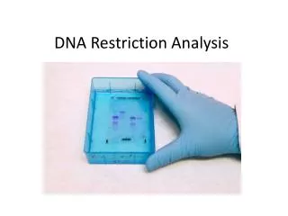
Part 3. Load your Samples and Run them by Electrophoresis • Pipette 10 μl from each tube (M, L, P, E, and H) into separate wells in the gel chamber • Important • Electrophorese at 100 V for 30–40 minutes
Restriction Fragment Length Polymorphism RFLP PstI EcoRI GAATTC GTTAAC CTGCAG GAGCTC Allele 1 1 2 3 CGGCAG GCGCTC GAATTC GTTAAC Allele 2 3 Fragment 1+2 Different Base Pairs No restriction site M A-1 A-2 Electrophoresis of restriction fragments M: Marker A-1: Allele 1 Fragments A-2: Allele 2 Fragments