Download
1 / 29
290 likes | 444 Views
HISTOLOGIC PATTERN OF LYMPH NODE BIOPSIES IN A TERTIARY HOSPITAL IN SOUTH EASTERN NIGERIA. MBATA G. C. 1,3 , NWEKE IG 2 , EGEJURU RO 2 , OMEJUA EG 1 , NWAKO OF 1 , CHIMA E I 3. INTRODUCTION Lymphadenopathy is a common clinical presentation in both medical and surgical clinics.

E N D
HISTOLOGIC PATTERN OF LYMPH NODE BIOPSIES IN A TERTIARY HOSPITAL IN SOUTH EASTERN NIGERIA. MBATA G. C.1,3, NWEKE IG 2, EGEJURU RO2, OMEJUA EG 1, NWAKO OF1, CHIMA E I3 .
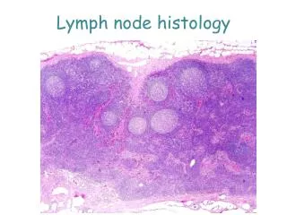
INTRODUCTION • Lymphadenopathy is a common clinical presentation in both medical and surgical clinics. • Causes are broadly divided into neoplasic and non neoplastic. • Non neoplastic causes predominate and range from infective to drug reaction, lipid storage disorders and inflammatory conditions. 1
Documented reports shows that non specific reactive hyperplasia are common in developed world while tuberculosis (TB) are common causes in developing world especially in Africa where HIV is quite common.2 • HIV apart from directly causing lymphadenopathy also contributes via several AIDS defining illnesses. 2, 3, 4.
Given the number of diseases causing lymphadenopathy, it is necessary to define the cause in a particular environment and age. • In children the cause is due to infective and reactive due to developing immune system. 5,6 • In elderly the cause is mainly due to malignancy4.
Clinical assessment of peripheral lymph node is easier. 7 • Assessment of visceral lymph node is more difficult since they require imaging assistance or laparotomy. 7 • Biopsy of lymph nodes in the upper part of the body is desired as they give better diagnostic yield. 7, 8 • Biopsy in the lower part of the body is less desired as they are characterized with nonspecific, inflammatory and fibrotic changes. 7,8.
Objective of the study • Data on the spectrum of diseases causing lymphadenopathy in the South eastern Nigeria are limited. • The study aims at investigating the causes and pattern of lymph node distribution in patients seen at FMC Owerri, Eastern Nigeria over a 4-year period.
Materials and methods • All cases of lymph node biopsies done from Jan 2010-Dec. 2013 were reviewed. • Clinical data regarding age, sex, anatomical sites of lymph node biopsies were obtained from request forms and case notes. • The relevant slides were retrieved from the archives of the Dept. of Pathology.
All slides were prepared from paraffin embedded blocks. • Routine stain done with eosin and haematoxylin. • Special stain done with ZN where necessary. • Cytogenetics, immunohistochemistry and molecular diagnostic technique like receptor genes rearrangement were not employed.
ETHICAL clearance was obtained from ethics committee of the institution. • Data analysis was done using SPSS version 16 Chicago IL.
RESULTS. • A total of 141 lymph node biopsies were done. • Constituting 6% of total histology during the 4-year period Jan.2010- Dec.2013 • Of the 141 cases; 60 males, 81 females. • M:F ratio was 1: 1.35. • Mean age 17.6 ± 8.5 years. see table 1.
TABLE IAGE DISTRIBUTION OF PATIENTS Mean Age = 17.6 ± 8.5years, Range = 68years (2-70 years).
TABLE 2 : • Shows the site, distribution and histological diagnosis and frequency of diff groups of lymph nodes. • Regional lymphadenopathy seen in 135 (95.7%). • Generilizedlymphadenopathy occurred 6(4.3%). • Cervical groups were frequently affected 64(45.4%). • Axillary groups 28(19.9%). • Supraclaviclar groups 12(8.5%).
Reactive hyperplasia most common cause 46(32.6%) • Tuberculous adenitis seen in 40 (28.3%). • Metastatic deposits seen in 27(19.1%). • Non hodgkins lymphoma 17(12.1%). • Hodgkins lymphoma 7(5%). • Onchocerciasis3(2.1%). • Rosai – Dorfman’s syndrome 1(0.7%).
TABLE 2SITE DISTRIBUTION AND HISTOLOGIC DIAGNOSIS TB – Tuberculosis. METAS -Metastasissis. NHL - Non Hodgkins Lymphoma. HL- Hodgkins Lymphoma. ONCHO- Onchocerciasis.
TABLE 3. • Shows histologic diagnosis, sex distribution and sex ratios of the patients. • Lymphadenopathy was more common in females. • Female ratios were higher in most conditions except non hodgkins lymphoma.
TABLE 3 HISTOLOGIC DIAGNOSIS, SEX DISTRIBUTION AND SEX RATIOS OF PATIENTS WITH LYMPHADENOPATHY
AFB were demonstrated in 12/40 (30%) of patients with TB adenitis. • All the patients with TB had voluntary testing and counseling for HIV. • 14/40 (35%) were HIV positive. • Of the 6 patients with generalized lymphadenopathy4/6 (66.6%) of them were HIV positive.
discussion • Palpable lymph nodes give an important clue to aetiologic diagnosis of disease condition. 2,9. • FNAC is commonly used but excision biopsy is the “gold standard’’. 2,9. • Biopsies were done on outpatient basis. • In line with most studies within and outside the sub region cervical lymph nodes were the most commonly biopsied, followed by axillary and supraclaviclar nodes. 2, 10.
The most common aetiologic factor in many studies was TB followed by reactive hyperplasia. 2, 3,4,7. • TB and reactive hyperplasia were seen to be more common in cervical lymph nodes. • Our study showed more female preponderance and more affectation in the young adults. • Documented evidence shows that TB is more common in the first three decades of life, reactive hyperplasia in the early years of life and malignancy in the elderly 2, 4,7.
Analysis of lymphadenopathy in the developing nations shows that infection remains an important cause. 5, 6. • TB remains an important cause in many developing nations 1,2,11. • Reactive hyperplasia and URTI (viral and bacterial) are also important cause in many developing areas 2,5,6. • Malignancy and reactive hyperplasia are more commoner in the developed world 12.
In our study of 141 cases, 46 (32.6%) had reactive hyperplasia. • TB which is the most common in many studies in our sub region was found to be the second aetiologic factor 40 (28.3%). • The percentage of TB found here is smaller than that recorded in many places. • Higher prevalence have been quoted in some series in India, Pakistan and Bangladesh 13 14.
The reason for this lower rate is because our study population included both adults and children. • Most studies were done in adult population only. • The reason for more children with reactive hyperplasia has been adduced to reaction to minor stimuli b/c of yet developing immune system 5,6,7. • In the United States reactive hyperplasia is more common cause 3, 12. • The reason being lower prevalence of TB and earlier detection of malignancy before onset of nodal metastasis 7.
Lymph node hyperplasia was also common in some studies done in India 15, South Africa 16 and Zimbabwe 9. • The hyperplasia appears to be a consequence of pathological process; an important factor is HIV 15. • Change in primary HIV lymphadenopathy ranges from mild follicular hyperplasia to diffuse to “burnt out” lymph node 15.
Lymphadenopathy due to metastasis was seen in 27(19.1%). • This is similar to other figures obtained in other Nigerian cities but significantly higher than those obtained from Zimbabwe 9 and Ethiopia 17. • In the US metastasis was found in 29% of cases 7, 12. • The most common cause of metastasis in our study was breast ca affecting the axillary nodes. This is in agreement with other studies done in Nigeria 14, 18.
Lymphoma constituted the most common malignancy causing lymphadenopathy 24(17%). • This is lower than values obtained in other Nigerian cities – Kano 23.6% 7, Ife 23.8% 19 and Jos 28.2% 4. • NHL was more common than HL supporting most other studies in the sub region and beyond 2, 20,21,22. • In the western world NHL was found to be 3-4 times more common than HL 23,24.
Onchocerciasis is a microfilarial infection that is common in the tropical sub saharan Africa. • Onchocerciasis was found in 3(2.1%) of our cases 13. • Found to be more common in females 13. • Also occurred predominantly in the inguinal lymph nodes 13.
CONCLUSION • Differential diagnosis of lymphadenopathy are many. • TB and reactive hyperplasia have remained the predominant cause in our environment, followed by metastasis and lymphoma. • Accurate diagnosis and early intervention is the key to good treatment outcome. • Definitive histological classification using modern technique like immunohistochemistry and cytogenetics should be made available in our tertiary institutions.
Rosai J. lymph nodes : in Ankerman’s surgical pathology, 2011. • Adeniji et al. peripheral lymphadenopathy in Nigeria. Afr. J of Med Sc. 2000. 3. Longo et al. lymphadenopathy. Harrison’s principles of Medicine 18th edition. 2012. • Obafunwa et al. primary lymphadenopathy in Jos. W. Afr. J of Med. 1992. • Lake et al. peripheral lymphadenopathy in childhood. Am J Dis. Child, 1978. • Narang et al. prevalence of TB lymphadenitis in Wardha district India. Int. J Tuberc. and L. Dis. 2005. 7. Ochicha et al. pathology of peripheral lymph node biopsies in Kano, Northern Nigeria. Ann. Afr, Med. 2007. 8. Ferrer R. Lymphadenopathy: differential diagnosis and evaluation. Am Fam. Med. 1998. • Sibanda et al. lymph node pathology in Zimbabwe. Q. J Med. 1993. 1 • Pindiga et al. Histopathology of primary peripheral lymph lymphadenopathy in Northern Nigeria. Nig. J of Surg. Research, 1999. 11. Bem et al. Importance of HIV –associated lymphadenopathy and TB adenitis in pts underoing lymph node biopsy in Zimbabwe.Br. J Surg. 1996. 12. Lee et al. Biopsy of peripheral lymph nodes. Am Surg. 1982. 13. Hemalath et al. TB adenitis in South-India. Tubercule 1972. • Chhadbra et al. A retrospective evaluation of non neoplastic superficial lympahenopathy. The Internet J. of Med 2005. 15. Mohan et al. Aetiology of peripheral lymphadenopathy in adults. Natl J. India; 2007. 16. Moore et al. diagnostic aspects of cervical lymphadenopathy in developing world. Paed. Surg. Int 2003. 17. Gestachew et al. pattern of histological diagnosis of cervical lymph node biopsies in Addis-Ababa. Ethiopian Med. J. 1999. 18. Obiora et al. pathology of lymphoreticular diseases in nigeria. Pion. Med. J; 2013. 19. Udani et al. TB. In Diseases of children in tropics and subtropics. London 1981. 20. Pindiga et al. histological types of lymphoma in North eastern Nigeria. Sahel Med. J (Nig.) 2002. 21. Adedeji. MO. Malignant lymphoma in Benin City, Nigeria. East Afr. Med;1989. 22. Obafunwa et al. Malignant lymphoma in Jos. Central Afr. J of Med. 1992. 23. Hartge et al. Cancer Surv.1994. 24. Croves et al. non Hodgkins lymphoma incidence in USA. J. Natl Cancer Inst. 2000.