Download
1 / 23
230 likes | 418 Views
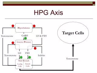
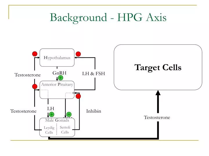
H ypothalamus. -. -. GnRH. LH & FSH. Testosterone. +. Anterior P ituitary. -. -. LH. Testosterone. Inhibin. +. +. Male G onads. Sertoli Cells. Leydig Cells. Background - HPG Axis. Target Cells. Testosterone. FSH. Micro. Albert Kwansa. encaps.

E N D
Hypothalamus - - GnRH LH & FSH Testosterone + Anterior Pituitary - - LH Testosterone Inhibin + + Male Gonads Sertoli Cells Leydig Cells Background - HPG Axis Target Cells Testosterone FSH
Micro Albert Kwansa
encaps Eric Lee John Harrison Client: Dr. Craig Atwood Advisor: Professor Murphy
ulation Yik Ning Wong
Background – Hypogonadism • Hypogonadism • General: Reduction or loss of gonad function • Target function: Testosterone production by leydig cells found in male gonads • Approach: Restore steroidogenic function of leydig cells
Challenges with traditional cell transplantation Immune Response Foreign Body Reaction Advantages of microencapsulation Cell entrapment Immunoisolation Selective transportation Sustained release of hormones from entrapped cells Micro-scale capsule size Cell Transplantation
Microcapsule Parameters Size exclusion via mesh size Microcapsule Size LH, FSH, O2, Nutrients Antibodies Testosterone, Wastes Biocompatibility Degradation
O Polyethylene glycol (PEG) • Synthetic polymer • Systematically variable mesh size • Non-biodegradable • Sustained cell protection • Bio-inert • Difficult for cells & proteins to adhere PEG PEGdA O O HO H O n n O
Previous Work • Used capsule size of 100µm diameter • Observed cell viability out to 8 days and detected negligible testosterone release • Current approach for improvements • Microcapsule size • UV exposure time • Adhesion peptide incorporation
Project Design Statement • Design PEGdA hydrogels for the encapsulation of Murine Leydig Tumor Cells in an effort to increase cell viability and testosterone secretion. • PEGDA hydrogels must provide immunoprotection and allow effective diffusion of oxygen, nutrients, hormones, and metabolic wastes.
Thickness (Micrometer) 0.0% 0 50 100 150 200 250 -10.0% -20.0% -30.0% Percent Change in Oxygen Concentration -40.0% -50.0% -60.0% Percent Change in Oxygen Concentration at Various Hydrogel Thicknesses as Compared to the Oxygen Concentration at the Site of Implantation Thickness Parameter • Testing Range = 25µm ~ 250µm • Tissue Implant size = 40µm ~ 200µm
Thickness Methodology Tape Spacers Liquid PEGdA Microscope Slide (Base) Microscope Slide (Top) Ready for UV Exposure
UV Exposure on Mechanical Properties of PEGdA • Cross Linking (Swelling Ratio) • Mesh Size • Stiffness of the PEGdA Network
Perfuse w/DI H2O, wait until equilibrium swelling is attained, and take second digital snapshot Fabricate thin gels under different UV times & take digital snapshot Compute change in volume via imaging software UV & PEGdA Hydrogel Swelling
Swelling Ratio Methodology Reduced Hydrogel Swollen Hydrogel
UV radiation on Cell Viability • PC3 cell culture in 10% serum media • Manual counting via hemacytometer • UV Time: 0 to 50 min @ 10 min intervals • Incubation for 18 hours at 37oC • Cell Titer-Blue Cell Viability Assay Fluorescence • Normalization of fluorescence to cell number
Expected RGD Results • RGD of different concentration and cells are injected into PEGdA • 0 – 2.5 mM RGD RGD effects on secretion
Expected RGD Results Macrophage Density adherence on RGD-PEG Adherent Macrophage Density
Pilot Study Conclusion • Mesh size: 4-5 nm • UV exposure time: <10mins • RGD concentration: <2.5mM
Future Work • Perform cell viability experiments up to 10 min at smaller increments • Cell counting via PicoGreen DNA Assay
References • Mellott. M, Searcy. K, Pishko. M (2001). Release of protein from highly cross-linked hydrogels of poly(ethylene glycol) diacrylate fabricated by UV polymerization. Biomaterials 22(9):929-41. • Muschler. G, Nakamoto C, Griffth L (2004). Engineering Principles of Clinical Cell-Based Tissue Engineering, The Journal of Bone and Joint Surgery (American) 86:1541-1558 • Yang. F, Williams. C, Wang. D, Lee. H (2004) The effect of incorporating RGD adhesive peptide in polyethylene glycol diacrylate hydrogel on osteogenesis of bone marrow stromal cells. Biomaterials. 2005 Oct;26(30):5991-8.
Acknowledgements • Dr. Craig Atwood, VA Hospital • Professor William Murphy • Professor Kristyn Masters • Professor John Kao • Dr. Daesung Lee • Amy Chung • Yi Jin Kim • Eun Jin Cho