Download

1 / 37
370 likes | 546 Views
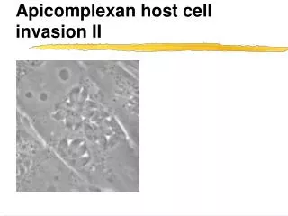
Apicomplexa host cell invasion. PV. Toxoplasma gondii - overview from last lecture. Sexual reproduction in the cat, very wide range of intermediate hosts Two asexual form, tachyzoites & bradyzoites , responsible for acute and chronic stages, infection is for life.

E N D
Toxoplasma gondii - overview from last lecture • Sexual reproduction in the cat, very wide range of intermediate hosts • Two asexual form, tachyzoites & bradyzoites , responsible for acute and chronic stages, infection is for life. • Toxoplasma is an opportunistic pathogen (severe pathology in immuno-compromised individuals) • T. gondii is an obligate intracellular parasite
Host cell invasion can be divided to different elements Attachments & PV formation (Kiss and spit) Gliding Penetration through the moving junction PV modifications
Host cell invasion can be divided to different elements Attachments & PV formation (Kiss and spit) Gliding Penetration through the moving junction PV modifications
Everyone has their own move • Flagella • Amoeboid movement • Toxoplasma - gliding motility
Different parasites use different mechanisms to invade cells • Trypansoma cruzi -- recruites lysosomes, then escape to replicate in the cytoplasm. • Leishmania -- invades macropahges by phagocytosis and is adjusted toextreme conditions of phagolysosome • Mycobacterium tuberculosis, -- enter by phagocytosis and blocks lysosomal maturation • Toxoplasma -- actively penetrate host and resides it own a specialized parasitophorous vacuole.
Invasion depends on parasite not host cell actin Salmonella • Cytochalasin D (CytD), a drug that prevents actin polymerization • Host cell invasion by Salmonella (which is taken up by phagocytosis) is inhibited by CytD. • Same goes for Toxoplasma. • In Salmonella, invasion can be rescued by using a drug resistant host cell mutant (Cyt1). • Toxoplasma invasion remains inhibited in the drug resistant host cell. • Generating a drug resistant parasite rescues invasion Toxoplasma
Motility element alone is dependent on actin polymerization • CytD treatment does not appear to inhibit attachment (bar graph in c shows number of parasites bound to cells at different drug doses) • CytD inhibits the movement of the parasite into the host cell. • A parasitophorous vacuole (PV) is still set up, however the parasite can not move in, and the moving junction remains at the apical tip of the parasite
A special myosin is required for T. gondii motility Normal myosin • Toxoplasma and other Apicomplexa have a parasite specific myosin (MyoA) • This myosin is localized right under the surface membrane of the parasite • Using genetic engineering a mutant parasite was constructed in which this myosin can be suppressed • Suppression of myosin result in loss of parasite motility (as seen for actin loss of motility also causes loss of host cell invasion) Suppressed myosin
The conveyor-belt model of gliding motility • Gregarines are a group of ‘primitive’ apicomplexans which parasitize invertebrates • In comparison to Toxoplasma or the malaria parasite these are fairly large cells, which makes them good subjects for microscopy studies
The conveyor-belt model of gliding motility • Beads attached to the surface of gregarines are ‘treadmilled’ to the end of the parasite cells • This suggests that the motility machinery moves little “hands” over their surface from the apical to the basal end of the parasite • Traction
Gliding parasites deposit proteins and lipid trails • Like a slug the parasite leaves behind a trail of surface proteins and lipids • A protease cleaves the little hands to disengage the parasite from the surface and allow for propulsion (sheddase)
Invasion depends on sequential protein secretion from three organelles Micronemes (Mn) Rhoptries (Rh) Dense granuoles (DGs)
Are micronemes the little hands acting during invasion? • Microneme proteins (MICs) can bind to a variety of carbohydrates found on the surface of cells • Secretion of micronemes brings MICs proteins to the parasite surface • Micronemes are secreted at the apical tip of the parasite and move to the posterior. This movement is blocked by cytD! • Are MICs the little hands?
MIC2 is required for T. gondii motility and invasion Using genetic engineering a mutant parasite was constructed in which the expression of a microneme protein can be suppressed
? ? ? ? ? ? Several molecules act together to create the gliding machinery - the glideosome
PM IMC MT The gliding machinery is anchored in the inner membrane complex
The conveyor-belt model • Motility depends of parasite actin/myosin • Myosin is anchored into the inner membrane complex membrane • Short actin filaments form and are moved towards the posterior end of the parasite by the myosin power • The short actin filaments are linked to microneme proteins by an adaptor -- movement of actin filaments results in movement of microneme proteins holding host cell receptors • The parasite glides over the substrate • Microneme proteins are shed at the back end
Host cell invasion can be divided to different elements Attachments & PV formation (Kiss and spit) Gliding Penetration through the moving junction PV modifications
Rhoptry proteins are involved at various points of the invasion process • Secretion of rhoptries is required for the formation of the parasitophorous vacuole • Like micronemes rhoptries are secreted at the apical tip of the parasite • One group of very interesting rhoptry proteins is injected into the host cell and manipulates gene expression in the host nucleus as well as start-up the PV -- ‘kiss and spit’ • Some rhoptry proteins make up (part) of the moving junction • Other rhoptry proteins are found throughout the membrane of the parasitophorous vacuole (PVM)
Rhoptry release upon attachment - kiss and spit • It appears that a variety of rhoptry proteins are directly injected into the host cell and that this is involved in the formation of the parasitophorous vacuole
Rhoptry proteins modify the function of the host cell nucleus • Several rhoptry proteins are injected into the host cell cytoplasm during invasion • They accumulate in the host cell nucleus • Interestingly, some of them are enzymes capable of changing the phosphorylation state of proteins (kinases & phosphatases) • Their precise function remains to be determined but it appears that they modulate gene expression in the host cell and that their activity is required for rapid growth and the ability to cause disease (virulence)
Second role of Rhoptry proteins during invasion - The moving junction
? ? ? PV Rhoptry proteins organize the moving junction
The moving junction skims proteins out of the membrane as the PV forms • The surface membrane of the host cell was labeled for protein (green) and lipid (red) prior to infection with Toxoplasma (blue) • Note that while the lipids are clearly visible in the vacuole the proteins are excluded
Where does the membrane for the PV come from? • Patch clamp cells and follow invasion by video microscopy • Certain electric properties of the cell (their capacitance) can be used as a measure of their total surface membrane • If membrane is parasite derived the surface area should grow during invasion if it is derived from the host cell surface the area should stay constant
Where does the membrane for the parasitophorous vacuole come from? • The PV membrane is derived from the host cell plasma membrane • The PV is provided by the parasite (e.g. by secretion from the rhoptries) • Both contribute to the PV
Rhoptry proteins are responsible for host cell mitochondria and ER recruitement to the PVM
Host cell invasion can be divided to different elements Attachments & PV formation (Kiss and spit) Gliding Penetration through the moving junction PV modifications
Dense granules proteins modify the PV environment - Toxo makes a home • Secretion of dense granules occurs after the vacuole is fully formed and continues throughout the intracellular growth of the parasites • Dense granule proteins likely play a role in modification of the vacuole into an environment supportive of parasite growth
Dense granule proteins are secreted into the PV • Certain dense granule proteins are soluble in the lumen of the PV others integrate into the membrane • These proteins are probably involved in modifying the vacuole
Dense granules are involved in establishing the intravacuolar network
Apicomplexan invasion • Active, parasite driven process • Depends on parasite actin/myosin motility (conveyor belt model) • Involves secretion of three major groups of proteins: • MICs from micronemes (motility, Moving-Junction). • ROPs and RONs from rhoptries (spit, parasitoforouse vacuole & moving Junction). • DGs from dense granules (makes PV into a suitable home).