Download
1 / 61
620 likes | 849 Views
Acute Kidney Injury No Good. Just Bad and Ugly. CM Yuan (with bits by KC Abbott) 3/2010. The Overall Approach To AKI. Determine if the patient actually has acute kidney injury, i.e., establish sudden loss of function. Determine etiology: Post-renal Pre-renal Intra-renal

E N D
Acute Kidney InjuryNo Good. Just Bad and Ugly. CM Yuan (with bits by KC Abbott) 3/2010
The Overall Approach To AKI • Determine if the patient actually has acute kidney injury, i.e., establish sudden loss of function. • Determine etiology: • Post-renal • Pre-renal • Intra-renal • Determine treatment and need for renal replacement therapy
What is AKI? “…Precipitous impairment of kidney function without regard to etiology or mechanism.” Rudnick, et al, Acute Renal Failure, 1988. Therefore, acute kidney injury (or acute renal failure) is a syndrome in which renal function is declining rapidly (ie: not in a steady state), and which can be the common manifestation of a wide variety of local or systemic mechanisms/diseases. Difficult to precisely define AKI “numerically”. Older literature didn’t clearly define AKI, and different studies used different definitions. Clinically it was a “I know it when I see it.” kind of diagnosis.
Clinical Features of AKI • Accompanied by urea retention (azotemia), failure to clear other waste products, and dysregulation of extracellular volume and electrolytes. • Usually marked by a rise in serum creatinine concentration or azotemia. • In early stages the sCr may be low even though the GFR is markedly reduced. • Don’t believe the reported eGFR derived from serum creatinine in a setting of AKI! The patient is NOT in a steady state, so the assumptions behind the eGFR calculation do not apply. • e.g., sCr rised from 0.8 to 1.3 to 2.3 mg/dL. eGFR reported as 40 ml/min/1.73m2, but rate of rise suggests GFR truly <10-15!
Rises in serum creatinine and BUN don’t necessarily mean AKI is present! • Serum creatinine is determined by both input (muscle mass and breakdown) and output (GFR and proximal tubular secretion). • sCr may increase without a GFR decline when medications (eg, cimetidine, trimethoprim) that inhibit the tubular Cr secretion are given. • sCr may also increase when there is muscle injury, as in rhabdomyolysis, over and above any decline in GFR. • ACE/ARBs can increase sCr due to glomerular hemodynamic effects, reducing GFRin the short term, but stabilizing it over the long term. They are often d/c’d in AKI or before high-risk procedures (surgery, contrast). • A rise in the BUN level can occur without renal injury, such as in GI or mucosal bleeding, steroid use, or protein loading.
So the Creatinine is elevated…AKI vs. CKD • Is it Acute? • This is the first step in assessing the “acute” loss of renal function! • Findings that suggest acute presentation of CKD or AKI complicating CKD: • Documented chronic increase in serum creatinine • Small kidneys on renal imaging • Findings c/w osteitis fibrosa cystica on x-ray • Relatively asymptomatic at very high levels of BUN and creatinine
RIFLE Criteria:An attempt to standardize the diagnosis of AKI for research purposes Risk: > 150-200% increase in s.creatinine; UO < 0.5 ml/kg/hr for > 6 hrs. Injury: > 200-300% increase in s. creatinine; UO < 0.5 ml/kg/hr for > 12 hrs. Failure: > 300% increase in s. creatinine or serum creatinine ≥ 4 mg%; UO < 0.3 ml/kg/hr for ≥ 24 hrs or anuria ≥ 12 hrs. Loss: Complete loss of renal function lasting > 4 weeks (need for RRT) ESRD: End-stage renal disease > 3 mo ADQI Workgroup; Critical Care. 2004
RIFLE: Validation • RIFLE was an attempt to standardize the definition of AKI, but it was originally conceived by committee, and NOT based on evidence. • Multiple studies have validated the RIFLE criteria as predictive of increasing mortality with increasing RIFLE class (Ricci, et al. KI. 2008. 73: 538.). • Compared to non-AKI, there is a stepwise increase in RR for death from Risk (RR=2.40) to Injury (RR=4.15) to Failure (RR=6.37); p<0.0001 for all.
AKINModifications to the RIFLE • The Acute Kidney Injury Network (AKIN) modified the RIFLE to include less severe AKI, and recommended volume correction and work-up for obstruction. • This is the group that recommended Acute Kidney Injury (AKI) replace the term ARF. • One problem with AKIN and RIFLE is that the underlying assumption is that AKI is ATN. Some AKI is NOT ATN, and a diagnostic work-up always should be done. • Renal Failure (RIFLE) or Stage 3 (AKIN) is not an indication for dialysis. It is a definition, NOT a decision threshold! • RIFLE and AKIN are epidemiologic and research tools (to define inclusion and endpoint criteria for clinical studies). Their clinical utility (except for prognosis, perhaps) is not clear.
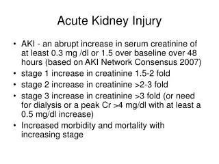
AKIN CriteriaModification to RIFLE (Mehta, et al. Crit Care. 2007) • Stage 1: Abrupt (within 48 hrs) increase in serum creatinine ≥ 0.3 mg/dL or increase to ≥ 150 to 200% from baseline or UO < 0.5 ml/kg/hr > 6 hrs. • Stage 2: Increase in serum creatinine to > 200 to 300% from baseline or UO < 0.5 ml/kg/hr > 12 hrs. • Stage 3: RRT or increase in serum creatinine > 300% from baseline (or serum creatinine ≥ 4.0 mg/dL with an acute increase of at least 0.5 mg/dL or UO < 0.3 ml/kg/hr for 24 hrs or anuria for 12 hours. Should only be used after volume status is optimized, and obstruction should be ruled-out if oliguria/anuria is the only diagnostic criteria. (Editorial comment: We’re only supposed to look for obstruction if there is oliguria alone? Lots of urinary obstruction presents with increased serum creatinine alone and polyuria!)
AKI: Acute Tubular Necrosis ATN (acute tubular necrosis) first described by Bywaters and Beal (BMJ, 1941) in patients who sustained crush injury during the London Blitz. Death was due to hypotension and hyperkalemia (the sine wave EKGs were included in the paper!). Autopsy showed necrosis of the renal tubular epithelial cells. Most AKI in the ICU-setting is due to ATN (acute tubular necrosis). Most patients with ATN will recover renal function spontaneously, IF they survive the period of renal failure, usually 7-21 days (unreferenced nephrology lore). This is usually heralded by an increase in UO, which was well-described in the days before RRT was available.
AKI: The course of ATN Teschan (Ann Intern Med, 1960) describes the course of ATN: Defined as acute decline in UO ≤ 400 ml/24 hr with compensated volume status, and no obstruction of renal vasculature or lower urinary tract. Oliguric phase: Continues until UO ≥ 1000 ml/24 hr (called “the day of diuresis). Early diuretic phase: Begins on day of diuresis, and continues until BUN reaches peak. Late diuretic phase: Begins on day of BUN peak, and continues until BUN < 30mg%. Recovery phase. “High output renal failure” exists when UO > 400 ml/24 hours.
Renal Tubular epithelial cells (RTE)’s are actually the sine qua non of ATN, shown above; A is a squamous epi, and B is an RTE: rounder, smaller and more symmetrical. Granular casts can be seen in hyperbilirubinemia without ATN
AKI--Prognosis • Despite 60 years of RRT, hospital-acquired AKI remains associated with about 40-60% mortality. • Study Group on Acute Renal Failure--BAMC (1957). Retrospective series of 1044 ARF patients. 49% mortality despite availability of HD for acute indications. • 66% mortality for post-traumatic ARF. • 30-32% mortality for obstetric- or nephrotoxin- associated ARF.
Clinical prognosis of AKI patients who require RRT is poor! • VA/NIH Acute Renal Failure Trial Network Study (NEJM, 2008) of subjects with AKI requiring RRT, 6 month overall mortality 52%. • Only 17% overall had recovery of renal function by Day 28. • Delannoy, et al (Int Care Med, 2009): prospective, observational, multi-center study of 205 adult patients undergoing RRT in ICU. 6 month mortality 62%. • 12% of survivors on RRT. • Lin, et al (Am J Surg, 2009): prospective, observational, multi-center study of 342 patients s/p post-op AKI requiring RRT (2002-2006). 90 day mortality 60%. • Independent predictors of mortality: older age, sepsis, higher SOFA score, CPR, TPN, need for CRRT.
Think About the Anatomy!The Black Box Approach to AKI Heart Renal a. Aorta Kidney--glomeruli, tubules, interstitium, intra-renal vessels Kidney Ureter Bladder Prostate (in some people...) Urethra
AKI: The Initial Differential • Pre-renal: due to decreased renal blood flow • Occurs when renal perfusion is decreased, without associated tissue injury • Most common cause of AKI in outpatients (50-70%) • 30-40% of hospital-acquired AKI • Function rapidly improves after perfusion is restored • Post-renal: due to obstruction of urine flow • Results from obstruction of urine flow at any site along the tubule/collecting system • Least common type of AKI, 2 - 10% • Reversible if obstruction is relieved in a timely manner
AKI: The Initial Differential (cont.) • Intra-renal: AKI due to an intrinsic, parenchymal renal insult • Wide differential includes ATN, but not exclusively ATN • 30-50% of all AKI • 60-70 % of inpatient AKI • Up to 80% of AKI in ICU • Combinations of the above
Pre-Renal Causes of AKI • Cardiac/CHF: Decrease in CO with resultant arterial under-filling--total body volume often expanded • Volume depletion Environmental, intake deficit, and fever GI (vomiting, diarrhea) Renal (especially over-diuresis) • Third Spacing/Peripheral Vasodilation/Hypotension Hypoalbuminemic states (cirrhosis, nephrosis) Sepsis, burns, crush injuries Anti-hypertensive drugs • Vascular Disasters: Aortic/Renal artery disruption
Using the FENa FENa = (UNa x Pcreat)/(Ucreat x PNa) x 100, where UNa = urine Na (meq/L); PNa = plasma Na (meq/L); Ucreat = urine creatinine (mg%); and Pcreat = plasma creatinine (mg%). In Nonoliguric States • FENa < 1% in pre-renal azotemia • FENa > 3% in oliguric ATN Miller, et al, Ann Intern Med, 89:47, 1978 Exceptions may be seen in acute GN, early obstruction, diuretic use, pre-renal states associated with metabolic alkalosis, with NSAIDs, early in rhabdo, iv contrast ATN.
Using FEUrea • In those who are receiving diuretics, a fractional excretion of urea (FEUrea) can be obtained since urea transport is not affected by diuretics. • With loop or thiazide diuretic use, urine Na excretion is increased distally due to inhibition of Na reabsorption. FEUrena = (Uurea x Pcreat)/(Ucreat x Purea) x 100,where Uurea = urine urea (mg/dL); Purea = plasma urea (mg/dl); Ucreat = urine creatinine (mg%); and Pcreat = plasma creatinine (mg%). • FEUrea < 35%is suggestive of a prerenal state.
Post-renal Causes of ARF • Need to Think Anatomically! -Renal Pelvis and Ureter Blood clot, stone/crystals, sloughed papilla, fungus ball, malignancy, retroperitoneal fibrosis. Iatrogenic obstruction (esp. in patients known to be obstructed, or post retroperitoneal surgery) -Bladder Prostate, stone, blood clots, malignancy, neuropathic (drugs, neurologic damage, systemic disease) -Urethra Strictures, iatrogenic (the obstructed Foley)
Intra-renal Causes of ARF • Glomerulonephritis Post-infectious, SLE, MPGN, RPGN, HSP With a “nephritic sediment”, it is useful to send C3/C4, which are depressed in acute nephritis (which has a very limited differential: post-strep GN, SLE, MPGN (including HepC), cryoglobulinemia, endocarditis • Systemic/Intra-renalVasculitis HUS, Wegener’s, Hypersensitivity angiitis, Scleroderma, PAN/microscopic PAN • Interstitial Nephritis Drugs, infection, infiltrative (tumor/sarcoid) • Acute Tubular Necrosis A diagnosis of exclusion! Multiple etiologies and (as yet) imperfectly understood pathophysiology!
Getting There….The Diagnostic Approach to AKI • History and Physical Examination “A careful history and physical examination almost always uncovers the most likely causes of ARF…” Rudnick, et al, Acute Renal Failure, (ed, Brenner & Lazarus) 1988 • Some Often Overlooked Parts of the History & Physical -Assessment of Volume Status: especially orthostatic pulse and BP, weight, edema -Historic or physical signs suggesting obstruction: sx of prostatism, history of a single kidney, pelvic exam -Medications: prescribed and OTC (esp. alternative tx)
Getting There….The Diagnostic Approach to AKI • The Flow Sheet Approach in complicated AKI • Non-invasive Tests -Urine Volume: anuria vs. oliguria vs. non-oliguria Anuria (100 cc/day or less) suggests strong possibility of urinary obstruction or vascular disaster. -BUN and Creatinine: ratio, rate of rise, U/P ratio -Fractional Excretion of Na (FENa) or Urea (FEUrea) -Urinalysis: may yield important diagnostic information in 70-80% of patients (Levinsky, et al, 1976).
Getting There….The Diagnostic Approach to ARF • Radiologic Studies -Renal Ultrasound and Doppler Flow Studies -Radioisotope studies (to establish renal arterial flow) -Renal arteriogram and venogram* • Other Invasive Tests -Central hemodynamic monitoring -Fluid challenge (only if you have reasonable evidence of volume depletion) -Renal Biopsy
Radiologic Rules to Live By…. • Don’t use iv contrast dye unless you have a very good reason. • To rule out cortical necrosis, renal arterial/aortic interruption, polyarteritis nodosa, or renal vein thrombosis. • NEVER use gadolinium. • The radiologist is your friend. Call and ask about the best way to get the job done. • Doppler US, radionuclide scanning, and CT and MR without contrast can sometimes be adequate.
The AKI Game Directions: 1. Read each case and answer the question. 2. Scoring will be done on the following scale. 3. You can score between 50 and -50 points. 40 to 50 points: Speak with me about a nephrology fellowship. -40 to -50 points: Hire a lawyer. All others: We all need to read more...
Case 1 24 y/o male presents with a 10 day history of myalgia, fatigue, anorexia, dark urine, decreased urine output, and lower extremity edema. PMH: + treatment with topical antibiotics for impetigo 4 weeks prior to admission. MEDS: None. PE: BP 172/106, P92 + bilateral lower extremity edema + 1 cm diameter crusted lesion with surrounding erythema on right upper arm, with tender right axillary adenopathy.
Case 1 (continued) LABS: BUN 94 mg%; creatinine 9.3 mg% CXR: wnl. UA: sp. gravity 1.010, pH 6.0, 2+ blood, 3+ protein. 15-20 rbc/hpf, + 1-2 granular cast/lpf. Mrs. Kiandoli calls you from the lab to have you look at this UA sediment finding
Case 1--The Question 1. What test would you get next to evaluate this patient? a) Renal ultrasound for renal size b) Renal arteriogram to r/o PAN c) ANCA panel/anti-GBM d) C3/C4 and ASO titer
Case 1--The Answer Is... d) C3/C4 and ASO titer (+10 points) This is post-streptococcal glomerulonephritis with ARF. Culture of skin lesion was + for group A beta hemolytic strep. C3 was depressed (29), ASO titer was v. elevated. Renal biopsy c/w clinical diagnosis.
Case 1--Scores for the Other Answers... a) Renal US: 12 cm bilaterally, wnl (0 points) b) A-gram to r/o PAN: Nl renal a-gram, patient sustains femoral arterial tear requiring surgical repair. (-10 points) c) ANCA and anti-GBM titre: Both return 2 weeks later, negative. (-2 points) The point of this case is to recognize that this is an “acute nephritis”, and that the proper work-up is to consider that differential for that syndrome. The combination of impetigo and a delayed development of AKI (without other systemic symptoms) suggests post-strep GN. The treatment was antibiotic therapy and RRT. The patient recovered.
Case 2 85 y/o male with a recent history of retinal toxoplasmosis presents with fatigue, malaise, anorexia, decreased urine output, and dark urine. PMH: + History of benign prostatic hypertrophy + History of hypertension x 35 years, with presumed secondary CKD, baseline creatinine of 2 mg% MEDS: Sulfadiazine 4 gm qid Cipro 500 mg bid Septra DS 1 tab po BID Captopril 25 mg qid
Case 2 (continued) PE: BP 122/70, P65, Tilt negative CV: +S4, no murmur. Abdomen benign. Prostate firm, large, with no nodules. No edema. LABS: BUN 70 mg%, creatinine 7.3 mg% UA: sp. gravity 1.011, pH 6.0, large blood, 100 mg% SSA, TNTC WBC and rbc/hpf and crystals shown.
Case 2: Crystals seen on urine microscopy …My! He’s on a lot of sulfa…
Case 2--The Question 1. What single test will best assist you in determining the etiology of ARF? a) Foley catheterization. b) Urine eosinophils. c) Renal ultrasound. d) CO2 arteriogram to r/o bilateral renal artery stenosis.
Case 2--The Answer Is... c) Renal ultrasound (+ 10 points) This demonstrated bilateral pelvocaliectasis with numerous echogenic foci c/w sulfadiazine stones. The crystal is a typical sulfa “wheat sheaf” crystal. The patient required acute dialysis, bilateral percutaneous nephrostomy and urinary tract lavage. He recovered, but was left with Stage 4 CKD.
Case 2--Scores for the Other Answers... a) Foley catheter: placed successfully, UO 20 cc/hr (+ 5 points) • Urine eosinophils: negative (0 points) • Renal a-gram: arteries with insignificant narrowing--patient has atheroembolic event, develops small bowel infarct, and dies. (-10 points) The point of this case--the urinalysis is always useful, and make sure to rule out obstruction!
Case 3 68 y/o male admitted to CCU with unstable angina. Stabilized with iv TNG/heparin/B-blocker. Cardiac catheterization and angioplasty within 48 hours of admission. Developed a large hematoma after sheath removal, and is at bedrest. Baseline s. creatinine--1.4 mg%. One day post procedure, creatinine was 1.9 mg%, and is 2.4 mg% today. ROS: + urinary frequency, hesitancy (has difficulty urinating supine, but has “managed”). + mild nausea since cath. Foley placed at time of cath, removed with no difficulty. PMH: HTN x 20 years, AODM (diet controlled x 5 years), Chronic insomnia.
Case 3 (continued) MEDS: Isordil 20 mg tid Atenolol 50 mg qd ASA 325 mg qd Lisinopril 10 mg qd Elavil 75 mg qhs Phenergan 25 mg prn nausea D5 1/2 NS at 100 cc/hr PE: BP 135/86, P 56 regular, Tilt negative. No skin changes c/w atheroemboli. CV: +S4. Lungs clear. No peripheral edema. Abdomen soft, + BS, mild suprapubic tenderness. Right groin hematoma resolving. UA: Sp. gravity 1.010, pH 6.0, trace protein, occ. fine granular/hyaline cast.
Case 3--The Question 1. What is the next step in evaluating this patient? a) Conservative management of iv contrast induced ATN. b) Foley catheter placement. c) Renal ultrasound. d) 24-hour urine for protein and creatinine.
Case 3--The Answer Is… b) Foley catheter placement. (+10 points) Acute/chronic bladder outlet obstruction/urinary retention. Post-void residual--400 cc. Prostate was enlarged. Contributing--Phenergan, Elavil, the need to urinate supine, and ? urethral trauma from previous Foley.
Case 3--Scores for the Other Answers… a) Conservative management: Renal function deteriorates. Sudden hyperkalemia, arrythmia, and death (-10 points). c) Renal ultrasound--mild pelvocalyceal dilation and a very enlarged bladder--Foley placed post (+5 points). • 24-hour urine: dropped in the lab--housekeeping called (0 points). Always assess for obstruction. Always….
Case 4 65 y/o male with squamous cell ca of the piriform sinus. 7 days prior to admission, he received 100 mg/m2 cisPlatin, followed by 5 days 5-FU. 3 days prior to admission he developed severe mucositis, fatigue, weakness, and poor oral intake. Now presents with above symptoms. PMH: HTN x 20 years. 60 pack year smoker. Baseline s. creatinine--1.1 mg%. MEDS: Maxzide 1 po qd Atenolol 100 mg qd
Case 4 PE: BP (standing) 98/54, P 72 BP (supine) 154/62, P 60 Weight 198 lbs (206 lbs at last follow-up). Severe oral mucositis. Lungs clear. CV no gallop, murmur, or rub. Abdomen soft, and non-tender. Prostate mildly enlarged. No edema. LABS : Na 136, K 3.7, Cl 97, HCO3 23 BUN/creat--60/3.6 mg%. WBC 2.6K, Plat 73K UA: Sp gravity 1014, pH 5.0, 0-1 fine granular cast/lpf.
Case 4--The Question 1. What is the next step in this patient’s evaluation? a) Urine eos--? Maxzide-induced AIN. b) Send urine for FENa to determine etiology and guide therapy. c) Volume replete with NS until tilt negative. d) Foley placement to monitor urine output.
Case 4--The Answer Is... c) Volume repletion until tilt negative (+10 points) Although this may be ATN due to cisPlatin, he is volume depleted, and must be resuscitated. Poor po intake, diuretics, tubular damage due to cisPlatin toxicity all may contribute. Creatinine came down to 2.1 mg% with volume repletion alone.
Case 4--Scores for the Other Answers... a) Urine eos: negative (0 points). b) FENa= 2%, not helpful. Nephrology staff abuses you at morning report for not volume repleting the patient promptly (-5 points). • Foley: UO 20 cc/hr. This immunocompromised patient develops urinary sepsis and multi-organ failure syndrome (-10 points). This is an example of pre-renal AKI. Urine eosinophils can be suggestive of AIN, but they are not diagnostic of it. FENa > 1% will not be helpful in a patient on diuretics. Foley catheters are dangerous due to the infection risk, and should only be used if indicated.
Case 5 21 y/o female s/p cadaveric kidney transplant 7 years ago for ESRD due to renal dysplasia. Maintained on CyA 100 mg bid and Medrol 10 mg qod with creatinine of 2.4 mg%, 6 months ago. Returns after 4 month work-study experience in Australia for medication refills and follow-up labs. LABS: BUN/creat--290/25.5 mg%, Na 131, K 5.0, Cl 97, HCO3 8, Ca 5.2, PO4 10.4. Hct 24%. ----------------------------------- You call her at home, and ask that she come back immediately for evaluation (with someone else driving…)