Download
1 / 54
730 likes | 2.43k Views
Mastication. Sitthichai Wanachantararak. Digestive System. Functions Prehension, ingestion Mastication Deglutition Digestion Absorption of nutrients Elimination of undigestible/undigested food products Other. From Mouth to Stomach. Mastication (chewing): Mixes food with saliva -

E N D
Mastication Sitthichai Wanachantararak
Digestive System • Functions • Prehension, ingestion • Mastication • Deglutition • Digestion • Absorption of nutrients • Elimination of undigestible/undigested food products • Other
From Mouth to Stomach • Mastication (chewing): • Mixes food with saliva - • Amylase = enzyme that can catalyze the partial digestion of starch. • Deglutition (swallowing): • Involves 3 phases: • Oral phase is voluntary. • Pharyngeal and Esophageal phases are involuntary. • Cannot be stopped. • Larynx is raised. • Epiglottis covers the entrance to respiratory tract
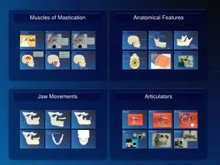
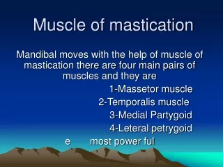
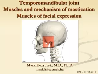
Muscles of Mastication Figure 10.7a
Muscles of Mastication Figure 10.7b
Action of muscles during masticatory movements • Opening / Depressor jaw muscles • mylohyoid / digastric / inferior lateral pterygoid • Closing / elevator jaw muscles • medial pterygoid / superficial masseter / tempolaris
Extrinsic Tongue Muscles Figure 10.7c
Tongue • Consist of 2 groups: intrinsic and extrinsic muscles. • Intrinsic muscle: change in tongue shape • Extrinsic muscle (eg. Genioglossus): response for protrusion and retrusion of the tongue Three major muscles that anchor and move the tongue • innervated by cranial nerve XII (hypoglossal nerve) • Complete tongue activity occurs in jaw movements and respiration, speech, taste, mastication, swallowing, and sucking.
Chewing • Activity of masticatory musclesduring chewingreflected • jaw-tracking devices and EMG • amplitude • onset timing • duration of the chewing cycle • Variation is related to occlusal contact relation and musculoskeletal morphology
Mastication : The crushing & grinding • 1 chewing cycle = opening + closing + power stroke • chewing sequence = numerous chewing cycle • chewing sequence could be divided into • preparatory series • reduction series • pre-swallow series
Mastication : The crushing & grinding • opening stroke • closing stroke / fast stroke • power stroke • puncture-crushing • tooth-tooth contact -buccal phase / phase I -lingual phase / phase II
Opening • Start from static intercuspal position, where jaw movement pauses for 194 ms in chewing cycle, • muscle activity begins in the ipsilateral inferior head of the lateral pterygoid muscle approximately half way through the period of tooth contact. • Follow closely by the action of the contralateral inferior lateral pterygoid muscles. • Both superior and inferior head of the lateral pterygoid muscle are active during the opening phase.
Opening • Early in the opening phase, digastric musclesbecome active and remain until maximum opening position • During the opening phase, masseter, temporalis, medial pterygoid, and superior head of lateral pterygoid muscles are inactive.
Closing • At initiation of jaw closing the inferior heads of the lateral pterygoid muscle ceases their functioning and activity • initiated in the contralateral medial pterygoid muscle
Closing • During early closing, contralateral medial pterygoid muscle • more active in wider strokes, • ceases activity during the intercuspal phase. • contralateral medial pterygoid controls the upward and lateral positions of the mandible
Closing • During early closing, contralateral medial pterygoid muscle • more active in wider strokes, • ceases activity during the intercuspal phase. • contralateral medial pterygoid controls the upward and lateral positions of the mandible
Closing • The ipsilateral and contralateral medial pterygoid muscles are active • in the onset of intercuspation when the chewing stroke is narrow, i.e., has a minimal lateral component • Activity increases in the anterior and posterior temporalis muscle, in the deep and superficial masseter muscles, and in the ipsilateral medial pterygoid muscle up to the peak 20 to 30 msbefore the onset of the intercuspal position
Closing • anterior and posterior temporalis muscle, in the deep and superficial masseter muscles, and in the ipsilateral medial pterygoid muscle activity declines in activity at the onset of intercuspation. • There appears to be reciprocal action between the inferior head of the lateral pterygoid muscle and the medial pterygoid muscle in same subject.
Clenching • In vertical affort (clenching in centric occlusion), most of the elevator muscles are activated maximally. • In some subjects the medial pterygoid muscle activity is low. • The variation between subjects related to occlusal contacts and musculoskeletal morphology. • The inferior head of the lateral pterygoid produces little activity or only 25 percent of maximum activity compared to the superior head.
Clenching • Muscle activity decreases when • less posterior teeth • only the incisors in contact • The digastric muscle • slightly active during vertical effort with intercuspal clenching • more active during vertical incisive clenching.
Trigeminal Sensory Pathway Primary Neurons • Nociceptor • Trigeminal nu. • Tactile • Motor nu. of V • Proprioceptive • Mesencephalic nu.
Rhythmic jaw movements in mastication • Chewing is more obviously complicated than alternating jaw-opening and jaw-closing reflexes. • Several models have been proposed to account for rhythmic jaw movements and sensory input interactions with proposed rhythm generators. • These reflexes perform useful functions when the body is in movement and during chewing but their characteristics change during the two situations.
Rhythmic jaw movements in mastication • Cyclic jaw movements are largely centrally programmed and require little in the way of proprioceptive control loop. • mouth is not merely a motor organ, but also a sensory perceptual system.
Jaw-opening reflex • A simple jaw-opening reflex (JOR) can be evoked experimentally by a brisk tap to a tooth • as well as • by noxious stimulation of the tooth pulp, facial skin, and widespread area in the oral cavity. • By stimulation of low-threshold afferents in the lips or oral mucosa • by light tactile stimulation of the peroral region in a fetus
Jaw-opening reflex • The jaw-opening reflex and the trigemino-neck reflexes are considered to protect the orofacial region against sudden contact with an unforeseen object when the body is in motion. • to protect the soft tissues and lips against being bitten during jaw closure • To against being damaged due to excessive occlusal forces if the teeth encounter a hard object.
Periodontal Sensory Pathway • Proprioceptive from periodontium has cell body in Mesencephalic nucleus of V • Pain in Trigeminal ganglion
Jaw-opening reflex • Neurons have cell bodies for mechanoreceptive afferents are located in the trigeminal gagnglion and in the mesencephalic nucleus of the trigeminal nerve. • The two cell groups appear to have similar thresholds for tooth displacement. • Central projections of primary afferents with cell bodies in trigeminal ganglion bifurcation and terminate on interneurons in the main sensory nucleus (MSN), • more rostral parts(nucleus oralis or interpolaris) of the V spinal nucleus (SpV) and on second order neurons in the spinal nucleus (SpV).
Jaw-opening reflex • These secondary neurons make synaptic connections directly or through interneurons with the motor neurons of jaw-closing muscles. • Axon terminals of the mesencephalic nucleus make synaptic connections with excitatory and inhibitoryinterneurons in the supratrigeminal area and in the trigeminal motor nucleus as well as making connections with the reticular formation (RF) and the upper cervical segment. • Intraoral mechanoreceptor pathways involve the trigeminal brain stem nuclei and the thalamus to the cortex.
Stretch or Myotatic reflex • So called Jaw-jerk reflexusually initiated experimentally by tapping on the chin. • Postural or antigravity reflex of jaw-closing muscles. • During locomotion the stretch reflex probably helps to maintain • position of the mandible relative to the maxilla • postural stability of the mandible
Stretch or Myotatic reflex • The reflex is activated when • muscles that elevate the mandible are stretched • activate muscle spindle afferents • conveyed through monosynaptic connections with the motoneurons of the trigeminal motor nucleus, • results in thejaw-closing reflex
Stretch or Myotatic reflex • Sensory feed back from the periphery may modulate the reflex and other afferent pathways • reticular formation in brain stem • V sensory nucleus in brain stem
Reflexes and chewing interactions • Simple jaw-opening and jaw-closing reflexes are adapted to perform useful functions in two different situations, they cannot continue to act the same way during mastication. • during movement of the whole body • during movements of the jaw • normal rhythmic jaw movements can take place without being interrupted by low threshold reflexes evoked by innocuous stimulation of the lips, teeth, and mucosa during chewing.
Reflexes and chewing interactions • The low-threshold input that can be evoked the JOR must be suppressed to allow normal jaw movements to occur during chewing. • The synaptic transmission at the terminals of low-threshold primary afferents appears to be tonically reduced by presynaptic depolarization during chewing.
Reflexes and chewing interactions • During jaw closure the amplitude of the JOR increases so that a strong stimulus in the periphery can interrupt jaw closure to avoid damage to the tissues if they are trapped between the teeth. • The protective potential of the JOR occurs in those phases pf chewing when injury is likely to occur.
Rhythmic jaw movements • Neuronal networks located in the brain are capable of generating rhythmic activity in trigeminal motor systems without peripheral feed back. • The site for the masticatory rhythm generator or central pattern generator (CPG) appears to be in the brain stem reticular formations (RF).
Rhythmic jaw movements • The CPG may modulate directly and indirectly the trigeminal motoneuron pool. • Rhythmic jaw movement (RJM) influence and are influenced by orofacial afferents has a differential effect on • the excitability of effector neurons • influences how information is transmitted.
Rhythmic jaw movements • Descending influence on RJM from cortical sites occurs. Input may activate the trigeminal motor pool during the initial phases of preparing and positioning of the food. • Such inputs also activate the CPG which modulated descending inputs from the motor cortex, and acts directly on the motor pool to drive RJM.
Rhythmic jaw movements • Peripheral input contributions to RJM are influences via the central motor program either • by modulation of motoneuronal excitability (stretch reflex) • by modulation of reflex circuits at the level of primary afferents or interneurons.
Neurological control during mastication • Coordination between • sensory feed back from peripheral organ • CPG :Central Pattern Generator neuron in brain stem • higher center • jaw reflexes
Motoneuronal Excitation • During the jaw-opening phase of mastication, • rhythmic inhibition occurs to inhibit the stretch reflex. • This postsynaptic hyperpolarization appears to be responsible for the phasic inhibition of the stretch reflex during jaw-opening • motoneuron pool is inhibited during chewing. • The muscle spindle feedback is mainly controlled by cyclical changes in the membrane potential of jaw-closing motoneurons.
Reflex modulation • neuron circuits are modulated at the level of primary afferent or interneurons. • modulation of sensory transmission occur through neurons in the trigeminal main sensory nucleus in the subnucleus oralis, and in the intertrigeminal area which lies between the sensory and motor nuclei.