Download
1 / 118
1.37k likes | 3.7k Views
BENIGN DISEASES OF THE UTERUS AND CERVIX. Rukset Attar, MD, PhD Depar t ment of Obstetrics and Gynecology. Benign Disorders of The Uterine Servix. Congenital Anomalies of the C ervi x Cervical agenesis İncomplete Mullerian fusion Failure of resorption Cervical Anomalies due to DES

E N D
BENIGN DISEASES OF THE UTERUS AND CERVIX Rukset Attar, MD, PhD DepartmentofObstetrics and Gynecology
Benign Disorders of The Uterine Servix • Congenital Anomalies of the Cervix • Cervical agenesis • İncomplete Mullerian fusion • Failure of resorption • Cervical Anomalies due to DES • Cervical Injuries • Laserations • Perforations • Ulcerations • Cervical stenosis • Annular detachment
Benign Disorders of The Uterine Servix • Cervical Infections • Acute cervicitis • Chronic cervicitis • Granulomatous Infections of the cerviks • Tbc • Rare Infections • Lymphogranuloma venereum • Cervical actinomycosis • Schistosomias of the cervix • Echinococcal cysts
Benign Disorders of The Uterine Servix • Cystic Abnormalities • Nabothian cyts • Mesonephric cysts • Cervical Stenosis • Benign Neoplasms • microglandular hyperplasia of the endocervical mucosa • cervical polyps • papillomas • leiomyomas
Congenital Anomalies of Cervix • Cervix develops from paramesonephric ducts in the sixth week of gestation • After fusion of the two Mullerian ducts in the midline there is resoption of the septum • In the absence of paramesonephric ducts there is agenesis of cervix and uterus • Cervical agenesis • Failure of Mullerian duct canalisation • Abnormal epithelial proliferation after canalisation • Usu with the absence of uterine corpus and upper vagina
Congenital Anomalies of Cervix • Incomplete Mullerian Fusion • Uterine didelphys ( 2 separate uterine horns) • Bicornuate uterus ( due to partial incomplete fusion) • Renal abnormalities are seen in 20-30% of women with Mullerian defects
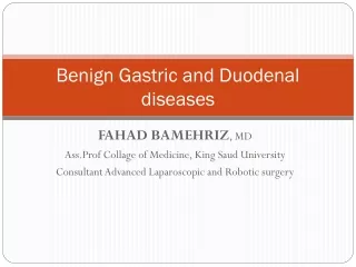
Cervical Abnormalities Due To DES • DES is a nonsteroid estrogen • Cervical abnormalities • Infertility • At risk for miscarriage, ectopic pregnancy, premature delivery • Cervical incompetency
Diethylstilbestrol-exposed uterus. Myometrial hypertrophy results in a T-shaped uterine cavity and cavity irregularity, which is pathognomonic for the anomaly.
Cervical Injuries • Lacerations • Vaginal Delivery (most common at the 3 and 6 o'clock positions ) • D&C • particularly on the postmenopausal patient • Preop use of laminaria or misoprostol may reduce it • Resecting LOOPin hysteroscopic surgery , rollerball (during hysterescopy) • Excessive traction of the ant lip of cervix • Perforations • Self induced abortion • Cervical dilatation • Insertion of radioactive sources • Conization
Cervical Injuries • Ulcerations • Due to vaginal pessary • Due to cervical stem pessary • May be due to uterine prolapse when the cervix protrudes through the vaginal introitus • Annular Detachment • Rare complication due to compression necrosis of the cervix during labor • Occurs when the ext os fails to dilateand the blood supply is compromised by pressure of the fetal head
Cervical Injuries • Cervical stenosis • usually occurs at the level of the internal os • In the premenopausal woman, it may be responsible for obstruction of menstrual flow, leading to amenorrhea and pelvic pain, and infertility • A postmenopausal woman with cervical stenosis may have pyometra, requiring evacuation of uterine contents and biopsy to rule out endometrial carcinoma. • Causes; • Cervical injury, surgical procedures such as cone biopsy, loop excision, or ablative techniques for treatment of dysplasia, and inflammatory • Excision by loop diathermy tends to remove less cervical stroma and therefore is less likely to cause cervical stenosis than a cold knife cone biopsy. • Radiation therapy, neoplasia, and atrophic changes are more common causes in the postmenopausal woman.
Cervical Infections • Acute Cervicitis • Chronic Cervicitis • Chlamydia trachomatis • N. gonorrhea • HSV • HPV • Trichomonas vaginalis • Candidial inf • Bacterial vaginosis
Cervical Infections • Chlamydia trachomatis • may infect the fetus during its passage through the birth canal, or • it may ascend via the endometrial cavity to the fallopian tubes causing salpingitis, pelvic and perihepatic peritonitis. • It has been implicated as the agent responsible for the Fitz-Hugh and Curtis syndrome (violin-string adhesions between the liver and the parietal peritoneum). • C trachomatis and N gonorrhoeae often are coinfecting agents in the etiology of acute and chronic cervicitis and salpingitis. • may be transmitted to the eyes, where it causes trachoma and inclusion conjunctivitis.
Cervical Infections • N. gonorrhea • common cause of cervicitis also infecting the columnar epithelium of the endocervix. • The mature squamous epithelium of the adult cervix and vagina is resistant to the invading organism. • As in the case of Chlamydia infections, the cervix acts as a nidus for ascending infection of the endometrium and the fallopian tubes, with upward invasion often occurring after a menstrual period and loss of the protective mucus plug.
Cervical Infections • HSV • produces cervical lesions similar to those found on the vulva. • The lesion is vesicular at first and then becomes ulcerative. • Primary infections may be extensive and severe, producing constitutional symptoms of low-grade fever, myalgia, and malaise lasting approximately 2 weeks. • Recurrences of lesser severity and duration are common. • HSV-2 in more than 90%, HSV-1 • After the initial infection has healed, the virus continues to reside in the epithelial cells of the cervix, and viral shedding occurs in asymptomatic patients. • Infection of infants during their passage through the birth canal has in women with active infection at term. • Women withantibodies to HSV-2 have a higher incidence of intraepithelial neoplasia as well as invasive malignancy (a direct etiologic link has not been established).
Cervical Infections • HPV • They are flatter and moister than the typical genital warts (condylomata acuminata) seen on the vulva and perianal skin. • In fact, they often are invisible to the naked eye, becoming visible only after application of a dilute solution of acetic acid (acetowhite epithelium) or by colposcopic examination (white epithelium, mosaicism, and coarse punctation). • More than 65 types of HPV have been identified. • Benign lesions of the cervix are associated with types 6, 11, 42, 43, 44, 53, 54, and 55, whereas types 16, 18, 31, 33, 35, 39, 45, and 56 are more often found in association with cervical intraepithelial neoplasia and invasive cancers. • Approximately one-third of women with HPV infection have coexistent cervicitis caused by other organisms. • The presence of cervicitis does not significantly affect the clinical course of HPV lesions.
Cervical Infections • HPV infection is characterized by squamous epithelial cell enlargement, multinucleation, and the perinuclear "halo" effect ofkoilocytosis. • The so-called "balloon cell" is almost pathognomonic of this condition. • Cellular changes of mild dysplasia (low-grade squamous intraepithelial lesion [SIL]), moderate or severe dysplasia (carcinoma in situ [CIS], high-grade SIL), and even invasive cancer may be associated findings. • Greatly enlarged, multinucleated cells with ground-glass cytoplasm and nuclei containing characteristic inclusion bodies are indicative of HSV infection
Cervical Infections • Complications • Cervical hemorrhage • Salpingitis • Leukorrhea • Cervical stenosis • Infertility
Granulomatous Infections Of The Cervix • Tuberculosis • Always sec to disease elsewhere usu pulmonary • Abdominal pain, irregular bleeding • Diagnosis made by biopsy • Histologically, the disease is characterized by tubercles undergoing central caseation • such lesions may be caused by other entities such as amoebiasis, schistosomiasis, brucellosis, tularemia, sarcoidosis, and foreign body reaction • Tbc bacillus must be demonstrated by acid-fast stain or culture • Medical therapy or surgery (ATH+BSO after a trial of CT) • Tertiary syphilis • Granuloma inguinale
Rare Infectious Diseases Of The Cervix • Lymphogranuloma venereum • A chlamydial inf and chancroid caused by Haemophilus Ducreii • Cervical actinomyces • Instruments • RIA • Schistosomiasis of the cervix • Involvement of pelvic and uterine veins by S.Haematobium • Echinococcal cysts
Cystic Abnormalities Of The Cerviks • Nabothian Cysts • Mesonephric Cysts
Benign Neoplasms of The Cervix • Microglandular Hyperplasia of the Endocervical Mucosa • Due to OC, pregnancy, inflammation • Cervical polyps • Arise as a result of focal hyperplasia of the endocervix • Due to chronic inflammation or abnormal responsiveness to hormonal stimulation or a localized vascular congestion of cervical blood vessels • Offen found in association with endometrial hyperplasia (hyperestrogenism may play a significant etiologic role) • malignant change is less than 1%, squamous cell carcinoma is the most common type; adenocarcinomas have been reported. • Endometrial cancer may involve the polyp secondarily. • Sarcoma rarely develops within a polyp.
Benign Neoplasms of The Cervix • Papillomas of the cerviks • Asympomatic • There are 2 types • Solitary ( cause unknown) • Condyloma accuminata • HPV • STD • Leiomyomas of the cerviks
Benign Disorders of The Uterine Corpus • Congenital Anomalies of the uterine corpus( Mullerian fusion anomalies) • Uterine Corpus Anomalies due to DES • Leiomyoma Uteri • Adenomyosis • Endometrial Polyps • Endometritis • Asherman Syndrome
Congenital Anomalies of Uterine Corpus • Incomplete Mullerian Fusion • Uterine didelphys ( 2 separate uterine horns) • Bicornuate uterus ( due to partial incomplete fusion) • Unicornuate uterus • Arcuate uterus • Renal abnormalities are seen in 20-30% of women with Mullerian defects
Congenital Anomalies of Uterine Corpus • Failure of Resorption • Septate uterus • If obstetric complications occur surgery has to be done
Uterine Corpus Abnormalities Due To DES • DES is a nonsteroid estrogen • Cervical abnormalities • Infertility • At risk for miscarriage, ectopic pregnancy, premature delivery • Cervical incompetency
Diethylstilbestrol-exposed uterus. Myometrial hypertrophy results in a T-shaped uterine cavity and cavity irregularity, which is pathognomonic for the anomaly.
Leiomyoma Uteri • Benign neoplasm arising from smooth muscle cells in the uterine wall • Pseudocapsule • 20-25% • More commonly multiple • Symptoms • Usually asymptomatic • Metrorrhagia • Menorrhagia • Pain • İnfertility
Leiomyoma Uteri • Etiology • Not known • Unicellular in origin • Estrogens are important in growth of myomas • Progestins increases mitotic activity • HPL may cause growth • Classification • Submucous • Intramural or interstitial • Subserous
Leiomyoma Uteri • Changes • Hyalinisation • Liquefaction • Calcification • Hemorrhage • Inflammation • Degeneration ( atrophic, hyaline, cystic, calcific, septic, carneous, fatty )
Leiomyoma Uteri • Clinical findings • Abnormal uterine bleeding 30% • Pain • Pressure effects • Intestinal obstruction • Compress to ureters, bladder or rectum • Pelvic venous compression • Infertility • Relationship is unknown • 27-40% of women with myomas are infertile • Spontation abortion • 2 times more
Leiomyoma Uteri • Complications • Myomas and Pregnancy • Rapid growth • Degeneration • Pain • Fetal malpresentation • Obstruction of the birth canal • Uterine inertia • Nonpregnant Women • Heavy bleeding • Urinary or bowel complication
Leiomyoma Uteri • Treatment • depends on the • patients age • parity • symptoms • general health • pregnancy status • reproductive plans • Expectant • Medical therapy • GnRHa • OC • Levonorgestrel releasing intrauterine device
Leiomyoma Uteri • Surgery • Myomectomi • Hysterectomi • Uterine fibroid embolisation • Endometrial ablation • Myolsis • Laparoscopic uterine artery occlusion • MR guided focused USG surgery
Adenomyosis • Is defined by the presence of endometrial glands and stroma within the myometrium • Can be diffuse or localised ( adenomyosis) • Incidence is 20% • Pathogenesis is unkown • Might be sec to postpartum endometritis because of the endom line break down • An arrest of Mullerian cells in the myometrium and later de nova endometrial gland development • Animal models suggest PRL and FSH stimulates growth
Adenomyosis • Symptoms and signs • Menorrhagia • Dysmenorrhea • Treatment • Hysterectomi • GnRHa
Endometrial Polyps • Is hyperplastic growth of the endometrium • Incidence increases directly with age, peaks in the fifth decade and declines with menopause • 10-24% • Risk factors • HT • Obesity • Tamoxifen therapy • Are considered to be estrogen sensitive • Rarely undergo malignant changes
Endometrial Polyps • Clinical Findings • Metrorrhagia • Prolapsus • Staining • Spotting • Treatment • Surgical excision • GnRHa • Hysterectomi
Endometritis • Symptoms • Uterine tenderness • Fever • Foul smelling discharge • May be chronic • Usu postmenopause • May be due to foreign body
Asherman Syndrome • Due to recurrent D&C, abortus • Sec amenorrhea, infertility, hypomenorrhea
BENIGN DISEASES OF THE OVARIES Rukset Attar, MD, PhD DepartmentofObstetrics and Gynecology
Benign Disorders of the Ovaries • Are common in reproductive ages • Operation only when rupture and hemorrhagia occurs • Physiologic Enlargement • Functional cysts • Hyperthecosis • PCO • Luteoma of pregnancy • Ovarian Neoplasms • Epithelial tumors • Sex cord stromal tumors • Germ cell tumors
Physiologic Enlargement • Functional cysts • Follicular cysts • 3-8 cm • Result from a failure of ovulation most likely sec to disturbances in the release of the pituitary gonadotropins • Typically asymp • May cause pain, dysparonia, AUB, bleeding and tortion • Expectant management or OC • Disappear within 60 days