Download
1 / 21
210 likes | 219 Views
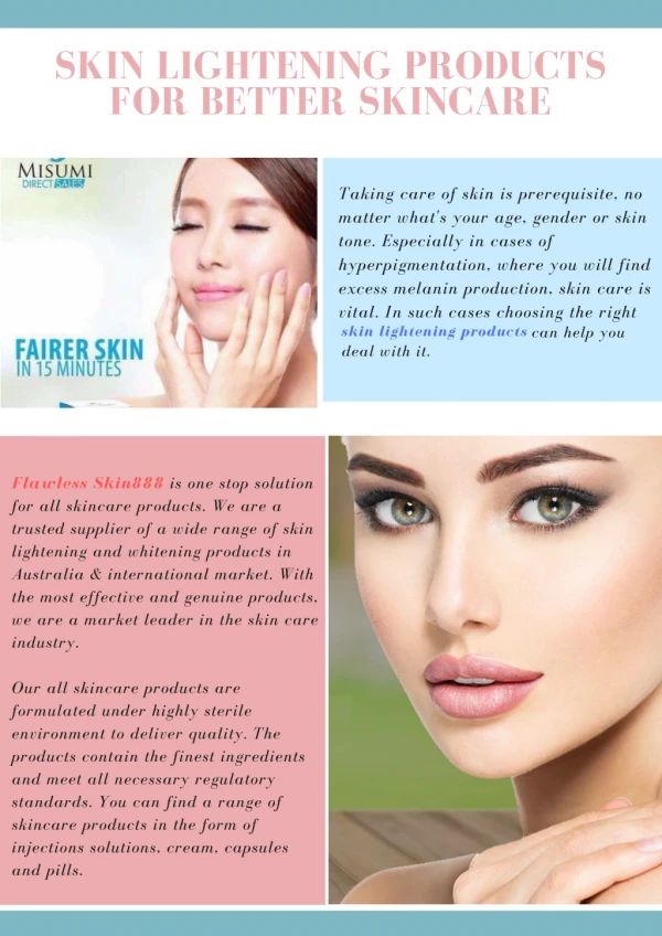
Skin lightening. Dr. Bassam Amro. Color of the skin?. The dermis and epidermal cells gives yellowish white background according to skin thickness. Superficial blood vessels gives red or blue tone. It varies according to state of nearness and dilation. Pigments carotene .

E N D
Skin lightening Dr. Bassam Amro
Color of the skin? • The dermis and epidermal cells gives yellowish white background according to skin thickness. • Superficial blood vessels gives red or bluetone. It varies according to state of nearness and dilation. • Pigments carotene. • Melanin responsible of racial color differences.
Color of the skin? • Melanin is synthesized in the melanocytes found in the epidermal basal layer, within the melanocytes melanin is bound to protein matrix to form melanosomes.
The melanocytes transfer their m.somes to surrounding keratinocytes and migrate upward through the epidermis. Figure1 Schematic drawing of an epidermal melanin unit showing a melanocyte and the group of keratinocytes with which it maintains functional contacts. I, endoplasmatic reticulum where tyrosinase is synthesized; II, premelanosome stage I; III, fully melanized melanosome; IV, passage of melanin granules from the melanocyte to keratinocytes; V, liberation of melanosomes free in the cytoplasm of keratinocytes. B.M., basal membranes; M, mitochondria; MS, melanosome; N, nucleus; E, endoplasmic reticulum; M.G., melanin granules.
Melanin, the skin pigment • Visible pigmentation in mammals results from the synthesis and distribution of melanin in the skin, hair bulbs, and eyes. • Melanin plays a crucial role in: - the absorption of free radicals generated within the cytoplasm. - shielding the houst from various types of ionizing radiations, including UV radiations. Thus melanin protects the skin against sunburn, actinic damage and cancer. - can act as a thermoregulator by absorbing different forms of energy and dissipate them as heat.
Melanins types : • Eumelanins; which are brown or black. • Phaemelanins; Which are red or yellow. Interestingly, it has the capacity to produce free radicals in response to UV radiation, inflicting cell injury, contributing to intensify UV induced skin damage, rather than to protect the skin.
Melanin biosynthesis • The essential enzyme in the melanin biosynthetic pathway is tyrosinase. • However, recent studies have shown that mammalian melanogenesis is not regulated solely by tyrosinase at the enzymatic level, and additional melanogenic factors have been identified, which can modulate pigmentation in either a positive or negative way.
Tyrosinase, the enzyme behind the tan • Tyrosinase is a multifunctional, glycosylated, copper containing oxidase. • Tyrosinase is synthesized by melanosomal ribosomes found on the rough endoplastic reticulum. • After synthesis, it is glcosylated. • It is subsequently delivered to melanosomes via coated vesicles in an inactive form. To date it remains unclear how tyrosinase is initially activated.
Tyrosinase, the enzyme behind the tan Tyrosinase is the rate-limiting, essential enzyme. As such it catalyes three different reactions in the biosynthetic pathway of melanin : 1-The hydroxlation of tyrosine to 3,4-dihydroxyphenylalanine (Dopa) 2-The oxidation of DOPA to DOPA-quinone 3-The oxidation of 5,6-dihydroxyindole (DHI) to indole quinone. The rate-limiting steps are 1 and 2. The quantity of melanin synthesized is thus proportional to the amount of tyrosinase activity present in the cell.
Additional melanogenic factors • There is now evidence that dopachrome can be converted to either 5,6-dihydroxyindole or the carboxy derivative 5,6-dihydroxyindole carboxylic acid (DHICA).This step appears to be catalyzed either by metal ions or by a recently discovered enzyme, dopachrometautomerase (occurs in melanosomes). • Also peroxidases can utilize DHI as a substrate and thus may play a potential role in melanogenesis.
Regulation of Melanogensis Is controlled at many different levels: • Genetic level : Melanocytes are initially derived from the neural crest and migrate throughout the embryo during development. These migration patterns are under strict gentic control and can lead to some interesting patterns when the final melanocyte distribution in the skin is not uniform, as in zebras and giraffes. • Cellular level by melanocytes synthesizing melanin within melanosomes; in varying sizes, numbers, and densities. • Subcellular level where the synthesis and expression of various melanogenic enzymes and inhibitors play a critical role.
These migration patterns are under strict gentic control and can lead to some interesting patterns when the final melanocyte distribution in the skin is not uniform.
Regulation of Melanogensis • Skin pigmentation depends upon the organization and functioning of epidermal melanin unit and several separate but related events : • Melanoblast migration from the neural crest. • Melanoblast differentiation into melanocytes. • The rate of synthesis and melanization of melanosomes. • The size of melanosomes. • Synthesis of melanin. • The efficacy of melanosome transfer into keratinocytes. • The rate of melanosome degradation within the keratinocytes. • The rate of synthesis, inhibition and decay of tyrosinase. • Activity of tyrosinase in melanosomes.
Differences in racial pigmentation • Skin color is a function of the size, number and distribution of melanosomes, and not of density of melanocytes. In fact, the number of melanocytes is the same in all races. However, melanocytes of darkly pigmented skin have thicker, longer, branched dendrites. • Melanocytes derived from black skin have up to ten times more tyrosinase activity and produce up to ten times more melanin than do melanocytes from white skin. The number of tyrosinase molecules present in white skin melanocytes is equal to the one found in black skin.
Differences in racial pigmentation • In black skin, melanin persists within the horny layer and leaves the skin in the end by natural desquamation, whereas in white skin the pigment is degraded in the granular layer, probably via specific enzymatic process. That means stratum corneum of white skin does not contain any melanin.
The control of melanin production • Direct effect of UV radiation • Melanocyte stimulatory hormone (MSH) secreted from pituitary gland • Oestrogens exert local effect during prgnancy.
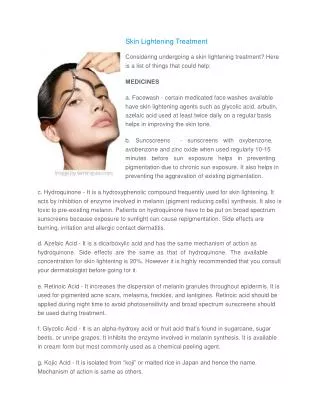
Mechanisms of depigmentation • Selectively destroy the melanocytes • Inhibit the formation of melanosomes • Inhibit the biosynthesis of tyrosinase • Inhibit the formation of melanin • Interfere with the transfer of melanosomes • Have a chemical effect on melanin