Download
1 / 70
760 likes | 1.37k Views
Exercise 7: Overview of the Skeleton. Bio 111 Deborah L. Huber. Lab Exam 2. ALL FILL IN! ALL identifying structures in photos or bones Spelling counts . Terms to know:. Periosteum Diaphysis Epiphysis Spongy bone Compact bone Medullary cavity Endosteum Lamellae

E N D
Exercise 7: Overview of the Skeleton Bio 111 Deborah L. Huber
Lab Exam 2 ALL FILL IN! ALL identifying structures in photos or bones Spelling counts
Terms to know: • Periosteum • Diaphysis • Epiphysis • Spongy bone • Compact bone • Medullary cavity • Endosteum • Lamellae • Osteocyte in lacuna • Canaliculi • Central canal (Haversian canal)
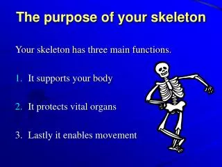
The functions of the skeletal system • Provides body an internal framework • Protects many of the body’s soft organs • Store lipids and many minerals • Provide a site for blood cell formation in their red marrow cavities
The functions of the skeletal system • Provides body an internal framework • Protects many of the body’s soft organs • Store lipids and many minerals • Provide a site for blood cell formation in their red marrow cavities
The functions of the skeletal system • Provides body an internal framework • Protects many of the body’s soft organs • Store lipids and many minerals • Provide a site for blood cell formation in their red marrow cavities
The functions of the skeletal system • Provides body an internal framework • Protects many of the body’s soft organs • Store lipids and many minerals • Provide a site for blood cell formation in their red marrow cavities
The functions of the skeletal system • Provides body an internal framework • Protects many of the body’s soft organs • Store lipids and many minerals • Provide a site for blood cell formation in their red marrow cavities
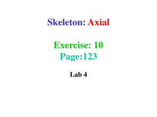
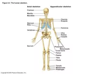
The two subdivisions of the skeleton: axial skeleton appendicular skeleton
The two subdivisions of the skeleton: axial skeleton appendicular skeleton
The two subdivisions of the skeleton: axial skeleton appendicular skeleton
Bone Markings • Bulges, depressions, and holes serve as • Sites of attachment for muscles, ligaments, and tendons • Joint surfaces • Conduits for blood vessels and nerves
Bone Markings • Bulges, depressions, and holes serve as • Sites of attachment for muscles, ligaments, and tendons • Joint surfaces • Conduits for blood vessels and nerves
Bone Markings • Bulges, depressions, and holes serve as • Sites of attachment for muscles, ligaments, and tendons • Joint surfaces • Conduits for blood vessels and nerves
Bone Markings • Bulges, depressions, and holes serve as • Sites of attachment for muscles, ligaments, and tendons • Joint surfaces • Conduits for blood vessels and nerves
Bone Markings: Projections • Sites of muscle and ligament attachment (Table 6.1) • Tuberosity—rounded projection • Crest—narrow, prominent ridge • Trochanter—large, blunt, irregular surface • Line—narrow ridge of bone • Tubercle—small rounded projection • Epicondyle—raised area above a condyle • Spine—sharp, slender projection • Process—any bony prominence
Bone Markings: Projections • Sites of muscle and ligament attachment (Table 6.1) • Tuberosity—rounded projection • Crest—narrow, prominent ridge • Trochanter—large, blunt, irregular surface • Line—narrow ridge of bone • Tubercle—small rounded projection • Epicondyle—raised area above a condyle • Spine—sharp, slender projection • Process—any bony prominence
Bone Markings: Projections • Sites of muscle and ligament attachment (Table 6.1) • Tuberosity—rounded projection • Crest—narrow, prominent ridge • Trochanter—large, blunt, irregular surface • Line—narrow ridge of bone • Tubercle—small rounded projection • Epicondyle—raised area above a condyle • Spine—sharp, slender projection • Process—any bony prominence
Bone Markings: Projections • Sites of muscle and ligament attachment (Table 6.1) • Tuberosity—rounded projection • Crest—narrow, prominent ridge • Trochanter—large, blunt, irregular surface • Line—narrow ridge of bone • Tubercle—small rounded projection • Epicondyle—raised area above a condyle • Spine—sharp, slender projection • Process—any bony prominence
Bone Markings: Projections • Sites of muscle and ligament attachment (Table 6.1) • Tuberosity—rounded projection • Crest—narrow, prominent ridge • Trochanter—large, blunt, irregular surface • Line—narrow ridge of bone • Tubercle—small rounded projection • Epicondyle—raised area above a condyle • Spine—sharp, slender projection • Process—any bony prominence
Bone Markings: Projections • Projections that help to form joints (Table 6.1) • Head • Bony expansion carried on a narrow neck • Facet • Smooth, nearly flat articular surface • Condyle • Rounded articular projection • Ramus • Armlike bar
Bone Markings: Projections • Projections that help to form joints (Table 6.1) • Head • Bony expansion carried on a narrow neck • Facet • Smooth, nearly flat articular surface • Condyle • Rounded articular projection • Ramus • Armlike bar
Bone Markings: Projections • Projections that help to form joints (Table 6.1) • Head • Bony expansion carried on a narrow neck • Facet • Smooth, nearly flat articular surface • Condyle • Rounded articular projection • Ramus • Armlike bar
Bone Textures • Compact bone • Dense outer layer • Spongy (cancellous) bone • Honeycomb of trabeculae
Bone Textures • Compact bone • Dense outer layer • Spongy (cancellous) bone • Honeycomb of trabeculae
Bone Textures • Compact bone • Dense outer layer • Spongy (cancellous) bone • Honeycomb of trabeculae
Spongy bone (diploë) Compact bone Trabeculae Figure 6.5
Structure of a Long Bone • Diaphysis (shaft) • Compact bone collar surrounds medullary (marrow) cavity • Medullary cavity in adults contains fat (yellow marrow)
Structure of a Long Bone • Diaphysis (shaft) • Compact bone collar surrounds medullary (marrow) cavity • Medullary cavity in adults contains fat (yellow marrow)
Structure of a Long Bone • Diaphysis (shaft) • Compact bone collar surrounds medullary (marrow) cavity • Medullary cavity in adults contains fat (yellow marrow)
Structure of a Long Bone • Epiphyses • Expanded ends • Spongy bone interior • Epiphyseal line (remnant of growth plate) • Articular (hyaline) cartilage on joint surfaces
Structure of a Long Bone • Epiphyses • Expanded ends • Spongy bone interior • Epiphyseal line (remnant of growth plate) • Articular (hyaline) cartilage on joint surfaces
Structure of a Long Bone • Epiphyses • Expanded ends • Spongy bone interior • Epiphyseal line (remnant of growth plate) • Articular (hyaline) cartilage on joint surfaces
Structure of a Long Bone • Epiphyses • Expanded ends • Spongy bone interior • Epiphyseal line (remnant of growth plate) • Articular (hyaline) cartilage on joint surfaces
Membranes of Bone • Periosteum • Outer fibrous layer • Inner osteogenic layer • Osteoblasts (bone-forming cells) • Osteoclasts (bone-destroying cells) • Osteogenic cells (stem cells) • Nerve fibers, nutrient blood vessels, and lymphatic vessels enter the bone via nutrient foramina • Secured to underlying bone by Sharpey’s fibers
Membranes of Bone • Periosteum • Outer fibrous layer • Inner osteogenic layer • Osteoblasts (bone-forming cells) • Osteoclasts (bone-destroying cells) • Osteogenic cells (stem cells) • Nerve fibers, nutrient blood vessels, and lymphatic vessels enter the bone via nutrient foramina • Secured to underlying bone by Sharpey’s fibers
Membranes of Bone • Periosteum • Outer fibrous layer • Inner osteogenic layer • Osteoblasts (bone-forming cells) • Osteoclasts (bone-destroying cells) • Osteogenic cells (stem cells) • Nerve fibers, nutrient blood vessels, and lymphatic vessels enter the bone via nutrient foramina • Secured to underlying bone by Sharpey’s fibers
Membranes of Bone • Periosteum • Outer fibrous layer • Inner osteogenic layer • Osteoblasts (bone-forming cells) • Osteoclasts (bone-destroying cells) • Osteogenic cells (stem cells) • Nerve fibers, nutrient blood vessels, and lymphatic vessels enter the bone via nutrient foramina • Secured to underlying bone by Sharpey’s fibers
Membranes of Bone • Endosteum • Delicate membrane on internal surfaces of bone
Membranes of Bone • Endosteum • Delicate membrane on internal surfaces of bone
Microscopic Anatomy of Bone: Compact Bone • Haversian system, or osteon—structural unit • Lamellae • Weight-bearing • Column-like matrix tubes • Central (Haversian) canal • Contains blood vessels and nerves
Microscopic Anatomy of Bone: Compact Bone • Haversian system, or osteon—structural unit • Lamellae • Weight-bearing • Column-like matrix tubes • Central (Haversian) canal • Contains blood vessels and nerves
Microscopic Anatomy of Bone: Compact Bone • Haversian system, or osteon—structural unit • Lamellae • Weight-bearing • Column-like matrix tubes • Central (Haversian) canal • Contains blood vessels and nerves
Microscopic Anatomy of Bone: Compact Bone • Haversian system, or osteon—structural unit • Lamellae • Weight-bearing • Column-like matrix tubes • Central (Haversian) canal • Contains blood vessels and nerves
Microscopic Anatomy of Bone: Compact Bone • Haversian system, or osteon—structural unit • Lamellae • Weight-bearing • Column-like matrix tubes • Central (Haversian) canal • Contains blood vessels and nerves
Microscopic Anatomy of Bone: Compact Bone • Perforating (Volkmann’s) canals • At right angles to the central canal • Connects blood vessels and nerves of the periosteum and central canal • Lacunae—small cavities that contain osteocytes • Canaliculi—hairlike canals that connect lacunae to each other and the central canal
Microscopic Anatomy of Bone: Compact Bone • Perforating (Volkmann’s) canals • At right angles to the central canal • Connects blood vessels and nerves of the periosteum and central canal • Lacunae—small cavities that contain osteocytes • Canaliculi—hairlike canals that connect lacunae to each other and the central canal
Microscopic Anatomy of Bone: Compact Bone • Perforating (Volkmann’s) canals • At right angles to the central canal • Connects blood vessels and nerves of the periosteum and central canal • Lacunae—small cavities that contain osteocytes • Canaliculi—hairlike canals that connect lacunae to each other and the central canal
Microscopic Anatomy of Bone: Spongy Bone • Trabeculae • Align along lines of stress • No osteons • Contain irregularly arranged lamellae, osteocytes, and canaliculi • Capillaries in endosteum supply nutrients