Download
1 / 104
1.06k likes | 1.24k Views
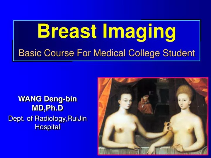
Breast Imaging Basic Course For Medical College Student. WANG Deng-bin MD,Ph.D Dept. of Radiology,RuiJin Hospital. Introduction.

E N D
Breast ImagingBasic Course For Medical College Student WANG Deng-bin MD,Ph.D Dept. of Radiology,RuiJin Hospital
Introduction “Breast cancer is one of the best studied human tumors, but it remains poorly understood” “ As in all medical endeavors, the practitioner should, whenever possible, use the results of scientific studies to guide clinical decision”
And the imaging modalities implemented in clinical practice for breast care must be served as the tools for detection and characterization of breast lesions. As we expect, they are very important for diagnosis and treatment.
1.5T MRI GE signa gemsow 0.5T MRI GE signa Sys#MRS Shanghai 2nd Medical University Rui Jin Hospital
X-ray ExaminationMammograpy • X-ray radiography (molybdenum X rays, rhodium X rays):MLO,CC,etc. • Galactography --demonstrates the ducts and ductule or their abnormalities.
USG • Ultrasonography (ultrasound) • B-mode US • Doppler US
CT • Computed tomography • plain scan, • enhanced scan (iodine)
MRI • Magnetic Resonance Imaging • high resolution for soft tissue • different tissue, different signal • enhanced scan
The others • Infrared thermal imaging • Computer diaphanography imaging,etc.
Imaging-Guided Percutaneous Biopsysupply specimens for pathologic examination • Fine needle aspiration biopsy (FNAB) • Needle core biopsy (NCB)
Interventional Therapy • Laser • Radiofrequency • Embolization • and so on
Accessory Breast Tissue • The most common site is axilla
Basic Imaging Signs of Breast Lesion • Mass/Lump • Calcification • Thickening and retraction of regional skin • Retraction of nipple • Enlargement or abnormality of blood vessels • Lymph nodes • Enhanced manifestations
Mass/Lump shape:round, oval, regular/irregular margin:clear or ambiguous, spiculation or smooth density or signal intensity:high/low/intermediate with or without calcification site:upper-outer quadrant breast, upper-inner quadrant breast, lower outer quadrant breast, lower-inner quadrant breast, nipple, central portion breast, axillary tail breast. Number:solitary or multiple
Calcification Size: large or micro Shape: ring-like, nodular or salt-like, branching Distribution: scattered or clustered with or without mass benign / malignant
Thickening and Retraction of Regional Skin • Frequently found in malignant tumors • Sometimes due to postsurgical scars.
Retraction of Nipple • Congenital-dysplasia • Acquired-malignant tumor
Enlargement or Abnormality of Blood Vessel • Mostly in malignant tumor due to increase of blood supply
Enlargement of Lymph Nodes • Axillary or intramammary lymph nodes
Administration of Contrast Agents for Breast Lesions • Implication of the lesion’s hemodynamics • washout type--malignant • linear--benign • plateau--malignant/benign