Download
1 / 32
330 likes | 738 Views
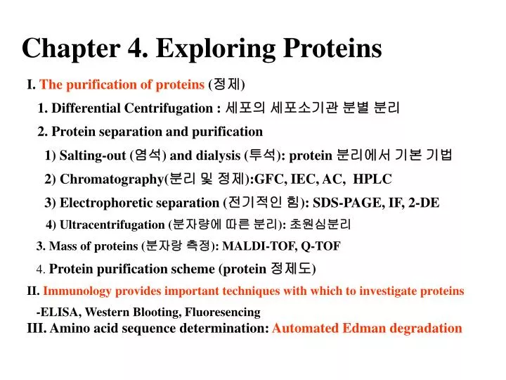
Chapter 4. Exploring Proteins. I. The purification of proteins ( 정제 ) 1. Differential Centrifugation : 세포의 세포소기관 분별 분리 2. Protein separation and purification 1) Salting-out ( 염석 ) and dialysis ( 투석 ): protein 분리에서 기본 기법 2) Chromatography( 분리 및 정제 ):GFC, IEC, AC, HPLC

E N D
Chapter 4. Exploring Proteins I. The purification of proteins (정제) 1. Differential Centrifugation : 세포의 세포소기관 분별 분리 2. Protein separation and purification 1) Salting-out (염석) and dialysis (투석): protein 분리에서 기본 기법 2) Chromatography(분리 및 정제):GFC, IEC, AC, HPLC 3) Electrophoretic separation (전기적인 힘): SDS-PAGE, IF, 2-DE 4) Ultracentrifugation (분자량에 따른 분리): 초원심분리 3. Mass of proteins (분자랑 측정): MALDI-TOF, Q-TOF 4. Protein purification scheme (protein 정제도) II. Immunology provides important techniques with which to investigate proteins -ELISA, Western Blooting, Fluoresencing III. Amino acid sequence determination: Automated Edman degradation
I. The purification of proteins. Is an essential first step in understanding their functions • Differential centrifugation (분별원심분리) - 세포의 protein 및 세포소기관을collection 가능 - 단계적으로 원심력을 높이면서 침전물과 상등액을 분리 하는 원심분리
2. Protein separation and purification - Can be achieved according to solubility, size, charge, and binding affinity • Salting-out and dialysis • (1) Salting-out (solubility에 의한 침전) • • 0.8M ammonium sulfate ; fibrinogen ( a blood-clotting protein ) • • 2.4M ammonium sulfate ; serum albumin • ∴ Need dialysis to remove excessive salts (2) Dialysis: 분자량에 따른 분리 • Semi-permeable membrane; pore-size 상이 (5,000-10,000 등) • 분자량에 따른 protein의 분리에도 이용되지만, 대량의 protein으로부터 무기염의 제거에 활용
2) Chromatography (1) Gel-filtration chromatography - Porous beads ( dextran, agarose, or polyacrylamide ) : Sephadex, sepharose, Biogel; 100µm in diameter -분자량에 따른 분리: 큰 것이 빠르고, 작은 것이 느림 Large molecules flow more rapidly through this column and emerge first because a smaller volume is accessible to porous beads.
(2) Ion-exchange chromatography (IEC) ① Cation exchange resin : CM cellulose • ”+” 띈 분자 부착 (정도는 +의 정도에 비례) • ”+” 또한 buffer의 pH에 따라 상이 ② Anion exchange resin : DEAE cellulose • ”-” 띈 분자 부착 (정도는 -의 정도에 비례) • ”-” 또한 buffer의 pH에 따라 상이
(3) Affinity chromatography (AC) ① Resin: 특수한 결합단이 부착된 resin ② Chromatographicic separation (결합친화력에 의한 분리) Addition of Glucose (G) Affinity chromatography of concanavalin A (shown in yellow) on a solid support containing covalently attached glucose residues (G) (4) High –Pressure Liquid Chromatography (HPLC) ① Column (solid phase): Normal phase (+, -: silica), reversed-phase (C18) ② Mobile phase: 유기용매, 물 등 사용; 주로 solid phase와 반대로 사용 ①Chromatic separation: 분자량, 전하, 친화력 등에 의한 분리 및 정량
3) Electrophoretic separation (1) SDS-PAGE (SDS-polyacrylamide gel electrophoresis) ① Formation of polyacrylamide gel
② SDS처리: protein을 SDS(detergent)로 처리하면 SDS는 변성된 protein을 결합하여 표면에 “-” 전하, 이는 분자량에 비례 ③Electrophoresis: 분자량이 작은 것이 멀리 내려감
④ Staining and destaining: Coomassie blue ⑤ Determination of molecular weight -x: log 분자량, y: 정상 scale (이동거리) -이동거리는 log분자량에 반비례
(2) Isoelectric focusing (IF) ① pI: protein에 따라 상이 ②pH gradient in a gel: gel상에 pH가 상이하도록 조제 ③ Electrophoresis: protein이 pI에 도달하면 더 이동 하지 않음 (A) The sample is loaded and voltage is applied. The proteins will migrate to their isoelectric pH, the location at which they have no net charge. (B) The proteins form bands that can be excised and used for further experimentation.
(3) Two-dimensional electrophoresis ① IF: 먼저 수행 ②SDS-PAGE: 나중 수행 ③ 활용: proteomics에 활용 (혼합물로부터 개개의 protein분리 가능) Isoelectric focusing can be combined with SDS-PAGE to obtain very high resolution separations
4) Ultracentrifugation - is valuable for separating biomolocules and determining their mass
3. Mass of a protein • Precisely determined by mass spectrometry • P mole 또는 f mol에서도 측정가능 • 가벼운 것이 먼저 flight하고, mass/charge로 표시 1) MALDI-TOF (Matrix-Assisted Laser Desorption-ionization Time of Flight) 2) Q-TOF The time of flight (TOF) These methods are called matrix-assisted laser desorption-ionization (MALDI)
4. Protein purification scheme • 정제에 대한 정량적인 평가가 가능 • 세포에 함유된 특수한 효소 기능을 갖고 glucose를 결합하는 HA pretein을 정제한다고 가정함
II. Immunotechniques to be investigated proteins 1. Antibodies 1) Antibody structure
2. Monoclonal antibodies preparation - Preparation of MAb Hybridoma cells
3. Assays 1) Enzyme-linked immunosorbant assay (ELISA) (a) in indirect ELISA, the production of color indicates the amount of an antibody to a specific antigen (b) in sandwich ELISA, the production of color indicates the quantity of antigen
2) Western blotting Very small quantities of a protein of interest in a cell or in body fluid can be detected by an immunoassay technique called Western blotting
3) Fluorescence-labeled antibody -Glucocorticoid receptor protein (hormone인 cortisone에 반응하는 전사인자)을 Green fluorescence protein과 반응하여 이들은 cytosil에 존재 (A). 여기에 cortisone을 처리하면 이것이 cytosol에서 핵으로 이동 (B)한 것을 현광현미경으로 관찰
III. Amino acid sequence determination 1. Edman degradation of peptides : Ala-Gly-Asn-Pro-Arg-Gly 1) Determination of amino acid composition (1) Acid hydrolysis: 6N HCl, 110 C°, 24 hr (2) Ion-exchange chromatography (sulfonated anion exchange resin) -Ninhydrin 반응으로 정색 first last
2. Edman degradation of huge peptide or proteins: TFVKAAWGK -Amino acid composition 결정 -N-terminal amino acid 결정 -Fragmentation하고, 각 fragmet에 대한 Edman degradation -Amino acid sequence 결정 (반드시 overlap 되어야 함) -S-S- 결합위치 결정
Amino acid composition 결정: 앞에서 설명한 방법에 준함 • Determination amino-terminal residue : Dabsyl-amino acid derivative
-Fluorescent derivatives of amino acids:아미노산 분리를 위한 발색단
3) Protein can be specifically cleaved into small peptides to facilitate analysis (1) Cleavage by CNBr: Met (2) Cleavage by trypsin: Lys and Arg (3) Cleavage by chymotrypsin: Bulky R group (aromatic rings and Leu etc)
4) Edman degradation of each fragment 5) Construction of overlapped peptides
6) Disulfide-bond reduction; diagonal electrophoresis (performic acid treatment) diagonal electrophoresis
3. Amino acid sequences are sources of many kinds of insight 1) Compared with all other known sequences to ascertain whether significant similarities exists: Protein 간의 유사성 검토 2) Comparison of sequences of the same protein in different species yields a wealth of information about evolutionnary pathways: 동일 단백질의 다른 종간의 진화과정 추정 3) Serves for signals designating their destinations or controlling their processing: Protein이 어디에서 왔는지에 대한 신호 (특수한 신호 peptide가 있음) 4) Searched for the presence of internal repeats: 반복 aa 서열 등은 protein의 기능 확인 (예, camadulin은 Ca을 결합하기 위한 4개의 반복 서열이 있음). 5) Provide a basic information for preparing Ab specific for a protein of interest: Ab 생산을 위한 정보 제공 6) Valuable for making DNA probes that are specific for the genes encoding the corresponding proteins: Probe (탐침) 합성