Download
1 / 36

E N D
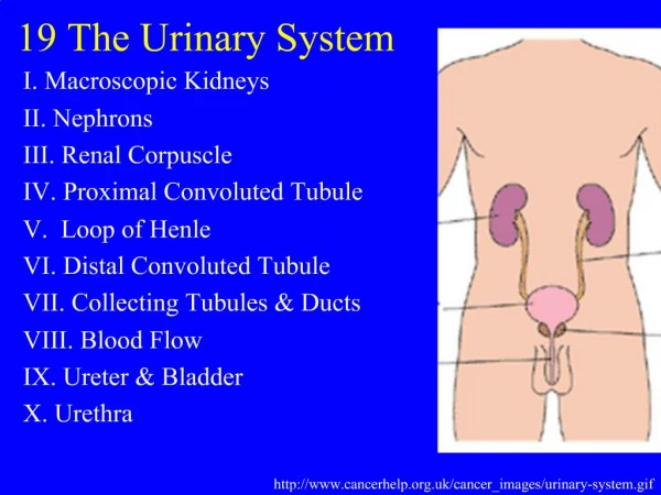
1. 19 The Urinary System I. Macroscopic Kidneys
II. Nephrons
III. Renal Corpuscle
IV. Proximal Convoluted Tubule
V. Loop of Henle
VI. Distal Convoluted Tubule
VII. Collecting Tubules & Ducts
VIII. Blood Flow
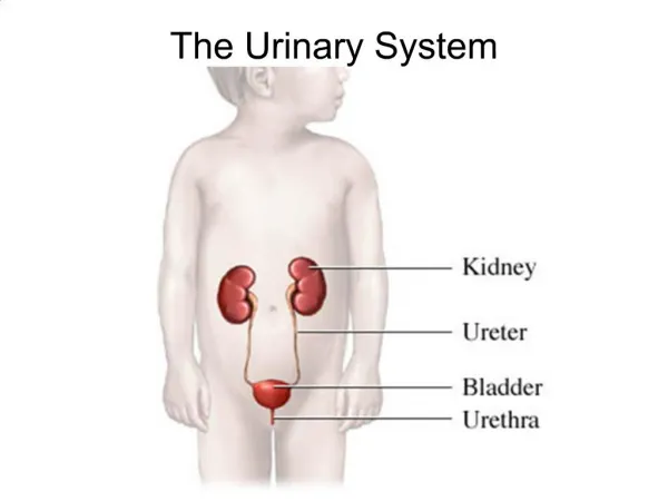
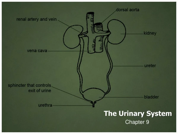
IX. Ureter & Bladder
X. Urethra
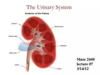
2. I. Macroscopic Kidneys A. Capsule & hilus (19.1)
B. Renal sinus
1. renal pelvis
2. major calyces
3. minor calyces
C. Renal medulla
1. renal pyramids
a. papilla
2. Renal column
D. Renal cortex
3. II. Nephrons A. Components (19.1)
1. renal corpuscle
2. PCT
3. loop of Henle
4. DCT
5. (collecting tubule & duct)
4. II. Nephrons B. Relationship to cortex and medulla (19.1)
5. III. Renal Corpuscle A. Glomerulus (19.3)
1. afferent arteriole
2. efferent arteriole
3. vascular pole
4. fenestrated capillaries (19.4)
6. III. Renal Corpuscle B. Bowman�s capsule
1. visceral layer
a. podocytes (19.3)
7. III. Renal Corpuscle B. Bowman�s capsule
1. visceral layer
a. podocytes
1) primary processes (19.5)
8. III. Renal Corpuscle B. Bowman�s capsule
1. visceral layer
a. podocytes
2) secondary processes (pedicels, foot processes) (19.5,19.6)
9. III. Renal Corpuscle Secondary processes interdigitate around glomerular capillaries. The narrow space between processes is the filtration slit. 19-5
10. III. Renal Corpuscle B. Bowman�s capsule
1. visceral layer
a. podocytes
3) filtration slits (19.7)
11. III. Renal Corpuscle B. Bowman�s capsule
1. visceral layer
a. podocytes
3) filtration slits
a) 25 nm wide (19.8)
4) basement membrane
12. III. Renal Corpuscle B. Bowman�s capsule (19.3)
2. urinary space
3. parietal layer
4. urinary pole
13. III. Renal Corpuscle B. Bowman�s capsule
5. EM view (19.4)
14. III. Renal Corpuscle C. Structure/function 19.7
1. glomerular filtrate
2. most blood components smaller than ~70 kDa
15. IV. Proximal Convoluted Tubule A. Overview (19.3,19.13)
1. begins at urinary pole
2. simple cuboidal epithelium
3. longer than DCT
16. IV. Proximal Convoluted Tubule B. Cell structure
1. brush border (19.14,19.15)
a. microvilli ~1 mm
17. IV. Proximal Convoluted Tubule B. Cell structure
2. basal border (19.4)
a. membrane invaginations
1) Na+/K+-ATPase
b. parallel mitochondria
(recall salivary glands)
18. IV. Proximal Convoluted Tubule C. Function 19.16
1. pinocytosis
2. active ion transport
3. Osmosis
4. exocytosis
19. IV. Proximal Convoluted Tubule C. Function 19.16
4. reabsorption
a. all glucose
b. amino acids
c. ~85% NaCl & H20
d. PO4, Ca++
e. protein
5. secretion/excretion
20. V. Loop of Henle A. Components (19.16)
1. thick descending limb
2. thin descending limb
3. thin ascending limb
4. thick ascending limb
21. V. Loop of Henle B. Cell structure
1. thick descending ~ PCT
2. thick ascending ~ DCT
3. thin limbs (19.18,19.19)
22. V. Loop of Henle C. Function 19-16
1. capability of forming hypertonic urine
a. water retention
2. thin descending limb
a. permeable to water
3. ascending limb
a. impermeable to water
23. VI. Distal Convoluted Tubule A. Location (19.16)
1. continuation of thick ascending limb
2. cortex
3. vascular pole of renal corpuscle
a. juxtaglomerular region
24. VI. Distal Convoluted Tubule B. Cell structure (19.16,19.19)
1. simple cuboidal epithelium
2. cells smaller than PCT
a. more nuclei visible/XS
3. lack brush border
25. VI. Distal Convoluted Tubule B. Cell structure (19.19)
4. less acidophilic than PCT
5. larger lumen than PCT
6. similar basal infoldings
26. VI. Distal Convoluted Tubule C. Function 19.16
1. under influence of aldosterone
2. absorb Na+ (and H2O)
3. secretes K+, H+, NH4++
27. VI. Distal Convoluted Tubule D. Juxtaglomerular region
1. macula densa of DCT (19.21)
a. DCT cells columnar
b. able to sense flow and ionic conc.
c. influences afferent arteriole constriction, glomerular filtration, renin secretion
28. VI. Distal Convoluted Tubule D. Juxtaglomerular region
2. JG apparatus (19.25)
a. modified smooth muscle of afferent arteriole
b. secretory granules
c. protein synthesizing organelles
d. synthesize renin
e. effect: increase Na+ and Cl- absorption � distal tubules
29. VII. Collecting Tubules & Ducts A. Cell structure
1. cuboidal to columnar (19.22)
2. weakly staining
3. few organelles
30. VII. Collecting Tubules & Ducts A. Cell structure
4. no basal striations (19.23)
5. clear intercellular borders
6. empty into minor calyx at renal papilla
31. VII. Collecting Tubules & Ducts B. Function 19-16
1. ADH (vasopressin)
a. causes epithelium to be permeable to H2O
b. reabsorption of H2O
32. VIII. Blood Flow A. Renal artery (19.26)
B. Interlobar arteries
C. Arcuate arteries
D. Interlobular arteries
33. VIII. Blood Flow E. Afferent arterioles (19.26)
F. Efferent arterioles
G. Peritubular capillary network
G. Vasa recta
34. IX. Ureter & Bladder A. Ureter 19-28
1. mucosa
a. transitional epithelium
b. lamina propria
2. muscularis
a. inner longitudinal
b. outer circular
3. adventitia
35. IX. Ureter & Bladder B. Urinary bladder
1. mucosa (19.27)
a. transitional epithelium
b. lamina propria
2. muscularis
a. fibers run in all directions
b. no layers
3. adventitia / serosa
36. X. Urethra A. Male urethra (22.1)
1. prostatic urethra
2. membranous urethra
3. pendulous urethra
a. pseudostratified / columnar epithelium
b. corpus spongiosum
4. bulbous urethra
37. X. Urethra B. Female urethra
1. pseudostratified columnar, 4-5 cm long
2. stratified squamous