Download
1 / 17
180 likes | 401 Views
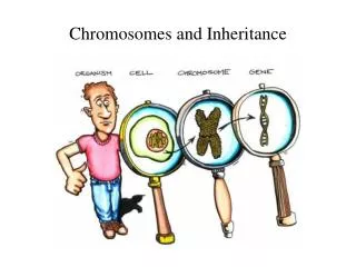
1. The laws of inheritance and the discovery of chromosomes. 18 66 : Gregor Mendel publishes his findings on the laws of inheritance based on experiments with pea plants. He develops three “principles” : Dominance , Segregation , and Independent Assortment.

E N D
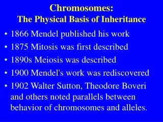
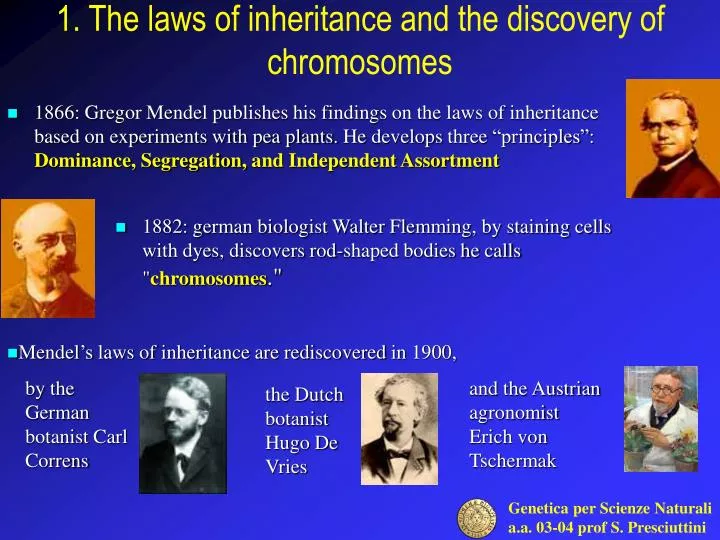
1. The laws of inheritance and the discovery of chromosomes • 1866: Gregor Mendel publishes his findings on the laws of inheritance based on experiments with pea plants. He develops three “principles”: Dominance, Segregation,and Independent Assortment • 1882:german biologist Walter Flemming, by staining cells with dyes, discovers rod-shaped bodies he calls "chromosomes." • Mendel’s laws of inheritance are rediscoveredin 1900, by the German botanist Carl Correns and the Austrian agronomist Erich von Tschermak the Dutch botanist Hugo De Vries
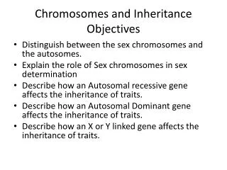
2. Mendel's principles • Dominance • each inherited characteristic is determined by two alternative hereditary factors, and one factor is dominant over the other. • Segregation • the sex cell of a plant or animal may contain one factor (allele) for different traits but not both factors needed to express the traits. • Independent assortment • Different characteristics are inherited independently from each other.
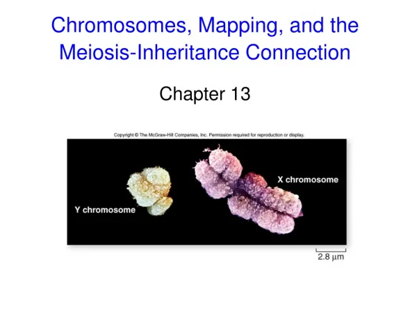
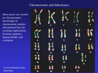
3. Where do genes reside? In 1902, Walter Sutton (an American who at the time was a graduate student) and Theodor Boveri (a German biologist) recognized independently that the behavior of Mendel's particles during the production of gametes in peas precisely parallels the behavior of chromosomes at meiosis: genes are in pairs (so are chromosomes); the alleles of a gene segregate equally into gametes (so do the members of a pair of homologous chromosomes); different genes act independently (so do different chromosome pairs). After recognizing this parallel behavior, both investigators reached the same conclusion that the parallel behavior of genes and chromosomes suggests that genes are located on chromosomes.
4. The word GENETICS is formulated • 1909: Danish botanist Wilhelm Johannsen proposes the term "gene" (from the Greek word "genos" which means "birth") to refer to a Mendelian hereditary factor. Johannsen also proposes two terms, genotype and phenotype, to distinguish between one's genetic make-up and one's outward appearance.
5. Genes are lined-up on chromosomes • 1915: Thomas Hunt Morgan, an American geneticist, publishes The Mechanism of Mendelian Heredity, in which he presents results from experiments with fruit flies that prove genes are lined up along chromosomes. He also describes the principle of "linkage" — that alleles located relatively close to one another on a chromosome tend to be inherited together. • By studying the frequency with which traits are inherited together, Morgan and co-workers create a "genetic map" of fruit fly chromosomes showing the relative locations of the genes responsible for dozens of traits, along with approximate distances between them on the chromosome. This work establishes the basis for gene mapping principles still used today.
6. Genes may cause quantitative variation • 1918: R.A. Fisher shows that continuous traits can be explainedby Mendelian segregation of genes: Mendel’s laws give basis for statisticalrelationships between parents and offspring. • His work reconciled the Darwinian view of evolution with the findings of the geneticists, posing the basis of the so-called “Modern Synthesis”
7. The first mutagenic agent • In 1927 Herman Muller reported that X rays could induce mutations in male fruit flies. In his investigations Muller found that the mutation rate among the male fruit flies was linearly related to the radiation dose. His results supplied the first experimental evidence of a mutagen, in this case, the X rays. Muller's work on mutation induction opened the door to the genetic technique of using mutations to dissect biological processes, which is still used extensively today
8. An early hypothesis on the function of genes • The first clues about the nature of primary gene function came from studies of humans. Early in the twentieth century, Archibald Garrod, a physician, made several observations about alkaptonuria (a form of arthrytis) and proceeded to propose the hypothesis that the information for producing specific enzymes in humans is inherited. He observed that inherited diseases reflect a patient's inability to make a particular enzyme, which he referred to as "inborn errors of metabolism“. • Garrod predicted that individuals affected with alkaptonuria would be deficient in one of the enzymes in a degradative, biochemical pathway. He had suggested that the specific enzyme was involved in the degradation of homogentisic acid, an intermediate in the breakdown pathway of phenylalanine and tyrosine. He came to this conclusion by feeding homogentisic acid to alkaptonuric patients and noting that the chemical was excreted in the urine in quantitatively similar amounts to what was administered.
9. The Beadle and Tatum experiment • Garrod's hypothesis was ahead of its time. Experiments that clarified the actual function of genes came from research in the 1940s on Neurospora by George Beadle and Edward Tatum, who later received a Nobel Prize for their work. • Beadle and Tatum analyzed mutants of Neurospora crassa, a fungus with a haploid genome. They first irradiated Neurosporacells to produce mutations and then tested cultures from ascospores for interesting mutant phenotypes. They detected numerous auxotrophsstrains (that cannot grow on a minimal medium unless the medium is supplemented with one or more specific nutrients). In each case, the mutation that generated the auxotrophic requirement was inherited as a single-gene mutation: each gave a 1:1 ratio when crossed with a wild type.
10. The life cycle of a fungus The fungus Neurospora spends most of its life cycle as a multicellular haploid organism, in which the cells are joined end to end to form hyphae, or threads of cells. The hyphae grow through the substrate and send up aerial branches that bud off haploid cells known as conidia (asexual spores). Conidia can detach and disperse to form new colonies The fungus has alternate mating strains, here called type A and type a. Mating can only take place between different mating strains and the result is a diploid cell in a long sac (ascus). The diploid cell undergoes meiosis producing four haploid cells. In the ascus, the results of segregation during metaphase 1 are kept in order. These haploid cells undergo one cycle of mitosis in the ascus leading to 8 spores(called ascospores) in order in the ascus.
11. Auxotroph mutants One set of mutant strains required arginine to grow on a minimal medium. These strains provided the focus for much of Beadle and Tatum's further work. They found that the mutations mapped into three different locations on separate chromosomes, even though the same supplement (arginine) satisfied the growth requirement for each mutant.
12. Inferring a metabolic pathway Beadle and Tatum discovered that the auxotrophs for each of the three loci differed in their response to the chemical compounds ornithine and citrulline, which are related to arginine. One mutant strain grew when supplied with ornithine, citrulline, or arginine in addition to the minimal medium, another grew on either arginine or citrulline but not on ornithine, and the third grew only when arginine was supplied.On the basis of the properties of the arg mutants, Beadle and Tatum and their colleagues proposed a biochemical model for such conversions in Neurospora
13. The one-geneone-enzyme hypothesis • Beadle and Tatum concluded that a mutation at a particular gene affects the functioning of a single enzyme. The defective enzyme, then, creates a block in some biosynthetic pathway. • This entire model was inferred from the properties of the mutant classes detected through genetic analysis. Only later were the existence of the biosynthetic pathway and the presence of defective enzymes demonstrated through independent biochemical evidence. • This model, which has become known as the one-geneone-enzyme hypothesis, was the source of the first exciting insight into the functions of genes: genes somehow were responsible for the function of enzymes, and each gene apparently controlled one specific enzyme. • Other researchers obtained similar results for other biosynthetic pathways, and the hypothesis soon achieved general acceptance. The one-geneone-enzyme hypothesis became one of the great unifying concepts in biology, because it provided a bridge that brought together the concepts and research techniques of genetics and biochemistry.
14. Which macromolecule carry the genes? • Chromosomes are composed of both DNA and proteins; early observations pointed to DNA as the molecule carring the genetic information, but many scientists were very reluctant to accept this idea. DNA was thought to be a simple and repetitive chemical. How could all the information about an organism's features be stored in such a simple molecule? How could such information be passed on from one generation to the next? Clearly, the genetic material must have both the ability to encode specific information and the capacity to duplicate that information precisely. What kind of structure could allow such complex functions in so simple a molecule?
15. An experiment with bacteriophages Resolution was provided in 1952 by Alfred Hershey and Martha Chase with the use of the phageT2(a virus specific to bacteria). The phage is relatively simple in molecular constitution. Most of its structure is protein, with DNA contained inside the protein sheath of its "head.“ The phage attaches to a bacterium's cell wall via its "tails" (shown in green) and injects its genes into the bacterium through its syringe-like blue column. It commandeers the bacterium's cellular machinery to make new phages. The cell eventually becomes so crowded, it bursts, releasing the new phages that head off to invade other cells.
16.32P and 35S • Phosphorus is not found in proteins but is an integral part of DNA; conversely, sulfur is present in proteins but never in DNA. • Hershey and Chase incorporated the radioisotope of phosphorus (32P) into phage DNA and that of sulfur (35S) into the proteins of a separate phage culture. • They then used each phage culture independently to infect E. coli with many virus particles per cell. After sufficient time for injection to take place, they used centrifugation to separate the bacterial cells from the phage ghosts and then measured the radioactivity in the two fractions. • When the 32P-labeled phages were used, most of the radioactivity ended up inside the bacterial cells, indicating that the phage DNA entered the cells. 32P can also be recovered from phage progeny. • When the 35S-labeled phages were used, most of the radioactive material ended up in the phage ghosts, indicating that the phage protein never entered the bacterial cell (Figure 8-3). • The conclusion is inescapable: DNA is the hereditary material; the phage proteins are mere structural packaging that is discarded after delivering the viral DNA to the bacterial cell.
17. DNA is the genetic material The Hershey-Chase experiment, which demonstrated that the genetic material of phage is DNA, not protein.