Download
1 / 6
60 likes | 62 Views
This study explores the potential relevance of high resolution ultrasound in assessing cranial nerves, specifically the optic, facial, vagus, and spinal accessory nerves. The findings suggest that ultrasound can be a safe and reliable tool for evaluating cranial nerve morphology in various disorders.

E N D
EVALUATION OF CRANIAL NERVES WITH ULTRASOUND (HRUS): A POSSIBLE RELEVANCE IN CLINICAL PRACTICEVALUTAZIONE DEI NERVI CRANICI CON ECOGRAFIA AD ALTA RISOLUZIONE (HRUS): POSSIBILE RILEVANZA NELLA PRATICA CLINICA Ferdinando Sartucci 1,2,3, Michelangelo Bartolotta 1, Michela Santin1, Tommaso Bocci 1, 4, Saverio Vitali 5, Giacomo Aringhieri 5, Piergirogio Greco 5, Davide Caramella 5 1Section of Neurophysiopathology, Department of Clinical and Experimental Medicine, University of Pisa, Italy; 2 CNR Institute of Neuroscience, Pisa, Italy; 3 Integrated Care Department Medical Specialty, AOUP, Pisa, Italy 4Clinical Center for Neurotechnologies, Neuromodulation, and Movement Disorders, Fondazione IRCCS Ca‘ GrandaOspedale Maggiore Policlinico, Milano, Italy; 5 Department of Translational Research on New Technologies in Medicine and Surgery, Division of Diagnostic and Interventional Radiology, University of Pisa, Pisa Italy.
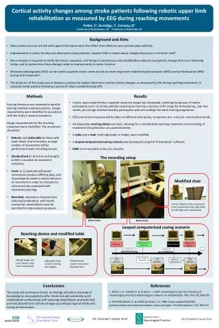
Introduction and aims Neuromuscular Ultrasounds (NMUS) refers to the use of high resolution ultrasounds (HRUS) of nerve and muscle to assess neuromuscular disorders. HRUS of cranial nerves is a recent novel subdomain, which may provide additional value in the assessment of cranial nerves. The most accessible and investigated cranial nerves are the optic (II), facial (VII), vagus (X), and spinal accessory(XI) nerves, even if data are available on others. We report our experience with HRUS in disorders involving these nerves aimed to help future investigations in the field. Materials and Methods We performed a retrospective study reviewing US recordings of cranial nerves, including 16 optic, 31 facial, 120 vagal and 8 spinal accessory nerves. All investigations were performed using an Esaote MyLabGamma or a Telemed Echo-wave II device in conventional B-Mode, using a standard protocol Esaote MyLabGamma Telemed Echo-wave II
RESULTS Optic nerve sonography resulted useful in measurement of optic nerve diameter (crossed section area, CSA), increased in intracranial pressure and in optic neuritis, and minimally reduced in ischemic disorders (fig. 1). Facial nerve, scanned during 1st week and after three months from Bell’s palsy, predicted poor recovery at 3 months in cases of enlarged facial nerve (fig. 2). Vagus nerve size resulted decreased in autonomic neuropathy with parasympathetic involvement; enlarged in demyelinating neuropathies and spared in motor neuron disease (fig. 3). Spinal accessory nerve was investigated in 8 cases of iatrogenic lesions, allowing to distinguish axonotmesis from neurotmesis (fig. 4).
OPTIC and FACIAL Nerves Tips: Measure of optic nerve diameter ( > 5.7 mm abnormal); allows assessment of nerve enlargement/atrophy. Relevance in: Intracranial hypertension in adults Intracranial hypertension in children Intracranial hypotension Optic Neuritis CIDP, sarcoidosis, CNS infections, mitochondrial disorders Tips: Measure of size (diameter), echogenicity and vascularity inside the parotid gland (tubular-like structure. Relevance in: Prognosis of Bell’s palsy: predict good and poor recovery at 3 months (100%positive, 77% negative) Thicked edematous perineurium, axon and myelin degeneration with intraneural inflammatory cells infiltration) Guillain-Barré syndrome, CIDP and hereditary neuropathies Figure 2 –Peripheral left facial palsy (case B.R., male, 54 yrs): the facial nerve is bigger than the contralateral one; it also shows a reduced echogenicity and a significant loss of the fascicular structure. Figure 1 –Orbital ultrasonography evaluation of the II cranial nerve in a patient with bilateral palpebral ptosis due to a pituitary macro-adenoma and intracranial hypertension (case M.V, male, 60 yrs) (normal value: < 5.7 mm).
VAGUS and ACCESSORY Nerves Tips: Measure of 0.76 + 0.12 mm Enlarged cross sectional area or reduced, focal swelling CSA (superior of HRUS over MRI). Relevance in: Preoperative mapping to avoid iatrogenic nerve transection Iatrogenic lesion, stretch injury Idiopathic Extrinsic causes of compression; serial follow-up of lesion Tips: Measure of Vagus nerve diameter (mean 5 + 2 mm); assessment of nerve enlargement/atrophy Relevance in: Vocal cord paralysis Charcot-Marie-Tooth (CMT) Laryngeal nerve palsies, Guillain-Barré syndrome, sarcoidosis, Lyme diseases, poorly understood autonomic disorders, diabetes, chronic alcoholism Figure 3 –Evaluation of the vagus nerve in a patient with recurrent episodes of syncope (R.M., female, 45 yrs). Note reduced dimensions at the right and the progressive loss of fascicular structure, thus suggesting axonal loss (normal value 5.8 + 1.3). Figure 4 –Evaluation of spinal accessory nerve rigth lesion in a patient with history of ipsilateral endoarteriectomy (G.M.P., female, 75 yrs).
Save the date: SINC Nat. Congr. Pisa, May 27-30, 2020. Conclusions Present data confirm that ultrasound scanning techniques are a safe, inexpensive, portable, non-invasive tool for rapid and reliable evaluation of morphology, and well-tolerated modality, useful complement of electrodiagnostic studies in the evaluation of above mentioned cranial nerves. Of possible future interest may be indirect evaluation of the oculomotor, trochlear, trigeminal, abducens and hypoglossal nerves through ultrasound examination of the muscles they innervate. Further investigation may add additional uses for HRUS in pre-operative mapping of nerves to avoid surgical injury, detection of intraneural blood flow in inflammatory disorders, and guidance of needle placement. Therefore, US should be included in the instrumental baggage in any Neurophysiological laboratory and physicians with experience in electrodiagnostic medicine should incorporate cranial nerve ultrasound into their practice. . Thank you for your kind attention Tawfik EA, Walker FO, Cartwright MS. Neuromuscular ultrasound of cranial nerves. J Clin Neurol 2015; 11(2): 109-21. For further informations: ferdinando.sartucci@med.unipi.it f.sartucci@ao-pisa.toscana.it