Download
1 / 50
550 likes | 796 Views
Protein Function. Myoglobin and Hemoglobin. O 2 Binding and Allosteric Properties of Hemoglobin. Hemoglobin binds and transports H + , O 2 and CO 2 in an allosteric manner

E N D
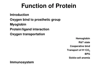
Protein Function Myoglobin and Hemoglobin
O2 Binding and Allosteric Properties of Hemoglobin • Hemoglobin binds and transports H+, O2 and CO2 in an allosteric manner • Allosteric interaction – of, relating to, undergoing, or being a change in the shape and activity of a protein (as an enzyme) that results from combination with another substance at a point other than the chemically active site • a regulatory mechanism where a small molecule(effector) binds and alters an enzymes activity
Protein Function O2 does not easily diffuse in muscle and O2 is toxic to biological systems, so living systems have developed a way around this. Physiological roles of: • Myoglobin • Transports O2 in rapidly respiring muscle • Monomer - single unit • Store of O2 in muscle high affinity for O2 • Diving animals have large concentration of myoglobin to keep O2 supplied to muscles • Hemoglobin • Found in red blood cells • Carries O2 from lungs to tissues and removes CO2 and H+ from blood to lungs • Lower affinity for O2 than myoglobin • Tetrameter - two sets of similar units (22)
x-ray crystallography of myoglobin • Made up of 8 helix A to H (proline near end) • very small due to the folding • hydrophobic residues oriented towards the interior of the protein • only polar AAs inside are 2 histidines • Red indicates Heme group
Structure of heme prosthetic group • Protoporphyrin ring w/ iron = heme • Four Pyrrole groups [A to D] linked by methane bridges • Fe+2 coordinated by prophyrin N atoms and a N from Histidine (blue) • This is known as His F8 (8th residue of the F helix • Iron is out of plane due to His 8 bond • A molecule of O2 acts as 6th ligand
Structure of heme prosthetic group • Two hydrophobic sidechains on O2 binding site of heme elp hold it in place • Valine E11 and phenylalanine CD1 • oxygenation changes state of Fe • Purple to red color of blood, Fe+3 – brown • Oxidation of Fe+2 destroys biological activity of myoglobin • Physical barrier of protein is to maintain oxidation state of Fe+2
free vs. bound heme - role of apoprotein • Apoprotein - the protein moiety of a molecule or complex • restricts heme dimers • keeps iron reduced • stabilizes transition state (O2 binding) • CO, NO and H2S binding - poison of O2 binding bind with greater affinity than O2 His E 7 decreases affinity of ligands (CO and O2 ) for Fe+2
Myoglobin and Hemoglobin • Hemoglobin is structurally related to myoglobin • very different primary sequence about an 18% homology in the primary sequence • 2 alpha subunits and 2 beta subunits • in adults there are very small amount of alpha 2delta 2 hemoglobin - significance of conserved amino acids between myoglobin and hemoglobin • these are the important aas which keep hemes in contact with the protein • Stabilizes helical arraignment • Interacts with heme/iron
There are two general structural states - the deoxy or T form and the oxy or R form. • One type of interactions shift is the polar bonds between the alpha 1 and the beta 2 subunits.
3D structure of hemoglobin and myoglobin • a1- b 1 units have 35 interactions • a1- b2 units have 19 interaction sites • similar units have few polar contacts • the two aand two b subunitsface each other through aqueous channels
Binding of oxygen dramatically alters the interactions and brings about a twisting of the two halves (alpha beta pairs) • Much of the quaternary changes takes place in the salt bonds between the C terminals of all four chains
Myoglobin O2 affinity MbO2 <-> Mb + O2 [Mb] [O ] 2 myoglobin -oxygen dissociation K = [MbO ] 2 [MbO ] 2 Y = [MbO ] + [O ] 2 2 2 [O ] 2 Y = [O ] + K 2 pO 2 Y = pO + P 2 50 Y = fraction of globin bound to O2 K = dissociation constant
Myoglobin vs. Hemoglobin O2 affinity • Note the hyperbolic vs sigmoidal nature of the two curves! • Sigmoidal curves indicate cooperativity
Hemoglobin -sigmoidal dissociation Hb(O ) =Hb +n O X = number of Hb subunits 2 n 2 n Y Y = fraction of globin bound to O2 log = n log pO - n log P • n = Hill constant - determined graphically by the - hill plot • n is the slope at midpoint of binding of log (Y/1-Y) vs log of pO2 if n = 1 then non cooperativity if n < 1 then negative cooperativity if n >1 then positive cooperativity 2 50 1 - Y • The experimentally determined slope does not reflect the number of binding sites however. It reflects, instead, the degree of cooperativity.
So how does this relate the biological effect of O2 affinity of Hb? • look at pO2 of alveoli vs. metabolically active tissue • What if oxygen dissociation were hyperbolic rather than sigmoidal?
effect of BPG effect of pH % O Sat % O Sat 2 2 pO pO 2 2 Alteration in oxygen binding by:- H+, CO2 2,3 bisphosphoglycerate (BPG) Bohr Effect (CO2 & pH effect on O2 binding) Simple rule of thumb- • High H+ conc and higher CO2 levels decrease O2 affinity for Hb • High O2 concentration decrease H+ and CO2 affinity for Hb • Hb acts as a H+ buffer in respiring tissue
Cooperative Binding of O2 to Hemoglobin No cooperativity pH = 7.4 pH = 7.2 %Saturation pH= 7.6 20 40 80 100 120 60 pO2 (torr)
BPG and CO2 Effects on O2 Binding Stripped Hb Hb + BPG Hb + BPG + CO2 %Saturation Hb + CO2 20 40 80 100 120 60 pO2(torr)
Functional Structure of Hemoglobin - allosteric regulations • Allosteric interaction - the binding of one ligand at one site in a protein that affects the binding of other ligands at other sites in the protein. This can be affect on binding can be cooperative (pos or neg). Allosterism is typically seen when sigmoidal binding / activity curves.
Simple definition of the concept of cooperativity.In its simplest meaning, cooperativity is a measure of communication between binding sites.Positive cooperativity describes a relationship in which the binding to one site facilitates the binding to the second, third, ect.No cooperativity puts all sites on equal footing and indicates no site to site influence. Negative cooperativity implies binding to one site hinders binding to subsequent sites.
Oxygen pressure in various fluids So why is this important.Look at the Hill plot and the plot of the of binding vs pO2 (figs 7-8 and 7-7 respectively). Think of when hemoglobin should be mostly saturated and when it would be best if it had a low saturation / affinity and thus "give up" its oxygen. Use the table below to help your thoughts. pO2 (Torr) 158 100 90 40 30 10 Region or fluid Inspired Air Aveolar Air Arterial Blood Capilary Insterstitual Fluid Cell Cytosol
Look at the pO2 pressure and think of where Hb will be loaded with oxygen and where it will deliver oxygen
How is this cooperation actually forced on hemoglobin? • The structures in one subunit effects the others • Structural differences of bound and unbound Hb • Two structures - T and R form depending on the oxygenation of Hb • T is the deoxy form, R is oxy form • allosteric modifiers shift or stabilize one particular conformation over the other • Monomer interactions, most chain interaction occurs between and chains • through mostly hydrophobic residues • and interactions are few and polar • and contact act as a switch
oxy and deoxy quaternary structures are different • change takes place between 1 - 2 and 2 - 1 • amino acids between 1 and 2 help to stabilize each forms
Oxygen binding shifts quaternary structure at long distances • binding of O2 ligand at 6th coordinate position pulls Fe into heme • moves proximal histadine (F8) and the alpha helix it is attached to. • shift in the helix is transmitted throughout of molecule
The T form finds the terminals in several important H bonds and salt bridges. • In the T form the C terminus of each subunit are "locked" into position through several hydrogen and ionic bonds. • Shifts into the R state break these and allow an increased movement throughout the molecule. • Note that binding of one or more oxygen can have a dramatic affect on the other subunits that have not yet bound an O2.
How Do Myoglobin (Hemoglobin) Bind to Oxygen Move down upon O2 binding His E7
How Do Myoglobin (Hemoglobin) Bind to Oxygen Move down upon O2 binding His E7
T and R State of Hemoglobin - Review • Below are the two major conformations of hemoglobin as predicted by the models for allosteric activation. • Oxygen will bind to hemoglobin in either state; however, it has a signficantly higher affinity for hemoglobin in the R state. • In the absence of oxygen, hemoglobin is more stable in the T state, and is therefore the predominant form of deoxyhemoglobin. R stands for relaxed, while T stands for tense, since this is stabilized by a greater number of ion pairs. • Upon a conformational change from the T state to the R state, ion pairs are broken mainly between the a1b2 subunits.
Bottom Line: CO2, BPG and H+ all help to stabilize Hb in R or T form.
Bohr Effect Continued The Bohr effect is the reversible shift in Hb affinity for O2 with changes in pH. H+ Transport (effect) - O2 binding to Hb releases H+ due to conformational changes in Hb - deoxyform (T form) brings Asp 94 close to His 146 (fig 7-11 (b)) - the proximity of an acidic amino acid increases the pK of histidine (pKa is now above the pH) and results in H+ “binding” to deoxyHb - in other words the His becomes protonated where it normally would be ionized • increasing pH stimulates Hb to bind to O2 • Bottom line - when O2 binds Hb, H+ is released from several amino acid's functional groups. When O2 is released, the amino acids become protonized and then "picks" up a H+. • So when the H+ is high (acidic conditions) the H+ is driven onto the terminal amino acids driving it into the T conformation
CO2 effects - In the red blood cells the picture is even more complicated. CO2 is removed by converting CO2 to bicarbonate CO2 + H2O <-> HCO3- + H+
bicarbonate (HCO3-) formed by the enzyme carbonic anhydrase • H+ produced by this enzyme is removed by the Hb as described above • This allows more CO2 to be removed in the form of bicarbonate
CO2 binding aids in reducing O2 affinity by changing conformation by the production of more H+ (R to T change)
This all aids the function of Hb.In active tissues respiration, (glycolysis) results in lactic acid formation. These tissues need more O2. Without the H+ effect Hb would hold on to more of the O2. The H+ induces Hb to dump 10% more of it's O2. • CO2 reversibly binds to N term (carbamate) to remove remaining CO2 R - NH2 + CO2 <-> R - NH - COO- + H+ R is the Hb N term amide The carbamide increases the T formation - deoxy form. • The reverse occurs in the lungs. This results in 1/2 of CO2 removal from tissues.
2,3 bisphosphoglycerate (BPG) Effects • Purified Hb has a different O2 affinity than it does in blood • 26 fold decrease change in affinity is due to 2,-3 diphosphoglycerate BPG
diphosphoglycerate BPG • present in human red blood cells at approximately 5 mmol/L. • It binds with greater affinity to deoxygenated hemoglobin • In bonding to partially deoxygenated hemoglobin it allosterically upregulates the release of the remaining oxygen molecules bound to the hemoglobin, thus enhancing the ability of RBCs to release oxygen near tissues that need it most
2,3 bisphosphoglycerate (BPG) • Purified Hb has a different O2 affinity than it does in blood • 26 fold decrease change in affinity is due to 2,-3 diphosphoglycerate BPG • (BPG replaced by nucleotides IHP and ATP in fish and birds) • - 1 BPG per Hb - binds in central cavity of Hb • - binds preferentially to deoxy Hb • - hydrophobic bonds with Lys and salt bridge with His • - O2 binding changes conformation and “kicks out” BPG • change in altitude increases concentration of BPG • Fetal F Hb has replaced His 143 with Ser - What might the consequences be?
Cooperative interactions between subunits Both models do not fully account for the effects of allosteric effectors • sequential model (D Koshland) • binding of one O2 induces T-R conformation change • 1st change is most difficult due to influence by 3 other subunits • binding of next three subunits happens sequentially, with higher affinity (easier T-R changes) • kinetics increase to the fully oxy Hb state as more O2 is bound
Concerted model (J Monod) • All R or all T no in between as in the Koshland model • concerted model means as more O2 binds, the R conformation is favored until all units are in the R conformation regardless of the total units bound to O2 • Affinities do NOT change until conformation changes • 1 O2 - all T; 2 O2 - nearly even equilibrium; 3 O2 mostly R; 4 O2 - mostly R form • energy from O2 binding causes the change in equilibrium • this model best fits O2 dissociation curve but with limits.
Sickle-Cell Anemia, a Molecular Disease One of the first “molecular” diseases found - sickle cell anemia • sickle cell - blood cell is elongated , mis-shaped (sickle) • occurs at low O2 concentration • caused by hemoglobin aggregates • inflammation in capillaries and pain • red blood cells break down - anemia • between 10% of American blacks and 25% of African blacks are heterozygous for sickle cell anemia • homozygous usually do not survive into adult hood • heterozygous individuals usually have no problem except when in severe oxygen deprivation
Single amino acid (point mutation) HbS vs. HbA changes structure • sickle cell b chains have a valine in place of glutamate • leads to more Hb S (sickle cell) has 2 more + charges than normal hemoglobin • Glu -Val occurs on exterior of protein - does not change O2 dissociation/allosteric properties of protein
Deoxy HbS precipitates • oxyHb phenylalanine b85 and leucine b88 interior • phe and leu shift to exterior • create a sticky patch with valine (hydrophobic bonding) • nucleation (cluster of aggregate) occurs logarithmically • homozygous - 1000 times faster than heterozygous • that means mixed genes can re-oxygenate faster than polymerization can occur
how can such a disease occur? • highest concentration of gene mutation occurs where there is high incidence of malaria • heterozygous individuals survive this disease better than those without • malaria causing parasite lives in red blood cells during part of its life cycle • partial sickling must interrupt life cycle of malaria parasite