Download
1 / 47
510 likes | 543 Views
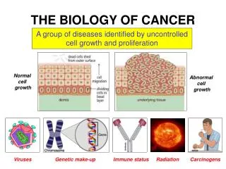
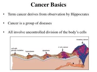
The Basics of Cancer Biology. Lucio Miele, M.D., Ph.D. Part II: “Partners in Crime -1”. Tumor Suppressors, Oncogenes, Enablers and Turncoats. Cancer is a genetic disease. “Cancer is, in essence, a genetic disease. Although cancer is complex, and environmental and other

E N D
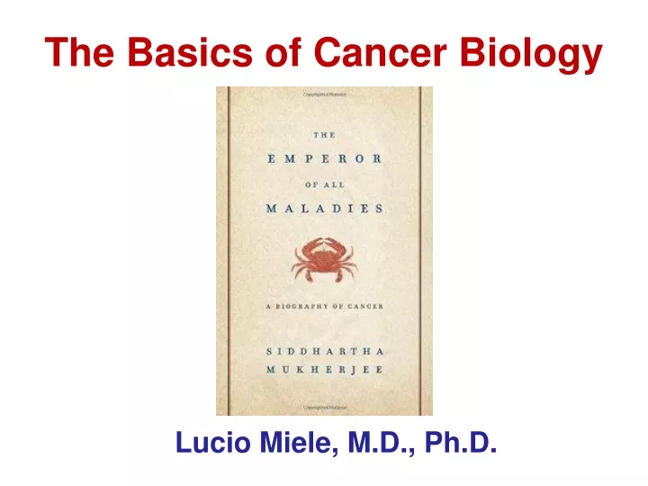
The Basics of Cancer Biology • Lucio Miele, M.D., Ph.D.
Part II: “Partners in Crime -1” Tumor Suppressors, Oncogenes, Enablers and Turncoats
Cancer is a genetic disease “Cancer is, in essence, a genetic disease. Although cancer is complex, and environmental and other nongenetic factors clearly play a role in many stages of the neoplastic process, the tremendous progress made in understanding tumorigenesis in large part is owing to the discovery of the genes, that when mutated, lead to cancer.” Bert Vogelstein (1988) NEJM 1988; 319:525-532
Abstract Cancer is a distinct type of genetic disease in which not one, but several, mutations are required. Each mutation drives a wave of cellular multiplication associated with gradual increases in tumor size, disorganization and malignancy. Three to six such mutations appear to be required to complete this process. Vogelstein B and Kinzler KW Trends Genet.1993
What are Cancer Genes? • From a genetic standpoint: Any gene which, when mutated, increases the risk of cancer is a cancer gene. • From a phenotypical or functional standpoint: Any gene which, when altered, causes the transformation of normal cells into cancerous cells is a cancer gene. • Note: a gene that increases cancer risk when mutated does not NEED to be transforming! Its effect could be indirect
Traditional Types of Cancer Genes Cancer genes include those whose products: • Directly regulate cell proliferation or survival a. Tumor suppressor genes (cell proliferation inhibitory) b. Oncogenes (cell proliferation promoting) • Are involved in the repair of damaged DNA a. DNA repair genes b. DNA recombination genes (homologous recombination, non-homologous end-joining) • Regulate TSGs or oncogenes a. Non-coding RNAs (miRNAs, lnRNAs) b. Epigenetic modifiers
Oncogenes and Tumor Suppressors • There are two broad categories of genes to think about when considering cancer-forming mutations: • Oncogene - An oncogene is a gene whose normal activity promotes cellular proliferation or division. • Tumor Suppressor - a gene that inhibits events leading towards cancer. (This type of gene will be discussed in tomorrow’s lecture.) Oncogene = gas pedal Tumor Suppressor gene = brakes
Sporadic Small % of cases Relative large % of cases
Oncogenes: • Mutation in one copy of the gene - Dominant • promote cell proliferation Tumor suppressor genes: • Mutations in both copies of the gene - Recessive • promote cell proliferation DNA repair genes: Usually recessive, loss-of-function mutations that increase spontaneously and environmentally induced mutation rates
Wellcome Trust Sanger Institute at http://cancer.sanger.ac.uk/cancergenome/projects/census Cancer Gene Census • Almost 2% of protein coding genes in humans are cancer genes • 15% of cancer genes bear germline mutations • 90% of them have somatic mutations • 10% of them show both germline and somatic mutations
Tumor Suppressor Genes • Tumor suppressor genes (TSG) are genes that normally slow down cell division, repair DNA damage, or cause cells to die (by apoptosis or programmed cell death, by necrosis, by autophagy or by or by mixed cell death mechanisms). • When tumor suppressor genes are damaged, deleted or epigenetically silenced, cells can grow out of control, which can lead to cancer. • More than 30 tumor suppressor genes have been identified, including p53, BRCA1, BRCA2, APC, and RB1. • An important difference between oncogenes and TSGs is that oncogenes result from the activation (turning on) of proto-oncogenes, but TSGs cause cancer when they are inactivated (turned off). • Another major difference is that while the oncogenes develop from mutations in normal genes (proto-oncogenes) during the life of the individual (acquired mutations), abnormalities of TSGs can be inherited as well as acquired.
Tumor suppressor gene A tumor suppressor gene is like the brake pedal on a car. It normally keeps the cell from dividing too quickly, just as a brake keeps a car from going too fast. When something goes wrong with the gene, such as a mutation, cell division can get out of control.
Tumor suppressors are mutated in both inherited and non-inherited cancers
Examples of Tumor Suppressor Genes • BRCA 1, BRCA 2: • Both proteins involved in DNA repair pathway (usually double strand break repair). • Loss of these proteins can increase the acquisition of subsequent mutations and chromosomal instability. • Mutations greatly increase the susceptibility to develop breast or ovarian cancer in women. • Wilms’ Tumor Suppressor (WT1): • This gene encodes a transcription factor with multiple functions • Mutation is associated with Wilms’ tumor (a renal cancer called nephroblastoma). • Von Hippel‐Lindau (VHL): • VHL protein degrades protein important for promoting angiogenesis, thereby inhibiting the ability to form new blood vessels. • Mutation is associated with renal cell carcinoma and other types of cancers.
Functions of Tumor Suppressor genes • 1. Regulator of cell cycle: • Serve as genes that insure proper progression through the cell cycle to mediate proper cell division. • Rb gene, INK-4 gene, P53 • 2. Inducer of apoptosis: • Proteins that promote apoptosis in response to improper signals, presence of DNA damage, etc. • Bim, a pro-apoptotic member of the Bcl-2 family of genes • 3. Transcription factors: • Repressor transcription factors: WT1 is a repressor that appears to suppress a factor (Insulin like growth factor) that can contribute to the development of a tumor. • Activator transcription factors: Members of the SMAD family are activated by TGF-β, leading to inhibition of cell proliferation • 4. Regulator of angiogenesis response (VHL)
Hereditary cancer susceptibility syndromes About 10% of the most common cancers are due to a hereditary predisposition • Examples: Breast/ovarian cancer Colorectal Retinoblastoma
RB - the first tumor suppressor gene cloned (1986) Loss of both alleles of RB leads to the development of Retinoblastoma in humans RB is seldom mutated in sporadic human cancers Inhibits proliferation, promotes differentiation but also inhibits apoptosis
Children with RB1 mutations will develop retinoblastoma very early in life because retinoblastoma originates from neuroblasts during retinal development. Therefore, retinoblastoma is not observed in adults
Current model of Rb in G1 cell cycle regulation Narasimha AM et al., eLife, 2014
Rb functions as a protein adaptor with at least 4 mechanisms of action: multitasking proteins Burkhart D et al., Nat Rev Can 2008
Two-Hit Hypothesis • The relatively high frequency of the second hit explains the early age of onset of tumors and that they are often bilateral. • In sporadic cancers two independent rare events within the target tissue must occur to form a tumor; hence, the later age of onset. • Loss of Heterozygosity (LOH) analysis can help identify tumor suppressor genes.
LOH LOH D10S107 D17S855 T/T T/C C/C 1st hit 2nd hit 1st hit Knudson two-hit model
Mm Mm Mm mm Mm mm Mm Mm Mm Mm Mm Mm Mm mm Mm Mm Mm Mm Mm Mm Mm Mm mm Mm Mm mm mm Mm Mm mm Mm mm Mm Mm Mm Mm Mm Mm mm Mm Mm mm Mm Mm Mm Mm mm Mm Mm mm Mm Mm Mm Mm Mm Mm Mm mm mm Mm mm mm Mm mm mm mm mm mm mm mm mm mm mm mm mm mm Mm mm mm mm Loss of Heterozygosity (LOH)
P P P p53 Active Cell cycle arrest Bax PUMA p21 Apoptosis Noxa Pigs p53: the most commonly mutated TSG in human cancers GADD45 DNA repair DNA damage Oncogenic signals p53R2 p53 post-translational modifications Inactive
p53 - a Classic Tumor Suppressor • Point mutations account for nearly 75% of all of the mutated forms of p53 found in tumors. (Compare to the other commonly mutated genes found in cancer.) • These mutations almost always lead to amino acid substitutions. • A majority of the identified point mutations (from over 15,000 mutated p53 genes sequenced in cancers) are present in the DNA-binding domain of the gene. • Therefore, these mutations inhibit the ability of p53 to bind to DNA and therefore inhibit its ability to activate (or repress) the genes listed in the previous table.
p53 - a Classic Tumor Suppressor • In addition to loss of or mutation to p53, p53 activity can be altered through deletion or mutations to other genes. • MDM2 binds to p53, inhibits the ability of p53 to act as a transcription factor, and targets it for ubiquitylation and subsequent degradation. • The action of MDM2 insures the 20 minutes half life of p53 in normal cells. • The amplification of the MDM2 gene in cancer would therefore interfere with the normal ability of p53 to respond to extracellular stimuli and prevent the required cell cycle arrest and promotion of apoptosis.
The effects of p53 are DOSE-DEPENDENT • When activated at low levels, p53 binds differentially to promoters that encode genes that suppress cell cycle progression and allow DNA repair (e.g., p21) • When DNA damage is not promptly repaired and active p53 accumulates, it binds lower affinity chromatin sites that contain the promoters of genes that cause apoptosis (e.g., NOXA, DAXX) • This mechanism permits DNA repair and survival if the damage is not irreparable, and causes the cell to commit suicide if the damage is irreparable • Hence, p53 has been called “the guardian of the genome”
DNA Damage E2F (1) (2) 1) Wt-p53binds to p21 promoter; 2) Elevated p21 inhibits CDK2/4 kinases, reducing Rb1 phosphorylation; 3) unphosphorylated Rb1 forms complex with E2F; 4) E2F cannot activate genes important for G1/S transition - no tumor. 2) Mut-p53 is unable to bind to p21 promoter, causing reduced p21 level; 2) CDK2/4 kinases induce Rb1 phosphorylation; 3) more E2F free from Rb1 complex; 4) more E2F will promote cell cycle progression - Tumor formation.
Dominant-negative mechanism WTp53 WTp53 Transcriptional activation WTp53 WTp53 Tetramer formation WTp53 Mutant p53 WTp53 Mutant p53 WTp53
p53 mutations • More than 50% of human tumors contain p53 mutations. • 80% are missense mutations in the DNA binding domain. • “Hot spot” mutations at pp175, 248, 249, 273, and 281 • Mutations of p53 are more frequent events and show worse prognosis compared to deletion of p53 gene. 248 273 175 281 249 II I III IV V C N Tetramerization domain Activation domain DNA binding domain
Some mutations turn p53 from a TSG into an oncogene: a Turncoat! • Over 50% of human cancers either have lost p53 protein or have mutations in the gene. • About 70% of Li-Fraumeni syndrome patients carry p53 alterations (heterozygous). • Loss of p53 in mice causes tumors dependent on gene dosage. • Mutant p53 accumulates as a nuclear phosphoroprotein. • The mutant p53 functions as dominant-negative that inactivates wild-type p53 function through a tetramer formation. • However, some forms of mutant p53 show unexpected oncogenic functions which can not be explained simply by loss of p53. These mutants activate transcription at some chromatin sites • Therefore, p53 (mutant p53) was originally believed to be an oncogene.
Identification of TSG • Before 2007 - Linkage analysis of familial cancer families and positional cloning of TSG - Loss of heterozygosity analysis to clone TSG • After 2007 - Exome sequencing of familial cancer family members and bioinformatics analysis - Mutation co-segragates with phenotype in family
Cancer gene (TSG) discovery by Exome sequencing Exclude variants in control
miRNAs and lncRNAs can be TSGs • By downregulating the expression of oncogenes • mRNA stability • mRNA translation arrest • By regulating the expression of cancer risk genes that are not themselves oncogenes • E.g., genes in the immune system that control cancer immune surveillance • By regulating the splicing of transcripts or the activity of regulatory elements in DNA