Download
1 / 41
410 likes | 589 Views
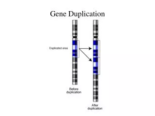
Folds Evolution through micro-gene duplication. Structural Evidence. Gene Duplication - Summary. Intro: A few examples Aspartyl Proteases [b.50] Double Psi beta barrels DNA Dependent RNA Polymerases Trypsin-like Serine Proteases [b.47] Unexpected Structural homologies. Hemerythrin.

E N D
Folds Evolution through micro-gene duplication Structural Evidence
Gene Duplication - Summary • Intro: A few examples • Aspartyl Proteases [b.50] • Double Psi beta barrels • DNA Dependent RNA Polymerases • Trypsin-like Serine Proteases [b.47] • Unexpected Structural homologies
Hemerythrin ....*....|....*....|....*....|....*....|....*....|....*....| 1I4Y_A 10 vwdpsfrtfYSIIDDEHKTLFNGIFHLAIDd~nADNLGELRRCTGKHFLNEQVLMQasqy68 consensus 3 kwdesfrtfYEVIDDEHKTLFNGINDLSEAnnrADNLKELVDYTVKHFKDEEALMEaagy62 consensus 56 lmeaagypdYEEHKKIHEDFVEKVGGLKAPv~gQADLKYLKDWLVNHIKTEDFKYKgkl~113 2MHR 9 vwdesfrvfYEQLDEEHKKIFKGIFDCIRDn~sAPNLATLVKVTTNHFTHEEAMMDaaky67 2MHR 61 mmdaakyseVVPHKKMHKDFLEKIGGLSAPv~dAKNVDYCKEWLVNHIKGTDFKYKgkl~118
Ferredoxin 1JNR_B1 ~~~~~~~~~~~~~~~~~~~~~~~~~~~~~~~~~~~~~~~~~~~~mpSFVnp~ekCDGCka15 1JNR_D1 ~~~~~~~~mpsfvnpekcdgckalertaceyicpndlmtldkekmkAYNrepdmCWEC~~50 ....*....|....*....|....*....|....*....|....*....|....*....| 1JNR_B16 lerTACEYICPNDLMTLDKekmkaynrepdmcwecyscvkmcpqgaidvrgyvdysplgg75 1JNR_D51 ~~~YSCVKMCPQGAIDVRGyvdysplggacvpmrgtsdimwtvkyrngkvlrfkfairtt107
LRV Repeats 70 8090 100 110120 ....*....|....*....|....*....|....*....|....*....|....*....| 1LRV 36 vavESGRQIdrFFRNNPhl~aVQYLADPFWERRAIAVRYSPVEALTPLIRDSDEVVRRAV94 1LRV 61 dpfWERRAI~~AVRYSPvealTPLIRDSDEVVRRAVAYRLPREQLSALMFDEDREVRITV118 130140 150 160170 180 ....*....|....*....|....*....|....*....|....*....|....*....| 1LRV 95 AYRLPREQLSALMFDEDREVRITVADRLPLEQLEQMAADRDYLVRAYVVQRIPPGRLFRF154 1LRV119 ADRLPLEQLEQMAADRDYLVRAYVVQRIPPGRLFRFMRDEDRQVRKLVAKRLPEESLGLM178 REPEAT PATTERN: [PReWY][DEQVLT][RV]RXX[IAV][AV]XRX[PR]XXXL-x(5)-[DE]X[DE]
OB Fold Repeats in BRCA2 OB2-OB3 Tandem – OB1-OB2 head-2-head pseudo-C2 (Yang et al. Pavletich 2002)
BRCT repeats in 53BP1 and BRCA1 Figure 1. (Ekblad et al. 2003) Schematic representation of the structures of the BRCT domains of BRCA1 and 53BP1. (A) BRCA1-BRCT with the linker region in black. (B) An overlay of the BRCT domains of BRCA1 (red) and 53BP1 (blue); the linker regions are shown in black and in turquoise, respectively. (C) 53BP1-BRCT (blue) bound to p53 core domain (yellow). The binding interface is concentrated on the L2 and L3 loops of p53, which interacts with the C terminus of the BRCT-N and the linker region of 53BP1. Interacting residues are shown in green (53BP1) and in red (p53).
Self associating motif - SerProt GSSDLYLVTRHADVIp~VRRrg~~~~~~dsRGSLL GSSGGPLLCPSGHAVgiFRAavctrgvakaVDFVP
Double PSI Beta Barrel (DPPB) (a) barwin; (b) EGV; (c) DMSO; reductase; (d) FDHH; (e) N-terminal lobe of endothiapepsin; (f) C-terminal lobe of endothiapepsin (Richard M Castillo1, Kenji Mizuguchi1, Venugopal Dhanaraj1, Armando Albert2,Tom L Blundell1* and Alexey G Murzin3 1999)
DNA-Dependent-RNA-Polymerase Figure 4m (Lakshminarayan M Iyer, Eugene V Koonin and L Aravind* 2003) Structure of the catalytic cleft of DDRP formed by interacting DPBB domains of the â and â' subunits. The metal-coordinating DbDGD motif of the â' subunit and the functionally important lysines projecting into the catalytic cleft of the â subunit are shown in ball and stick representation. The two double psi-barrels are juxtaposed in an asymmetric head to tail configuration.
Figure 5 (Lakshminarayan M Iyer, Eugene V Koonin and L Aravind* 2003) A hypothetical scheme of evolution of two types of 6-stranded â-barrels from 3-stranded units. The scheme was derived as the most parsimonious explanation for the phyletic patterns and structural peculiarities of each lineage of 6-stranded barrels. The emergence of particular properties or characters characteristic of a given clade is indicated by horizontal bars.
Trypsin-like Serine Protease D1-D2 Structural Alignment – 38 residues GSSDlylvtrhadvipvrRRGD----SRGSLLsprpvsylk QVVStat--------qsfLATCvngvCWTVYHga------- gssgGPLLcpsghaVGIFRAAVCtrgVAKAVDFVPVEsme gs--KTLAgpkg--PITQMYTNV---DQDLVGWQAPPgar
Trypsin-like Serine Protease –D2 d1/d2 Optimised localsequence alignment framed by structural alignment GSSDLYLV~~TRHADVIpVRRrgdsRGSLL GSSGGPLLcpSGHAVGI~FRAavctRGVAK
Trypsin-like Serine Protease – D1-D2 D2a> GSSDLYLVTRHADVIp~VRRrg~~~~~~dsRGSLL D2b> GSSGGPLLCPSGHAVgiFRAavctrgvakaVDFVP D1a>QVVStat--------qsFLATCvngv~~~CWTVYH
Ser vs Asp Proteases subdomains 1 &2 ....*....|....*....|....*....|....*....|....*....|....*....| 1NS3_A 61 sktlagpkgpitqmytnvdqdlvgwqappgarsltpctcgsSDLYLVTRhADVIPVRRrg120 1MER_A 1 ~~~~~~~~~~~~~~~~~~~~~~~~~~~~~~~~~xqvtlwqrPLVTIKIG~GQLKEALLdt26 ....*....|....*....|....*....|....*....|....*....|....*....| 1NS3_A121 dsrgsllsp~~~~~~~~~~~~~~~~rpvsylkgssGGPLLCpsGHAvGIFRAavctrgva164 1MER_A 27 gaddtvleemslpgrwkpkmiggiggfikvrqydqILIEIC~~GHK~AIGTVlvgptpvn83
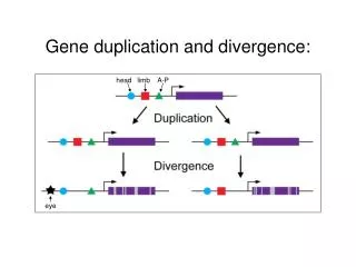
Detecting SubFold Gene Duplication • If a protein has a duplicated fold in tandem, head on tail, and/or intertwined or else, most likely the fold itself has internal symmetry and might come from gene duplication • Why? • Answering why is one goal • How? • Evolution mechanism …
Originality: • It is not be the first to see new things that that sets apart original minds, but to see with a new eye the old thing that every one has seen without seeing it The first discoverer is often the banal and odd … chance.