Download
1 / 25
320 likes | 630 Views
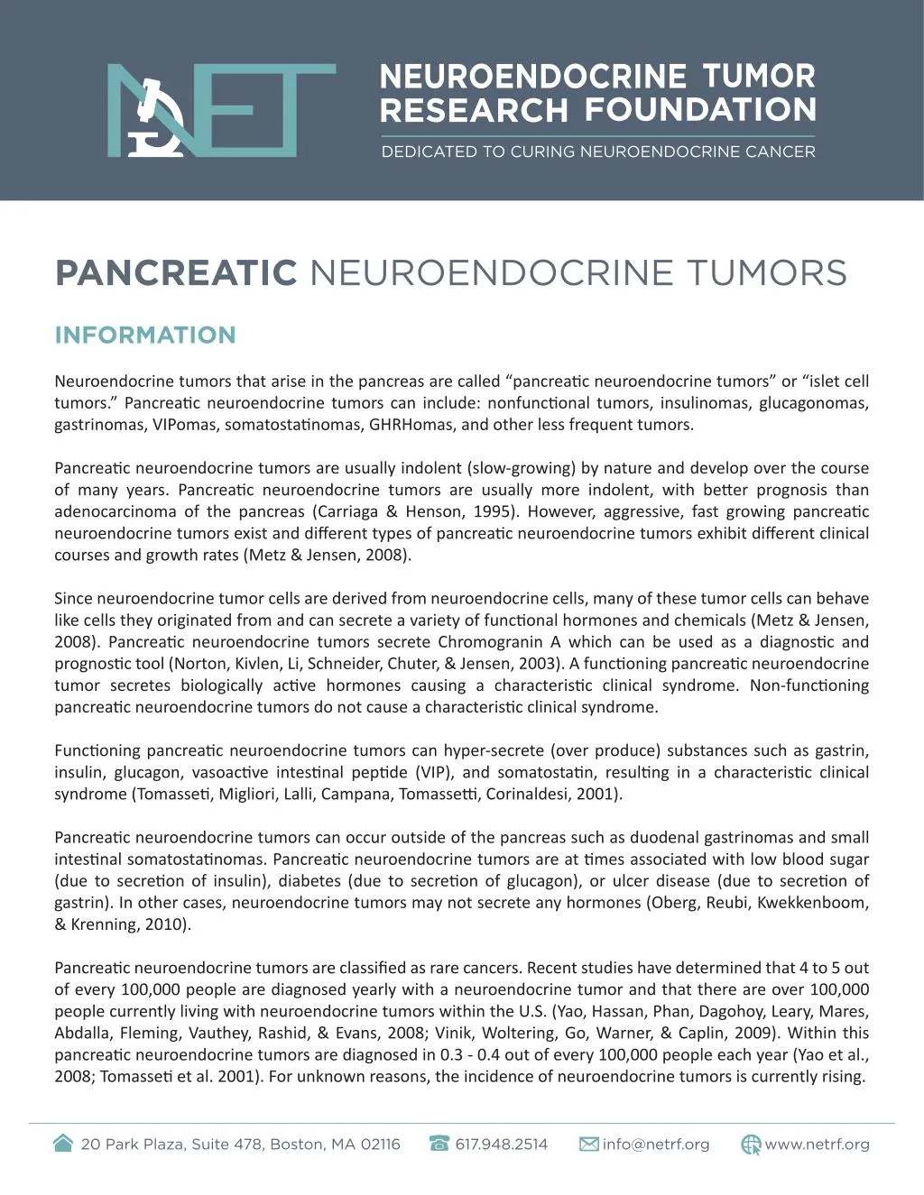
<br>https://netrf.org/pancreatic-nets<br>Neuroendocrine tumors that arise in the pancreas are called “pancreatic neuroendocrine tumors” or “islet cell<br>tumors.”

E N D
PANCREATIC NEUROENDOCRINE TUMORS INFORMATION Neuroendocrine tumors that arise in the pancreas are called “pancreatic neuroendocrine tumors” or “islet cell tumors.” Pancreatic neuroendocrine tumors can include: nonfunctional tumors, insulinomas, glucagonomas, gastrinomas, VIPomas, somatostatinomas, GHRHomas, and other less frequent tumors. Pancreatic neuroendocrine tumors are usually indolent (slow-growing) by nature and develop over the course of many years. Pancreatic neuroendocrine tumors are usually more indolent, with better prognosis than adenocarcinoma of the pancreas (Carriaga & Henson, 1995). However, aggressive, fast growing pancreatic neuroendocrine tumors exist and different types of pancreatic neuroendocrine tumors exhibit different clinical courses and growth rates (Metz & Jensen, 2008). Since neuroendocrine tumor cells are derived from neuroendocrine cells, many of these tumor cells can behave like cells they originated from and can secrete a variety of functional hormones and chemicals (Metz & Jensen, 2008). Pancreatic neuroendocrine tumors secrete Chromogranin A which can be used as a diagnostic and prognostic tool (Norton, Kivlen, Li, Schneider, Chuter, & Jensen, 2003). A functioning pancreatic neuroendocrine tumor secretes biologically active hormones causing a characteristic clinical syndrome. Non-functioning pancreatic neuroendocrine tumors do not cause a characteristic clinical syndrome. Functioning pancreatic neuroendocrine tumors can hyper-secrete (over produce) substances such as gastrin, insulin, glucagon, vasoactive intestinal peptide (VIP), and somatostatin, resulting in a characteristic clinical syndrome (Tomasseti, Migliori, Lalli, Campana, Tomassetti, Corinaldesi, 2001). Pancreatic neuroendocrine tumors can occur outside of the pancreas such as duodenal gastrinomas and small intestinal somatostatinomas. Pancreatic neuroendocrine tumors are at times associated with low blood sugar (due to secretion of insulin), diabetes (due to secretion of glucagon), or ulcer disease (due to secretion of gastrin). In other cases, neuroendocrine tumors may not secrete any hormones (Oberg, Reubi, Kwekkenboom, & Krenning, 2010). Pancreatic neuroendocrine tumors are classified as rare cancers. Recent studies have determined that 4 to 5 out of every 100,000 people are diagnosed yearly with a neuroendocrine tumor and that there are over 100,000 people currently living with neuroendocrine tumors within the U.S. (Yao, Hassan, Phan, Dagohoy, Leary, Mares, Abdalla, Fleming, Vauthey, Rashid, & Evans, 2008; Vinik, Woltering, Go, Warner, & Caplin, 2009). Within this pancreatic neuroendocrine tumors are diagnosed in 0.3 - 0.4 out of every 100,000 people each year (Yao et al., 2008; Tomasseti et al. 2001). For unknown reasons, the incidence of neuroendocrine tumors is currently rising. 20 Park Plaza, Suite 478, Boston, MA 02116 617.948.2514 info@netrf.org www.netrf.org
Pancreatic neuroendocrine can be difficult to diagnosis with the average time between tumor development and diagnosis being between 5 and 10 years (Vinik, Feliberti, Perry & Nakave, 2008; Vinik et al., 2009). Survival rates for individuals with pancreatic neuroendocrine tumors vary and depend upon tumor type, the location of the tumors, the size of the tumors, the extent and growth rate of liver and bone metastases, proliferative indices, presence of clinical syndromes and many other factors (Metz & Jensen, 2008). Currently, surgery is the only option that offers hope for a cure (Ramage, Ahmed, Ardill, Bax, Breen, Caplin, Corrie, Davar, Davies, Lewington, Meyer, Newell-Price, Poston, Reed, Rockall, Steward, Thakker, Toubanakis, Valle, Verbeke, Grossman, and UK and Ireland Neuroendocrine Tumor Society, 2012; Metz & Jensen, 2008). Pancreatic neuroendocrine tumors can be associated with genetic syndromes such as Multiple Endocrine Neoplasia Type 1 (MEN1), Von Hippel-Lindau Disease (VHL),Tuberous Sclerosis Complex and Neurofibromatosis Type 1 (NF1) (Metz & Jensen, 2008). MEN1 is the most significant genetic syndrome - over 80% of patients with MEN1 develop pancreatic neuroendocrine tumors, over 40% of patients develop gastrinomas and smaller percentages develop other types of pancreatic neuroendocrine tumors (Metz & Jensen, 2008; Gibril & Jensen, 2004; Brandi et al., 2002). What is a Tumor? Cells are the building blocks of all life. All cells have highly specific functions, but not all cells have the same function. When cells that have similar functions are grouped together they form a tissue. Tissues when grouped together to perform a specific function are called organs. All cells of the human body have the same DNA (genetic language). Cell growth and replication is highly controlled and is encoded in each cell’s DNA. However if there are enough mutations (changes) within a cell’s DNA, a cell can grow and replicate uncontrollably. A tumor is a mass formed by an abnormal growth of cells within the body. A tumor can be non-cancerous (benign) or cancerous (malignant). A tumor is considered cancerous when it has uncontrolled proliferation (abnormal growth) and can invade and destroy surrounding tissue. Malignant tumors can also have the ability to metastasize (spread to other organs of the body). What is a Neuroendocrine Tumor? The neuroendocrine system consists of highly specialized neuroendocrine cells which act as an interface or junction between the nervous system and the endocrine system. The endocrine system is made up of cells whose function is to produce and secrete hormones into the bloodstream. Hormones are biochemical messengers that help to regulate many different processes within the body. The nervous system is composed of specialized cells (neurons) that control the activities of all body parts. A neuroendocrine cell is a cell which receives neuronal input (a signal from a nerve cell) and releases hormones in response to this signal. A neuroendocrine tumor can develop anywhere there are neuroendocrine cells. The most common sites from which neuroendocrine tumors arise are the lungs, appendix, small intestine, rectum and pancreas (Yao, Hassan, Phan, Dagohoy, Leary, Mares, Abdalla, Fleming, Vauthey, Rashid, & Evans, 2008). Neuroendocrine tumors that arise in the pancreas are called pancreatic neuroendocrine tumors or islet cell tumors. When neuroendocrine tumors originate in other areas, they are often classified as carcinoid tumors. 20 Park Plaza, Suite 478, Boston, MA 02116 617.948.2514 info@netrf.org www.netrf.org
Since neuroendocrine tumor cells are derived from neuroendocrine cells, many of these tumor cells can behave like cells they originated from and can secrete a variety of hormones. A functioning neuroendocrine tumor is one that secretes biologically active hormones causing a clinical syndrome. Non-functioning neuroendocrine tumors do not cause clinical syndromes. Carcinoid tumors and pancreatic neuroendocrine tumors share similarities including often indolent behavior, ability to secrete biologically active hormones, and well-differentiated histology (Reidy, Tang & Saltz, 2009). INFORMATION REFERENCES • Carriaga, M. and Henson, D. (1995). Liver, gallbladder, extrahepatic bile ducts, and pancreas. Cancer, 75, 171-190. • Jensen, R., Berna, M., Bingham, D. and Norton, J. (2008). Inherited pancreatic endocrine tumor syndromes: advances in molecular pathogenesis, diagnosis, management, and controversies. Cancer, 113(7), 1807-1843. Retrieved from: http://www.ncbi.nlm.nih.gov/pubmed/18798544. • Metz, D. and Jensen, R. (2008). Gastrointestinal neuroendocrine tumors, pancreatic endocrine tumors. Gastroenterology, 135(5), 1469-1492. Retrieved from: http://www.ncbi.nlm.nih.gov/pubmed/18703061. • Norton, J., Kivlen, M., Li, M., Schneider, D., Chuter, T., and Jensen, R. (2003). Morbidity and mortality of aggressive resection in patients with advanced neuroendocrine tumors. Archives of Surgery, 138(8), 859- 866. Retrieved from: http://www.ncbi.nlm.nih.gov/pubmed/12912744. • Oberg, K. E., Reubi, J. C., Kwekkeboom, D. J., Krenning, E. P. (2010) Role of somatostatins in gastroenteropancreatic neuroendocrine tumor development and therapy. Gastroenterology, 139(3), 742-753. Retrieved from: http:// www.ncbi.nlm.nih.gov/pubmed/20637207. • Ramage, J., Ahmed, A., Ardill, J., Bax, N., Breen, D.J, Caplin, M.E., Corrie, P., Davar, J., Davies, A.H., Lewington, V., Meyer, T., Newell-Price, J., Poston, G., Reed, N., Rockall, A., Steward, W., Thakker, R.V., Toubanakis, C., Valle, J., Verbeke, C., and Grossman, A.B., and UK and Ireland Neuroendocrine Tumor Society (2012) . Guidelines for the management of gastroenteropancreatic neuroendocrine (including carcinoid) tumours (NETs). Gut, 61(1):6-32. Retrieved from: http://www.ncbi.nlm.nih.gov/pubmed/22052063. • Tomasseti, P., Migliori, M., Lalli, S., Campana, D., Tomassetti, V., Corinaldesi, R. (2001). Epidemiology, clinical features and diagnosis of gastroenteropancreatic endocrine tumours. Annals of Oncology, 12(2), S95-S99. Retrieved from: http://www.ncbi.nlm.nih.gov/pubmed/11762360. • Vinik, A. I., Feliberti, E., Perry, R., Nakave, A. (2008). Diffuse Hormonal Systems and Endocrine Tumor Syndromes. Retrieved from: http://www.endotext.org/guthormones/index.htm/. • Vinik, A. I., Silva, M. P., Woltering, E. A., Go, V., Warner, R., Caplin, M. (2009). Biochemical Testing for Neuroendocrine Tumors. Pancreas, 38(8), 876-899. Retrieved from: http://www.ncbi.nlm.nih.gov/ pubmed/19855234. • Yao J. C., Hassan, M., Phan, A., Dagohoy, C., Leary, C., Mares, J. E., Abdalla, E. K., Fleming, J. B., Vauthey, J. N., Rashid, A., Evans, D. B. (2008). One hundred years after “carcinoid”: epidemiology of and prognostic factors for neuroendocrine tumors in 35,825 cases in the United States. Journal of Clinical Oncology, 20(26), 3063- 3072. Retrieved from: http://www.ncbi.nlm.nih.gov/pubmed/18565894. 20 Park Plaza, Suite 478, Boston, MA 02116 617.948.2514 info@netrf.org www.netrf.org
LOCATIONS & CLASSIFICATIONS Pancreatic Neuroendocrine Tumor Histological Features Since neuroendocrine tumors, including pancreatic neuroendocrine tumors, represent a heterogeneous group of tumors, the World Health Organization in 2000 updated the classification system for them based upon their clinical pathological criteria (Brandi, Gagel, Angeli, Bilezikian, Beck-Peccoz, Bordi, Conte-Devolx, Falchetti, Gheri, Libroia, Lips, Lombardi, Mannelli, Pacicni, Ponder, Raue, Skogseid, Tamburrano, Thakker, Thompson, Tomassetti, Tonelli, Wells, and Marx, 2002). Each category includes both functioning and non-functioning tumors. • Well-differentiated endocrine tumors, with benign or uncertain behavior. • Well-differentiated endocrine carcinomas, with low-grade malignant behavior. • Poorly differentiated endocrine carcinomas, with high-grade malignant behavior. • Endocrine/exocrine carcinomas with characteristic of both endocrine and exocrine tumors. Carcinoid tumors and pancreatic neuroendocrine tumors fall within the classification of well-differentiated endocrine tumors. Tumors classified as poorly differentiated endocrine carcinoma or endocrine/exocrine carcinoma with characteristics of both endocrine and exocrine tumors are not covered on the NET Research Foundation’s website. For these types of tumors please contact your physician for more information. For our purposes, the terms carcinoid tumors and pancreatic neuroendocrine tumors refer to well-differentiated endocrine tumors and the information provided should not be applied to any tumors not characterized as well- differentiated. Presence of Clinical Syndrome Pancreatic neuroendocrine tumors are classified as functioning or non-functioning. Functioning A functioning pancreatic neuroendocrine tumor secretes biochemically active substances such as hormones which cause specific clinical syndromes such as Carcinoid Syndrome, Zollinger-Ellison Syndrome, or symptoms from hormone over secretion such as insulin or glucagons. Non-functioning A non-functioning pancreatic neuroendocrine tumor secretes specific substances but these substances are either inactive and/or do not cause any clinical syndrome. Pancreatic Neuroendocrine Tumor Classification Pancreatic neuroendocrine tumors vary in clinical course, location, and hormone secretion. Pancreatic neuroendocrine tumors can occur outside of the pancreas such as the duodenum and small intestine. 20 Park Plaza, Suite 478, Boston, MA 02116 617.948.2514 info@netrf.org www.netrf.org
Functioning Pancreatic Neuroendocrine Tumors Insulinoma characterized by excess secretion of insulin and pro-insulin which causes confusion, sweating, dizziness, weakness and unconsciousness (Ramage, Ahmed, Ardill, Bax, Breen, Caplin, Corrie, Davar, Davies, Lewington, Meyer, Newell-Price, Poston, Reed, Rockall, Steward, Thakker, Toubanakis, Valle, Verbeke, Grossman, and UK and Ireland Neuroendocrine Tumor Society, 2012; Tomasseti, Migliori, Lalli, Campana, Tomassetti, Corinaldesi, 2001). Prolonged hypoglycemia (low blood sugar) can have permanent impact on brain function. Insulinomas tend to be small and rarely metastasize (Metz & Jensen, 2008; Modlin, Oberg, Chung, Jensen, de Herder, Thakker, Caplin, Delle Fave, Kaltsas, Krenning, Moss, Nilsson, Rindi, Salazar, Rusziniewsku, & Sundin, 2008; Kulke, Anthony, Bushnell, de Herder, Goldsmith, Klimstra, Marx, Pasieka, Pommier, Yao, and Jensen, 2010), many report metastases in about 10% of patients (Ramage et al., 2012). Insulinomas can be associated with MEN1. (Tomasetti, et al., 2001). Gastrinoma Characterized by Zollinger-Ellison Syndrome caused by excess secretion of gastrin (Tomasetti et al., 2001). Zollinger-Ellison syndrome causes diarrhea and peptic-ulcers. Patients with Zollinger-Ellison syndrome may also develop gastric carcinoid as a result of prolonged gastrin hypersecretion (over production). Patients frequently have liver and lymph node metastasis at diagnosis (Tomasetti et al., 2001). Gastrinomas occur mainly in the duodenum and pancreas (Tomasetti et al., 2001). When Gastrinomas occur in the duodenum they are frequently associated with MEN1. (Tomasetti et al., 2001; Ramage et al., 2012). All patients with Zollinger-Ellison Syndrome should be screened for MEN1 (Mamikunian, Vinik, O’Dorisio, Woltering, & Go, 2009). Glucagonoma Characterized by hypersecretion of glucagon which causes skin rash, diabetes, and weight loss (Tomasetti et al., 2001; Ramage et al., 2012; Metz & Jensen, 2008). Glucagonomas occur mainly in the pancreas, are typically large, and are highly metastatic. Glucagonoma is rarely associated with MEN1 (Tomasetti et al., 2001). VIPoma Characterized by hypersecretion of Vasoactive Intestinal Peptide (VIP) which causes Verner-Morrison Syndrome. Symptoms of Verner-Morrison Syndrome include severe watery diarrhea, which can be life threatening. Somatostatinoma –Somatostatinomas are primarily or exclusively composed of somatostatin producing cells. Somatostatinomas are found in the pancreas and duodenum. (Tomasetti et al., 2001; Metz & Jensen, 2008; Mamikunian, 2009). Somatostatinomas may not provoke clinical symptoms or they may be characterized by clinical symptoms of gallbladder stones, diabetes, weight loss, and diarrhea (Metz & Jensen, 2008). 20 Park Plaza, Suite 478, Boston, MA 02116 617.948.2514 info@netrf.org www.netrf.org
Non-Functioning Pancreatic Neuroendocrine Tumors Non-functioning pancreatic neuroendocrine tumors are not associated with characteristic clinical symptoms from secretion of hormones. Non-functioning pancreatic neuroendocrine tumors occur in the pancreas. They do not cause hormonal symptoms but can cause pain, weight loss and jaundice. Because there is no characteristic clinical syndrome associated with non-functioning pancreatic neuroendocrine tumors they can be difficult to diagnose and are often diagnosed after presenting with large tumors and liver metastases (Metz & Jensen, 2008; Kulke et al., 2010). Inherited Versus Sporadic Pancreatic neuroendocrine tumors can be sporadic or familial, when the patient has genetically inherited mutations, which predispose development of tumors. Sporadic Cancer causing mutation arise randomly Inherited Cancer causing mutations are inherited such in the case of MEN-I. Pancreatic neuroendocrine tumors can be associated with genetic syndromes such as Multiple Endocrine Neoplasia Type 1 (MEN1), Von Hippel-Lindau Disease (VHL),Tuberous Sclerosis Complex and Neurofibromatosis Type 1 (NF1) (Metz & Jensen, 2008). MEN1 is the most significant genetic syndrome - over 80% of patients with MEN1 develop pancreatic neuroendocrine tumors, over 40% of patients develop gastrinomas and smaller percentages develop other types of pancreatic neuroendocrine tumors (Metz & Jensen, 2008; Gibril & Jensen, 2004; Brandi et al., 2002). MEN1 is an autosomal dominant inherited syndrome characterized by multiple endocrine and non-endocrine tumors (Brandi et al., 2001). The tumors most frequently observed in patients with MEN1 include parathyroid adenomas, pituitary adenomas, and pancreatic endocrine tumors. VHL is an autosomal dominant inherited syndrome caused by a mutation in the VHL gene. Pancreatic neuroendocrine tumor patients should always have a clinical examination including family history to exclude genetic syndromes like MEN1 (Ramage et al., 2012). Presence of MEN1 or another clinical syndrome may change the optimal treatment course and so it is important to rule out. If a patient has a genetic syndrome it may be advised that certain family members are tested as well. 20 Park Plaza, Suite 478, Boston, MA 02116 617.948.2514 info@netrf.org www.netrf.org
LOCATIONS & CLASSIFICATIONS REFERENCES • Brandi, M., Gagel, R., Angeli, A., Bilezikian, J., Beck-Peccoz, P., Bordi, C., Conte-Devolx, B., Falchetti, A., Gheri, R., Libroia, A., Lips, C., Lombardi, G., Mannelli, M., Pacicni, F., Ponder, B., Raue, F., Skogseid, B., Tamburrano, G., Thakker, R., Thompson, N., Tomassetti, P., Tonelli, F., Wells, S., and Marx, J. (2002). Guidelines for diagnosis and therapy of MEN type 1 and type 2. The Journal of Clinical Endocrinology and Metabolism, 86(12): 5658- 5671. Retrieved from: http://www.ncbi.nlm.nih.gov/pubmed/11739416 • Gibril, F. and Jensen, R. (2004). Zollinger-Ellison syndrome revisited: diagnosis, biologic markers, associated with inherited disorders, and acid hypersecretion. Current Gastroenterology Reports, 6(6), 454-463. Retrieved from: http://www.ncbi.nlm.nih.gov/pubmed/15527675. • Kulke, M., Anthony, L., Bushnell, D., de Herder, W., Goldsmith, S., Klimstra, D., Marx, S., Pasieka, J., Pommier, R., Yao, J., and Jensen, R. (2010). NANETS Treatment Guidelines: Well-Differentiated Neuroendocrine Tumors of the Stomach and Pancreas. Pancreas, 39, 735-752. • Mamikunian, G., Vinik, A., O’Dorisio, T., Woltering, E., and Go, V. (2009). Neuroendocrine Tumors: A Comprehensive Guide to Diagnosis and Management, Fourth Edition. Retrieved from: http://www. interscienceinstitute.com/docs/Neuroendocrine-Tumors-4th-Edition.pdf. • Metz, D. and Jensen, R. (2008). Gastrointestinal neuroendocrine tumors, pancreatic endocrine tumors. Gastroenterology, 135(5), 1469-1492. Retrieved from: http://www.ncbi.nlm.nih.gov/pubmed/18703061. • Modlin, I., Oberg, K., Chung, D., Jensen, R., de Herder, W., Thakker, R., Caplin, M., Delle Fave, G., Kaltsas, G., Krenning, E., Moss, S., Nilsson, O., Rindi, G., Salazar, R., Rusziniewski, P., Sundin, A. (2008). Gastroenteropancreatic neuroendocrine tumors. Lancet Oncology, 9(3), 203. Retrieved from: http://www. ncbi.nlm.nih.gov/pubmed/18177818. • Ramage, J., Ahmed, A., Ardill, J., Bax, N., Breen, D.J, Caplin, M.E., Corrie, P., Davar, J., Davies, A.H., Lewington, V., Meyer, T., Newell-Price, J., Poston, G., Reed, N., Rockall, A., Steward, W., Thakker, R.V., Toubanakis, C., Valle, J., Verbeke, C., and Grossman, A.B., and UK and Ireland Neuroendocrine Tumor Society (2012) . Guidelines for the management of gastroenteropancreatic neuroendocrine (including carcinoid) tumours (NETs). Gut, 61(1):6-32. Retrieved from: http://www.ncbi.nlm.nih.gov/pubmed/22052063. • Tomasseti, P., Migliori, M., Lalli, S., Campana, D., Tomassetti, V., Corinaldesi, R. (2001). Epidemiology, clinical features and diagnosis of gastroenteropancreatic endocrine tumours. Annals of Oncology, 12(2), S95-S99. Retrieved from: http://www.ncbi.nlm.nih.gov/pubmed/11762360. 20 Park Plaza, Suite 478, Boston, MA 02116 617.948.2514 info@netrf.org www.netrf.org
SYMPTOMS AND SIDE EFFECTS Pancreatic neuroendocrine tumors can cause life-threatening symptoms from both hormone hypersecretion (over production) as well as tumor growth and invasion. They may also be asymptomatic, however: as the tumors grow they can cause obstructive symptoms or symptoms from growth and invasion of surrounding tissue. Obstructive Symptoms Individuals with pancreatic neuroendocrine tumors may experience symptoms such as abdominal pain, nausea, and vomiting, even though diagnostic scanning shows nothing. Many individuals diagnosed with liver metastases have reported having undiagnosed abdominal pain for several years prior to their diagnosis. Carcinoid Syndrome Pancreatic neuroendocrine tumors can secrete a variety of hormones which can cause many clinical symptoms such as flushing and diarrhea. Symptoms occurring together may be classified as a syndrome. Typical Carcinoid Syndrome Typical Carcinoid Syndrome is the most common form of Carcinoid Syndrome and is most often caused by midgut carcinoids that have metastasized to the liver. Excess serotonin is the hormone most frequently related to Carcinoid Syndrome. The syndrome is characterized by brief episodes of flushing, diarrhea, cough, wheezing, shortness of breath, heart disease, and in rare cases, pellagra. Flushing and diarrhea are the two main symptoms that are associated with Carcinoid Syndrome. Diarrhea can be mild to severe which may lead to weight loss and life style changes. The flushing may be light pink to a deep red and occurs in the face and in the nipple-line. It may be triggered by stress, alcohol, exercise and certain types of foods. Carcinoid Crisis Individuals with Carcinoid Syndrome can also experience Carcinoid Crisis which can occur spontaneously or be stress induced. A Carcinoid Crisis can be a life-threatening event that requires careful monitoring. Symptoms of a Carcinoid Crisis may include severe hypotension or hypertension, irregular and/or rapid heartbeat, wheezing, prolonged flushing, severe dyspnea (shortness of breath), and peripheral cyanosis (lack of oxygenated blood). Carcinoid Heart Disease Pancreatic neuroendocrine tumors can secrete a variety of hormones and vasoactive substances such as serotonin. When these substances are released from liver metastases, the right side of the heart is exposed to them. As a result, patients may experience Carcinoid Heart Disease characterized by plaque lesions in the right side of the heart. Carcinoid Heart Disease can cause right-sided heart failure (Moller, Connolly, Rubin, Seward, Modesto, & Pellikka, 2003). Carcinoid Heart Disease is most common on the right side of the heart but can also occur on the left side (Tomassetti, Migliori, Lalli, Campana, Tomasetti, Cordinaldesi, 2001). While serotonin production is related to development of Carcinoid Heart Disease, there is evidence of increased cardiac lesions during somatostatin analog therapy (Moller et al., 2003). All neuroendocrine tumor patients should be familiar with Carcinoid Heart Disease and discuss appropriate monitoring with their physician. 20 Park Plaza, Suite 478, Boston, MA 02116 617.948.2514 info@netrf.org www.netrf.org
Cushing’s Syndrome Certain pancreatic neuroendocrine tumors can secrete adrenocorticotropic hormone (ACTH) causing Cushing’s Syndrome. Cushing’s Syndrome is characterized by excessive upper body weight gain, skin disorders (bruising and poor healing), baldness, and psychological disorders such as depression and anxiety. Zollinger-Ellison Syndrome Gastrinomas hypersecrete (over produce) gastrin causing Zollinger-Ellison Syndrome. Symptoms of Zollinger- Ellison Syndrome include diarrhea and peptic-ulcers. Patients with Zollinger-Ellison Syndrome may also develop gastric carcinoid as a result of prolonged gastrin hypersecretion. Verner Morrison Syndrome VIPomas hypersecrete Vasoactive Intestinal Peptide (VIP) which causes Verner-Morrison Syndrome. Symptoms of Verner-Morrison Syndrome include severe watery diarrhea, which can be life threatening. Insulinoma Syndrome Insulinomas hypersecrete insulin and pro-insulin which causes confusion, sweating, dizziness, weakness and unconsciousness (Ramage, Ahmed, Ardill, Bax, Breen, Caplin, Corrie, Davar, Davies, Lewington, Meyer, Newell- Price, Poston, Reed, Rockall, Steward, Thakker, Toubanakis, Valle, Verbeke, Grossman, and UK and Ireland Neuroendocrine Tumor Society, 2012; Tomassetti et al., 2001). Prolonged hypoglycemia (low blood sugar) can have permanent impact on brain function. Glucagonoma Syndrome Glucagonomas hypersecrete glucagon which causes skin rash, diabetes, and weight loss. SYMPTOMS AND SIDE EFFECTS REFERENCES • Kulke, M. H. (2007). Clinical Presentation and Management of Carcinoid Tumors. Hematology/Oncology Clinics of North America, 21, 433-455. Retrieved from: http://www.ncbi.nlm.nih.gov/pubmed/17548033. • Moller, J. E., Connolly, H. M., Rubin, J., Seward, J. B., Modesto, K., Pellikka, P. A. (2003). Factors associated with progression of carcinoid heart disease. New England Journal of Medicine, 13(348), 1005-1015. Retrieved from: http://www.ncbi.nlm.nih.gov/pubmed/12637610. • Ramage, J., Ahmed, A., Ardill, J., Bax, N., Breen, D.J, Caplin, M.E., Corrie, P., Davar, J., Davies, A.H., Lewington, V., Meyer, T., Newell-Price, J., Poston, G., Reed, N., Rockall, A., Steward, W., Thakker, R.V., Toubanakis, C., Valle, J., Verbeke, C., and Grossman, A.B., and UK and Ireland Neuroendocrine Tumor Society (2012) . Guidelines for the management of gastroenteropancreatic neuroendocrine (including carcinoid) tumours (NETs). Gut, 61(1):6-32. Retrieved from: www.ncbi.nlm.nih.gov/pubmed/22052063. • Tomasseti, P., Migliori, M., Lalli, S., Campana, D., Tomassetti, V., Corinaldesi, R. (2001). Epidemiology, clinical features and diagnosis of gastroenteropancreatic endocrine tumours. Annals of Oncology, 12(2), S95-S99. Retrieved from: http://www.ncbi.nlm.nih.gov/pubmed/11762360. 20 Park Plaza, Suite 478, Boston, MA 02116 617.948.2514 info@netrf.org www.netrf.org
DIAGNOSIS Pancreatic neuroendocrine tumors, like many neuroendocrine tumors, can be very difficult to diagnose. It is common for individuals with pancreatic neuroendocrine tumors to remain asymptomatic until the tumors have metastasized or grow large enough to affect normal bodily functions. After an individual develops symptoms, diagnosis can be problematic since the symptoms of neuroendocrine tumors can mimic other diseases. If your physician suspects you have a pancreatic neuroendocrine tumor, there are specific biochemical tests which measure tumor markers and imagining tests that can help confirm a diagnosis or pancreatic neuroendocrine tumor and potentially determine the tumor type, location, load and prognosis. A tissue biopsy of a suspected tumor is, in most cases, the only definitive test to diagnosis a pancreatic neuroendocrine tumor. If you have already been diagnosed with a pancreatic neuroendocrine tumor, biochemical and imaging tests are very important tools for disease staging and clinical management. Pancreatic Neuroendocrine Tumor Biochemical Testing Pancreatic neuroendocrine tumors produce a variety of substances which include hormones, proteins, and biogenic amines. Some tumors are termed functioning since they are able to secrete an active form of these substances, which can cause a characteristic clinical syndrome such as Carcinoid Syndrome, Zollinger-Ellison Syndrome, and Cushing’s Syndrome. Biochemical tests are necessary to diagnosis a functional pancreatic neuroendocrine tumor (Vinik, Woltering, Warner, Caplin, O’Dorisio, Wiseman, Coppola, Go, 2010). However most pancreatic neuroendocrine tumors are non-functioning and are not associated with a characteristic syndrome either because the substances secreted are biologically inactive or because they do not cause any specific symptoms (Metz & Jensen, 2008). The substances secreted by a pancreatic neuroendocrine tumor can be measured by biochemical tumor markers. Biochemical tumor markers can be divided into two categories: those which are specific to a particular type of pancreatic neuroendocrine tumor and those which are general. The most common tumor marker for pancreatic neuroendocrine tumors is Chromogranin A (CgA). Chromogranin A is a secretory protein that is common to most neuroendocrine tumor cells, and is a general tumor marker for neuroendocrine tumors. CgA is a useful marker for diagnosing, staging and monitoring pancreatic neuroendocrine tumors (Metz & Jensen, 2008; Tomasetti, Migliori, Lalli, Campana, Tomassetti, & Corinaldesi, 2001). Since it is secreted into the blood stream it can be measured by a simple blood test. Blood plasma levels of CgA have been shown to relate to prognosis ( Kulke, 2007a). Somatostatin analogs can reduce CgA levels and so CgA should be used with caution among patients using somatostatin analogs (Vinik, Woltering, Warner, Caplin, O’Dorisio, Wiseman, Coppola, Go, 2010). Drugs such as PPI’s can also affect CgA levels, please discuss medications that may affect CgA levels with your physician. For patients with confirmed or suspected Zollinger-Ellison Syndrome there can be risks to discontinuing PPI’s and patients should speak with a physician regarding appropriate use of PPI’s prior to CgA testing (Vinik et al., 2010). 20 Park Plaza, Suite 478, Boston, MA 02116 617.948.2514 info@netrf.org www.netrf.org
Other general markers for pancreatic neuroendocrine tumors include neuron specific enolase, pancreatic polypeptide, and pancreastatin (Metz & Jensen, 2008). Specific tumor markers for functioning pancreatic neuroendocrine tumors include: gastrin, insulin, pancreatic polypeptide, glucagon, somatostatin, and vasoactive intestinal peptide. (Tomasetti, Migliori, Lalli, Campana, Tomassetti, & Corinaldesi, 2001) All neuroendocrine tumors, including pancreatic neuroendocrine tumors, secrete hormones. However, what they secrete depends upon the type of tumor and the tumor location. The following table depicts pancreatic neuroendocrine tumors by type, secreted substances, and symptoms associated with secreted substances. TUMOR TYPE All pancreatic neuroendocrine tumors Non-functioning TUMOR MARKER CgA CHARACTERISTIC SYMPTOMS None CgA, Pancreatic Polypeptide Abdominal pain, weight loss, jaundice Gastrinoma Insulinoma CgA, Gastrin CgA, Insulin, Pro-insulin, C-peptide Cga, Glucagon Cga, Vasoactive Intestinal Peptide CgA, Somatostain Gall-bladder stones, diabetes, diarrhea, steatorrhea, anemia CgA Zollinger-Ellison Syndrome Hypoglycemia Glucagonoma VIPoma Rash, diabetes, weight loss Verner Morrison syndrome Somatostatinoma Pancreatic neuroendocrine tumor causing Carcinoid Syndrome Pancreatic neuroendocrine tumor causing Cushing’s Syndrome Carcinoid Syndrome CgA, ACTH Cushing’s Syndrome (Tomasetti et al., 2001; Metz & Jensen, 2008; Ramage et al., 2012; Vinik, Strodel, Eckhauser, Moattari, & Lloyd, 1987; Kulke, Anthony, Bushnell, de Herder, Goldsmith, Klimstra, Marx, Pasieka, Pommier, Yao, Jensen, 2010) Depending upon the tumor markers your physician is testing for there are certain foods and medications to avoid prior to testing. Speak with your physician to discuss foods and medications to discontinue prior to testing. 20 Park Plaza, Suite 478, Boston, MA 02116 617.948.2514 info@netrf.org www.netrf.org
Pancreatic Neuroendocrine Tumor Imaging Along with biochemical testing, there are several imaging techniques which are useful to help determine a tumor(s) location, size, and extent of metastases. The imagining technique used and the combination thereof depend upon the primary tumor type, location, presence or absence of hormonal symptoms (functioning vs. non- functioning), and extent of the disease (Sundin, Garske, Orlefors, 2007). Imaging is especially important when liver metastases are suspected because liver function tests can be an unreliable predictor of liver metastases (Kulke, 2007a; Kulke, 2007b). Computed Tomography (CT) and Magnetic Resonance Imaging (MRI) Computed Tomography (CT) is an imaging technique that uses a highly specialized X-Ray machine and computers to create multiple cross sectional images of the body. CT can generate images of different body tissues as well as help detect tumors. Magnetic Resonance Imaging (MRI) uses radio waves, a powerful magnetic field and a computer to generate detailed (2 or 3 dimensional) images of the body. These images are very useful in contrasting different types of tissue as well as detecting abnormal growths such as tumors within the body. MRI can create better images than CT, but is less commonly used.(Rockall & Reznek, 2007) CT/MRI can be useful to detect both functioning and non-functioning tumors in the pancreas and to define the extent of metastasis (particularly liver and lymph node metastasis). Effectiveness of CT/MRI for pancreatic neuroendocrine tumors is dependent upon tumor size and location with difficulties visualizing tumors smaller than 3 centimeters, and tumors in the tail of the pancreas and duodenum. (Rockall & Reznek, 2007). As gastrinomas, insulinomas, and duodenenal somatostinomas are frequently small they may be missed with CT/ MRI. (Metz & Jensen, 2008) Both CT and MRI are useful to define liver metastasis in pancreatic neuroendocrine tumors and some studies have shown that these modalities have better results in imaging liver metastases than Somatostatin Receptor Scintography. (Dromain, de Baere, Caillet, Laplanche, Boige, Ducreux, Duvillard, Elias, Schlumberger, Sigal, & Baudin, 2005). Somatostatin Receptor Scintigraphy (SRS) or Octreotide Scan Somatostatin Receptor Scintigraphy (SRS) is a type of radionuclide scan that uses the radionuclide (radioactive substance) Octreotide (111-In-DTPA-octreotide) and a highly specialized machine to detect Neuroendocrine tumors. When octreotide is injected into a patient’s vein, it can travel through the bloodstream and bind to pancreatic neuroendocrine tumors. Octreotide is a synthetic (man-made), radiolabled analogue of the naturally occurring hormone somatostatin. Over 90% (Kulke 2007a) of all neuroendocrine tumor cells have receptors for somatostatin (Reubi, Kvols, Waser, Nagorney, Heitz, Charboneau, Reading, & Moertel, 1990). Different types of pancreatic neuroendocrine tumors express different levels of somatostatin receptors. Insulinomas less frequently express somatostatin receptors than other types of pancreatic neuroendocrine tumors (Oberg, 2010). Octreotide, like somatostatin, is able to 20 Park Plaza, Suite 478, Boston, MA 02116 617.948.2514 info@netrf.org www.netrf.org
bind to two of the five receptors (receptors two and five) on pancreatic neuroendocrine tumors (Oberg, 2009). SRS is used to find the tumors which bind octreotide. If the octreotide binds to the tumors, doctors can visualize them through the use of an imaging machine. Scans can be done at different intervals following an octreotide injection: 4 hours, 24 hours and 48 hours. However a scan at 24 hours after octreotide injection is preferred (Sundin, Garske, & Orlefors, 2007; Mamikunian, Vinik, O’Dorisio, Woltering, & Go, 2009). SRS can be used to detect neuroendocrine tumors that bind octreotide and are greater than 1- 1.5 cm (Dromain, de Baere, Caillet, Laplanche, Boige, Ducreux, Duvillard, Elias, Schlumberger, Sigal, & Baudin, 2005). SRS can be used to find primary pancreatic neuroendocrine tumors as well as metastases to the liver, lungs and bones (Metz & Jensen, 2008). Patients who are being treated with a somatostatin analogue such as Sandostatin or Lanreotide are strongly encouraged to temporarily discontinue treatment before undergoing SRS because somatostatin analogues used for treatment and for the scan compete for the same receptor (Sundin, Garske, & Orlefors, 2007). Patients should speak with their physicians to determine when and for how long they should discontinue treatment to maximize SRS. SRS has varying effectiveness in detecting different types of pancreatic neuroendocrine tumors. SRS is useful in detecting gastrinomas, non-functioning tumors, somatostatinomas, and VIPomas. SRS is not as effective in detecting insulinomas because insulinomas express lower levels of somatostatin receptors and are frequently small. SRS is not only useful in imaging tumors but because it is a functional imaging technique SRS is also commonly used to predict response to somatostatin analogue therapy as well as peptide receptor radionuclide therapy (PRRT) (Kulke, Anthony, Bushnell, de Herder, Goldsmith, Klimstra, Stephen, Pasieka, Pommier, Yao, & Jensen, 2010). Positron Emission Tomography (PET) Positron Emission Tomography (PET) is another form of radionuclide scan that uses a radioactive material and a special scanning device to detect cancerous tumors. Most commonly, the radionuclide 18F-labelled deoxy- glucose (FDG) is used to detect many forms of cancer. However FDG is not effective in detecting most tumors with the exception of tumors with high proliferative activity and low differentiation (Adams, Baum, Rink, Schumm- Drager, Usadel, & Hor, 1998). Instead, 68Ga-DOTA-TOC is the radionuclide that is most commonly used with PET to detect pancreatic neuroendocrine tumors (Sundin, Garske, & Orlefors, 2007). PET with 68Ga-DOTA-TOC works in a similar fashion to octreotide in that like octreotide,68Ga-DOTA-TOC is able to bind to specific receptors on pancreatic neuroendocrine tumors tumors. Once bound, the tumors can be visualized with a PET scan. However,68Ga can be accumulated much faster by pancreatic neuroendocrine tumors and so the scan for the tumors can be done approximately one hour after the 68Ga has been administered (Sundin, Garske, & Orlefors, 2007) .68Ga-DOTA-TOC has been effective in detecting pancreatic neuroendocrine tumors that are greater in size than 0.5 cm (Sundin, Garske, & Orlefors, 2007) 68 GA DOTA-TOC is not currently FDA approved for use in the United States. Other radionuclides that are used with PET are: 11C-labelled L-dihydroxyphenylalanine,18F L-dihydroxyphenylalanine, and 5-Hydroxly-L-tryptophan. 20 Park Plaza, Suite 478, Boston, MA 02116 617.948.2514 info@netrf.org www.netrf.org
Endoscopy Endoscopy is a medical procedure that uses an endoscope to view the lining of multiple organs and tracts of the body. An endoscope is a flexible or rigid tube that has imaging capabilities and can enable small surgical procedures. Endoscopic ultrasound can be used to locate small tumors in the pancreas and duodenum including those such as small gastrinomas and insulinomas that are often missed by CT, MRI, and SRS (Metz & Jensen, 2008). DIAGNOSIS REFERENCES • Adams, S., Baum, R., Rink, T., Schumm-Drager, P., Usadel, K., and Hor, G. (1998). Limited value of fluorine-18 fluorodeoxkyglucose positron emission tomography for the imaging of neuroendocrine tumours. European Journal of Nuclear Medicine, 25(1), 79-83. Retrieved from: http://www.ncbi.nlm.nih.gov/pubmed/9396878. • Dromain, C., de Baere, T., Caillet, H. Laplanche, A., Boige, V., Ducreux, M., Duvillard, P., Elias, D., Schlumberger, M. Sigal, R., Baudin, E. (2005). Detection of liver metastases from endocrine tumors: a prospective comparison of somatostatin receptor scintigraphy, computed tomography, and magnetic resonance imaging. Journal of Clinical Oncology, 23(1), 70-78. Retrieved from: http://www.ncbi.nlm.nih.gov/pubmed/15625361. • Kulke, M. H. (2007). Clinical Presentation and Management of Carcinoid Tumors. Hematology/Oncology Clinics of North America, 21, 433-455. Retrieved from: http://www.ncbi.nlm.nih.gov/pubmed/17548033. • Kulke, M. H., (2007). Gastrointestinal neuroendocrine tumors: a role for targeted therapies? Endocrine Related Cancer, 14(2), 207-219. Retrieved from: http://www.ncbi.nlm.nih.gov/pubmed/17639038. • Kulke, M. H., Anthony, L., Bushnell, D., de Herder, W., Goldsmith, S., Klimstra, D., Marx, S., Pasieka, J., Pommier, R., Yao, J., Jensen, R. (2010). NANETS Treament Guidelines: Well-Differentiated Tumors of the Stomach and Pancreas. Pancreas, 39(6), 735-752. • Kvols, L. K., Brown, M. I., O’Connor, M. K., Hung, J.C., Hayostek, R. J., Reubi, J. C., Lamberts, S. W. J. (1993). Evaluation of a Radiolabeled Somatostain Analog (I-123 Octreotide) in the Detection and Localization of Carcinoid and Islet Cell Tumors. Radiology, 187(1), 129-133. Retrieved from: http://www.ncbi.nlm.nih.gov/pubmed/8383865. • Mamikunian, G., Vinik, A., O’Dorisio, T., Woltering, E., Go, V. (2009). Neuroendocrine Tumors: A Comprehensive Guide to Diagnosis and Management. InterScience Institute, Fourth Edition. Retrieved from: http://www. interscienceinstitute.com/docs/Neuroendocrine-Tumors-4th-Edition.pdf. • Metz, D. and Jensen, R. (2008). Gastrointestinal neuroendocrine tumors, pancreatic endocrine tumors. Gastroenterology, 135(5), 1469-1492. Retrieved from: http://www.ncbi.nlm.nih.gov/pubmed/18703061. • Oberg, K. (2009). Is it time to widen the use of somatostatin analogs in neuroendocrine tumors? Journal of Clinical Oncology, 27(28), 4635-4636. Retrieved from: http://www.ncbi.nlm.nih.gov/pubmed/19704053. • Oberg, K. E., Reubi, J. C., Kwekkeboom, D. J., Krenning, E. P. (2010) Role of somatostatins in gastroenteropancreatic neuroendocrine tumor development and therapy. Gastroenterology, 139(3), 742-753. Retrieved from: http:// www.ncbi.nlm.nih.gov/pubmed/20637207. • Ramage, J., Ahmed, A., Ardill, J., Bax, N., Breen, D.J, Caplin, M.E., Corrie, P., Davar, J., Davies, A.H., Lewington, V., Meyer, T., Newell-Price, J., Poston, G., Reed, N., Rockall, A., Steward, W., Thakker, R.V., Toubanakis, C., Valle, J., Verbeke, C., and Grossman, A.B., and UK and Ireland Neuroendocrine Tumor Society (2012) . Guidelines for the management of gastroenteropancreatic neuroendocrine (including carcinoid) tumours (NETs). Gut, 61(1):6-32. Retrieved from: http://www.ncbi.nlm.nih.gov/pubmed/22052063. 20 Park Plaza, Suite 478, Boston, MA 02116 617.948.2514 info@netrf.org www.netrf.org
• Rockall, A.G., Reznek, R.H. (2007). Imaging of neuroendocrine tumours (CT/MR/US). Best Pract Res Clin Endocrinol Metab. 21(1):43-68. Retrieved from: http://www.ncbi.nlm.nih.gov/pubmed/17382265 • Reidy, D. L., Tang, L. H., Saltz, L. B. (2009). Treatment of advanced disease in patients with well-differentiated neuroendocrine tumors. Nature Clinical Practice, Oncology, 6(3), 143-152. Retrieved from: http://www.ncbi. nlm.nih.gov/pubmed/19190591. • Reubi, J., Kvolz, L., Waser, B., Nagorney, D., Heitz, P., Chaboneau, J., Reading, C., Moertel, C. (1990). Detection of somatostatin receptors in surgical percutaneous needle biopsy samples of carcinoids and islet cell carcinomas. Cancer Research, 50(18), 5969-5977. Retrieved from: http://www.ncbi.nlm.nih.gov/pubmed/2168286. • Sundin, A., Garske, U., Orlefors, H. (2007). Nuclear imaging of neuroendocrine tumours. Best Practice & Research, Clinical Endocrinology & Metabolism, 21(1), 69-85. Retrieved from: http://www.ncbi.nlm.nih.gov/ pubmed/17382266. • Tomasseti, P., Migliori, M., Lalli, S., Campana, D., Tomassetti, V., Corinaldesi, R. (2001). Epidemiology, clinical features and diagnosis of gastroenteropancreatic endocrine tumours. Annals of Oncology, 12(2), S95-S99. Retrieved from: http://www.ncbi.nlm.nih.gov/pubmed/11762360. • Vinik, A., Strodel, W., Eckhauser, F., Moattari, A., Lloyd, R. (1987). Somatostatinomas, PPomas, neurotensinomas. Seminars in Oncology, 14(3), 263-281. Retrieved from: http://www.ncbi.nlm.nih.gov/pubmed/2820062. • Vinik, A., Woltering, E., Warner, R., Caplin, M., O’Dorisio, T., Wiseman, G., Coppola, D., Go, V. (2010). NANETS Consensus Guidelines for the Diagnosis of Neuroendocrine Tumor. Pancreas, 39(6), 713-734. TREATMENT Pancreatic Neuroendocrine Tumor Treatment Pancreatic neuroendocrine tumors can be very difficult to treat. Pancreatic neuroendocrine tumors can be benign to highly malignant, indolent (slow growing) to very aggressive in development, and range from asymptomatic to causing debilitating syndromes. As a result, a multi-disciplinary team consisting of specialist physicians in NETs (gastroenterologists, oncologists, and endocrinologists), surgeons, radiologists, nuclear medicine specialists, histopathlogists, and clinical nurse specialists is often recommended (Ramage, Ahmed, Ardill, Bax, Breen, Caplin, Corrie, Davar, Davies, Lewington, Meyer, Newell-Price, Poston, Reed, Rockall, Steward, Thakker, Toubanakis, Valle, Verbeke, Grossman, and UK and Ireland Neuroendocrine Tumor Society, 2012). Pancreatic Neuroendocrine Tumor treatment must be tailored to each patient’s tumor burden and symptoms. Treatments may be focused on inhibiting tumor growth or symptom relief. Often, this means that any given treatment plan may consist of a combination and/or series of several treatments. Be sure to discuss treatment options thoroughly with your physician(s). Ultimately, all treatment decisions should be made by the patient. Pancreatic Neuroendocrine Tumor Surgery The surgical treatment of pancreatic neuroendocrine tumors depends on the tumor type, location, extent of metastases, as well as other factors. For individuals who have metastases, surgery can often increase survival and provide palliative care depending on tumor size and location (Steinmuller, Kianmanesh, Falconi, Scarpa, Taal, Kwekkeboom, Lopes, Perren, Nikou, Yao, Dell Fave, O’Toole, & Frascati Consensus Conference participants, 20 Park Plaza, Suite 478, Boston, MA 02116 617.948.2514 info@netrf.org www.netrf.org
2008). For both local and metastatic tumors surgery can be helpful for reducing symptoms from tumor secretion (Akerstrom, Hellman, Hessman, & Osmak, 2005). Surgical resection of a functioning pancreatic neuroendocrine tumor should be considered when possible (Kulke, Anthony, Bushnell, de Herder, Goldsmith, Klimstra, Marx, Pasieka, Pommier, Yao, & Jensen, 2010). A multimodal approach combining surgery with embolization or other treatment methods may also be possible for patients with liver metastases (Steinmuller et al., 2005). For all patients who undergo surgery, continued and extensive follow up is recommended. Insulinomas Insulinomas are frequently small with indolent behavior. In these cases treatment is indicated to relieve the characteristic syndrome from hormone hypersecretion. The type and extent of the surgery depends on the nature, location and size of the tumor(s). For small benign insulinomas enucleation can be preformed. Pancreatic enucleation is an operation when the tumor is removed from the pancreas without removing any pancreatic tissue. For cases where tumors are larger than 1 or 2 cm, where aggressive behavior is suspected, or where there is suspected local, liver, or lymph node invasion pancreatoduodenectomy or aggressive pancreatic resection may be indicated. Pancreatic-duodectonomy also known as a Whipple Procedure is an extensive surgical operation of the pancreas, duodenum and other organs. Gastrinoma Since the advent of medications such as Proton Pump Inhibitor’s (PPI’s) to control symptoms surgery for patients with gastrinoma has been considered controversial and different opinions exist. In certain cases removal of these tumors can help to decrease hormonal syndrome, alleviate pain, prevent future metastases and increase survival (Norton, Fraker, Alexander, Gibril, Liewehr, Venzon, & Jensen, 2006; Kulke et al., 2010). The type and extent of the surgery depends on the nature, location and size of the tumor(s). Depending upon location and metastatic behavior gastrinoma may be removed by pancreatic enucleation, or more invasive pancreaticoduodenectomy. Pancreatic enucleation is an operation when the tumor is removed from the pancreas without removing any pancreatic tissue. Pancreaticduodectonomy also known as a Whipple Procedure is an extensive surgical operation of the pancreas, duodenum and other organs. Glucagonoma Glucagonomas are often large with metastasis to the liver. The type and extent of the surgery depends on the nature, location and size of the tumor(s). Surgical treatment may require distal pancreatectomy. A distal pancreatectomy is removal of the body and tail of the pancreas leaving the head intact. VIPoma The type and extent of the surgery depends on the nature, location and size of the tumor(s). Surgical treatment may require distal pancreatectomy. A distal pancreatectomy is removal of the body and tail of the pancreas leaving the head intact. 20 Park Plaza, Suite 478, Boston, MA 02116 617.948.2514 info@netrf.org www.netrf.org
Non-functioning Pancreatic Neuroendocrine Tumors These tumors do not causes characteristic hormonal symptoms but surgery may be indicated to prevent obstructive symptoms, manage symptoms of pain, jaundice, and weight loss or inhibit tumor growth. Because non-functioning neuroendocrine tumors are difficult to diagnosis they are usually large at diagnosis and may require pancreaticduodenectomy. Pancreaticduodectonomy also known as a Whipple Procedure is an extensive surgical operation of the pancreas, duodenum and other organs. Liver Metastasis The liver is the most common site for pancreatic neuroendocrine tumors to metastasize but it is rare for the liver to be the primary site of neuroendocrine tumor development (Reidy, Tang, & Saltz, 2009). The type and extent of surgery for liver metastasis is contingent upon tumor type, size, location, disease progression, site of origin and other factors. Liver resection, the surgical removal of part of the liver, is a common treatment protocol for individuals for whom a complete resection is possible (Norton, Warren, Kelly, Zuraek, & Jensen, 2003; Cho, Labow, Tang, Klimstra, Loeffler, Leverson, Fong, Jarnagin, D’Angelica, Weber, Blumgart, & Dematteo, 2008). For individuals for whom a complete resection is not possible, surgery, in combination with other treatment modalities, may be used to debulk (decrease) tumor burden. Resection and debulking (for individuals for whom the majority of tumor burden is removed) have resulted in increased survival and a decrease in disease symptoms (Norton et al., 2003). Presence of liver metastasis is a major prognostic factor with presence of liver metastasis indicating worse outcome (Metz & Jenson, 2008). In certain cases, a two-stage surgical resection can be done for patients with extensive liver metastases. The first phase of a two-stage resection involves the radical resection of a portion of the left side of the liver with right portal vein ligation to encourage the left side of the liver to regenerate. After the liver is allowed to regenerate, the right side of the liver is then removed. In a very small group of individuals with neuroendocrine tumor liver metastases, orthotopic liver transplantation (OLT) has been used. OLT is the process in which the diseased liver is completely removed and replaced with a healthy, donor liver (Steinmuller et al., 2008). Currently, there is little clinical evidence on the results of radical, two-part liver resections and orthotopic liver transplantation. Due to the lack of clinical evidence, the benefit of these procedures, in particular OLT, has yet to be determined. (Lang, Oldhafer, Weimann, Schlitt, Scheumann, Flemming, Ringe, & Pichlmayr, 1997; van Vilsteren, Baskin-Bey, Nagorney, Sanderson, Kremers, Rosen, Gores, & Hobday, 2006; Blonski, Reddy, Shaked, Siegelman, & Metz, 2005) Lymph Nodes Lymph nodes are often the site of neuroendocrine tumor metastases. When an individual is diagnosed with a pancreatic neuroendocrine tumor and is a surgical candidate, the lymph nodes surrounding the affected area should be examined for metastases and removed if affected. A lymphadenectomy is the surgical removal of one or more groups of lymph nodes (Akerstrom et al., 2005). 20 Park Plaza, Suite 478, Boston, MA 02116 617.948.2514 info@netrf.org www.netrf.org
Non-Surgical Therapies If curative surgery is not possible, other treatment options are available to individuals with pancreatic neuroendocrine tumors. Currently, there is no non-surgical curative treatment, but there are several non-surgical treatment options which can result in decreasing tumor bulk, halting tumor progression, and/or managing tumor symptoms. The type of treatment used is determined by tumor type, size, location, disease progression, as well as many other factors. While there is currently no non-surgical curative treatment, progress in pancreatic neuroendocrine tumor treatment is being made through clinical trials. Somatostatin Analogues The excess of hormones produced and secreted into the body by pancreatic neuroendocrine tumors can cause characteristic syndromes from hormonal hypersecretion. Most neuroendocrine tumors, have five highly specialized receptors for the naturally occurring hormone somatostatin. (Reubi, Kvolz, Waser, Nagorney, Heitz, Chaboneau, Reading, & Moertel, 1990). When somatostatin is bound to these receptors, especially receptors two and five, it inhibits the release of the various hormones that cause many of the symptoms associated with hormonal hypersecretion (Oberg, 2009). Synthetic analogues (man-made versions) of somatostatin can mimic somatostatin by binding to receptors two and five and inhibiting hormone secretion. Currently, there are two synthetic somatostatin analogue products available: octreotide (Sandostatin) and lanreotide (Somatuline Depot) (Oberg, 2009). These somatostatin analogues have been proven to control, decrease and prevent symptoms associated with pancreatic neuroendocrine tumors (Oberg, 2009). Somatostatin analogs can be used for initial management of some patients with glucagonomas, VIPomas, and somatistatinomas (Kulke et al., 2010). Somatostatin analogs may be used to manage hormone secretion from pancreatic neuroendocrine tumors and is approved for use in patients with VIPomas (Metz & Jensen, 2008). In patients with VIPomas somatostatin analogs can improve diarrhea but dose escalation may be necessary to maintain benefit (Metz & Jensen, 2008). Somatostatin analogs may be used to manage patients with insulinoma but patients should be monitored as somatostatin analogs can worsen symptoms of hypoglycemia. Somatostatin analogs can decrease glucagon levels and improve skin rash in patients with glucagonoma. For patients with symptoms that are unresponsive to somatostatin analogs breakthrough medication, increased dosage or more frequent dosage can be considered (Kulke et al., 2010). Side effects from somatostatin analogs include diarrhea, nausea, gallstones and glucose intolerance. In a recent study, octreotide also demonstrated possible antitumor effects when compared to a placebo in patients with well-differentiated carcinoid tumors of midgut origin, limited hepatic tumor mass and a resected primary tumor. Similar studies do not yet exist for pancreatic neuroendocrine tumors. Novel somatostatin analogs such as pasireteotide are currently in clinical trials to determine their role in treating characteristic syndromes from neuroendocrine tumors and/or for antitumor effects. Pasireotide is one somatostatin analog in clinical development which binds to somatostatin receptors one, two, three and five. 20 Park Plaza, Suite 478, Boston, MA 02116 617.948.2514 info@netrf.org www.netrf.org
Proton Pump Inhibitors (PPIs) Gastrinomas secrete excess gastrin causing Zollinger-Ellison Syndrome. Proton Pump Inhibitors can effectively control the clinical symptoms of Zollinger-Ellison Syndrome and is the preferred non-surgical treatment (Metz & Jensen, 2008). Proton Pump Inhibitors (PPIs) are a group of drugs used to reduce production of gastric acid by blocking an enzyme in the wall of the stomach. All patients with Zollinger-Ellison Syndrome should work with a physician to manage PPI usage – treating symptoms may not be sufficient to neutralize the gastrin hypersecretion and a physician should follow acid output (Metz & Jensen, 2008). PPIs are most commonly given as oral tablets but can be given intravenously in patients who cannot tolerate oral therapy (Metz & Jensen, 2008). In animal studies long-term exposure to high doses of PPIs has been shown to lead to development of gastric carcinoid tumors. However, in humans, there is no evidence of increased rates of gastric carcinoid tumors among patients with Zollinger-Ellison Syndrome on chronic PPI treatment (Metz & Jensen, 2008). Diazoxide Insulinomas can secrete insulin causing hypoglycemia. Diazoxide can be used to treat hypoglycemia associated with insulinomas. Diazoxide is a benzothiadiazide that inhibits insulin release. Side effects include sodium/fluid retention and nausea. Interferon-α Interferons are naturally occurring proteins that are secreted by specialized cells in the body to activate the body’s natural protective response to harmful substances including some tumors. There are many types of interferon produced by the body. A synthetic version of one type, interferon-α, can be used to stabilize tumor growth in pancreatic neuroendocrine tumors. (Metz & Jensen, 2008). However, interferon-α can have severe side effects, such as myelosuppression (the decrease in bone marrow activity resulting in lower blood cell levels), fatigue, depression and changes in thyroid function. Cytotoxic Chemotherapy Cytotoxic chemotherapy is the use of anticancer drugs that target and kill rapidly proliferating (dividing) cells. Evidence has shown that well-differentiated pancreatic neuroendocrine tumors are more responsive to chemotherapeutic drugs than well-differentiated carcinoid tumors. Poorly differentiated neuroendocrine tumors have been shown to respond to chemotherapeutic drugs, especially the combination of cisplatin and etoposide (Moertel, Kvols, O’Connell, & Rubin, 1991; Metz & Jensen, 2008; Grabowski & Baum). Pancreatic neuroendocrine tumors may respond to chemotherapeutics such as streptozocin. Streptozocin is approved by the Food and Drug Administration (FDA) to treat pancreatic neuroendocrine tumors. Temozolomide has also shown effectiveness in treatment pancreatic neuroendocrine tumors; in particular among patients with deficiency of the MGMT gene (0^6 methylguanine DNA methyl transferace deficiency and respnse to temozolomide based therapies in patients with neuroendocrine tumors (Kulke, Hornick, Frauenhoffer, Hooshmand, yan, Enzinger, Meyerhardt, Clark, Stuart, Fuchs, & Redston, 2009). MGMT is a DNA repair enzyme. Several chemotherapeutics are currently being investigated in combination with other chemotherapeutics and/ or targeted agents to determine their effects on neuroendocrine tumors. 20 Park Plaza, Suite 478, Boston, MA 02116 617.948.2514 info@netrf.org www.netrf.org
Ablative Therapies Hepatic Artery Embolization All cells require an adequate blood supply to survive. The human liver has two main sources of blood: the portal vein and hepatic artery. The portal vein supplies blood to most liver cells while tumor cells mostly depend on the hepatic artery for their blood supply. A hepatic embolization is a non-surgical procedure which involves the blockage of selective branches of the hepatic artery that supply tumor cells with blood. This blockage is made possible by the injection of embolic particles (specialized particles that cause a blockage) which travel to and cut off tumor blood supply. There are two types of embolization of the hepatic arteries: 1) bland embolization – the injection of just embolic particles, and 2) chemoembolization – the injection of embolic particles and chemotherapeutic agent (drug). Individuals with liver metastases may be considered candidates for hepatic embolization or hepatic chemoembolization if they have non-resectable liver metastases, uncontrolled growth of liver metastases and/ or uncontrolled symptoms (Reidy, Tang, & Saltz, 2009). However, other factors such as physical health and the extent of tumor growth must also be taken into consideration. These procedures can have very positive but short-term results of: a decrease in tumor size, a decrease in tumor symptoms, and a halt in tumor progression. Duration of response is highly variable (Reidy, Tang, & Saltz, 2009). Individuals who are candidates may undergo more than one embolization. Common side-effects of either procedure can include fever, fatigue, abdominal pain, nausea and vomiting. The severity of these varies for each individual. For liver-directed therapies the order of treatments may matter and the decision to have one therapy may affect treatment options down the road (Kulke et al., 2010). Please discuss any limitations that embolizations may place on future treatment options with your physician. Radioembolization Radioembolization is a form of selective internal radiation therapy (SIRT). It is a minimally invasive procedure that combines embolization and radiation therapy to target liver metastases. Radioembolization involves the injection of millions of radioactive microspheres (microscopic beads) into a branch of the hepatic artery which supplies blood to the tumor. From there, the microspheres travel to the tumor site where they inhibit the blood supply to the tumor and emit radiation effectively killing tumor cells. Currently, there are two radioactive microsphere products available for patients with metastatic tumors to the liver, one made of glass and the other resin. Both products use Yttrium-90 (90Y), a beta emitting radionuclide. Individuals with liver metastases may be considered candidates for hepatic embolization or hepatic chemoembolization if they have non-resectable liver metastases, uncontrolled growth of liver metastases and/ or uncontrolled symptoms (Saxena, Chua, Bester, Kokandi, & Morris, 2010). Other factors such as physical health, extent of tumor burden and prior treatment therapies must also be taken into consideration. These procedures can have very positive but short-term results of: a decrease in tumor size, a decrease in tumor symptoms, and a halt in tumor progression. Currently, the role of radioembolization in combination with other therapies is not well understood. 20 Park Plaza, Suite 478, Boston, MA 02116 617.948.2514 info@netrf.org www.netrf.org
Common side-effects of radioembolization can include fever, abdominal pain, fatigue, nausea and vomiting. The severity of these varies for each individual. For liver-directed therapies the order of treatments may matter and the decision to have one therapy may affect treatment options down the road (Kulke et al., 2010). Please discuss any limitations that these procedures may place on future treatment options with your physician. Radiofrequency Ablation (RFA) Radiofrequency ablation (RFA) is a minimally invasive procedure that uses a high frequency electrical current to destroy tumor cells. RFA involves placing a small probe into a tumor. Electrical currents (which are at the same range of radiofrequency) are sent through the probe. This effectively raises the temperature of the tumor tissue and destroys it. RFA can be done laparoscopically but is more commonly done in combination with liver resection. Individuals with inoperable neuroendocrine tumors may be candidates for RFA. RFA has been shown to temporarily decrease tumor burden, stall tumor progression and temporarily relieve tumor symptoms. There are many limitations to RFA, including tumor size and tumor location. Tumors that are greater in diameter than 3 cm are difficult to eradicate and RFA cannot be used in tumors that are greater in diameter than 5 cm (Steinmuller et al., 2005). Peptide Receptor Radionuclide Therapy (PRRT) Most neuroendocrine tumors, including functioning and non-functioning pancreatic neuroendocrine tumors, have five highly specialized receptors that bind to the naturally occurring hormone somatostatin. Octreotide is a synthetic analogue (a man-made version) of somatostatin that is able to attach to two of these five somatostatin receptors. Peptide receptor radionuclide therapy (PRRT) combines octreotide with a radionuclide (a radioactive substance) to form highly specialized molecules called radiolabeled somatostatin analogues or radiopeptides. These radiopeptides can be injected into a patient and will travel throughout the body binding to carcinoid tumor cells that have receptors for them. Once bound, these radiopeptides emit radiation and kill the tumor cells they are bound to. There are three radionuclides that are attached to octreotide to create radiopeptides: indium 111 (111In), yttrium 90 (90Y) and lutetium 177 (177Lu). These radiopeptides differ in the type of radiation they emit as well as the depth of tissue into which they penetrate (Kwekkeboom, D., de Herder, W., van Eijck, C., Kam, B., van Essen, M., Teunissen, J., Krenning, E., 2010). Tissue penetration is an important factor since a certain range of radiation is necessary to kill tumor cells but not damage surrounding, healthy tissues.111In emits both Auger electrons and γ-radiation and has the shortest range of tissue penetration (10 µm), 90Y emits β-radiation and has a range of 12 mm, and 177Lu emits both β-radiation and γ-radiation and has a range of 2 mm (Grabowski & Baum). Studies have shown that in certain individuals, the short-term results of PRRT with 177Lu and 90Y (and 111In to a much lesser degree) are: a decrease in tumor size, a decrease in symptoms, and a halt in tumor progression (Bushnell, O’Dorisio, O’Dorisio, menda, Hicks, Van cutsem, Baulieu, Borson-Chazot, Anthony, Benson, Oberg, 20 Park Plaza, Suite 478, Boston, MA 02116 617.948.2514 info@netrf.org www.netrf.org
Grossman, Connolly, Bouterfra, Li, Kacena, LaFrance, & Pauwels, 2010). Among pancreatic neuroendocrine tumors Gastrinomas and glucagonomas seem to have the best results (Grabowski, & Baum). Common side effects of radiopeptide therapy are nausea, vomiting and abdominal pain. Other less common side-effects are bone, liver and kidney toxicity, and mild hair loss (Bushnell et al., 2010). Individuals whose tumors can be visualized by somatostatin receptor scintigraphy (SRS) or 68 GA –DOTATE PET/ CT and have inoperable neuroendocrine tumors that are growing or individuals whose symptoms are not well managed by somatostatin analogues may be candidates for PRRT (Bushnell et al., 2010). However, the extent of tumor growth, kidney function, liver function, prior treatments, and many other factors must also be considered. Molecular Targeted Therapies to Treat Pancreatic Neuroendocrine Tumors Pancreatic neuroendocrine tumors are formed by an abnormal growth of cells within the body. Normally, the growth and replication of all cells within the body is strictly regulated at a molecular and genetic level. However, tumors are made up of cells that have undergone multiple mutations in their genetic code, which allow them to grow and replicate without the normal controls. By understanding what molecular and genetic mutations have occurred, scientists can develop drug therapies that target these mutations (targeted therapies) effectively stopping tumor cell growth and even promoting tumor cell death. At this time, there are two molecular pathways for which novel targeted therapies are being developed. Vascular Endothelial Growth Factor (VEGF) Inhibitors All cells require an adequate blood supply to survive. Cancer cells, since they tend to replicate faster than normal cells, require an even greater blood supply. In order to achieve this, many tumors, including pancreatic neuroendocrine tumors, undergo angiogenesis, the development of new blood vessels. Vascular endothelial growth factor (VEGF) is a highly specialized chemical signal that cells produce in order to stimulate new blood vessel growth. In pancreatic neuroendocrine tumors, this signal is over expressed (Kulke, 2007). Several targeted therapies called angiogenic inhibitors are currently being investigated to see if they can effectively suppress VEGF in pancreatic neuroendocrine tumors or inhibit pathways that would disrupt its production or effects. One angiogenic inhibitor, Sunitinib malate (SUTENT), is approved by the US FDA for patients with progressive, well-differentiated pancreatic neuroendocrine tumors that are unresectable locally advanced, or metastatic (Raymond, 2011). The approval was based on a double-blind, randomized, placebo-controlled, phase III trial with progression free survival as the primary endpoint. This phase III trial was conducted at institutions worldwide and the results were published in the New England Journal of Medicine. Common side effects of sunitinib included diarrhea, nausea, asthenia (weakness), vomiting and fatigue (Raymond, 2011). SUTENT is only the second therapy approved by the FDA to treat pancreatic neuroendocrine tumor patients since 1982. 20 Park Plaza, Suite 478, Boston, MA 02116 617.948.2514 info@netrf.org www.netrf.org
Mammalian Target of Rapamycin (mTOR) Inhibitors Normally, cells that have unfixable mutations in the genetic code will undergo apoptosis (programmed cell death). Neuroendocrine tumors cells, like other cancer cells, do not do this. Instead, their growth and death is unregulated (Kulke, 1999). The mammalian target of rapamycin (mTOR) is a protein that is involved in many cellular pathways including cell growth and death (Reidy, 2005). In neuroendocrine tumors, mTOR is not regulated and consequently promotes tumor cell growth. Targeted therapies called mTOR inhibitors deactivate mTOR and prevent cellular growth and replication. Everolimus (Afinitor) is drug therapy that inhibits mTOR. NET Research Foundation-funded scientists at Johns Hopkins University identified mutations in the mTOR signaling pathway among patients with non-functioning pancreatic neuroendocrine tumors (Jiao, 2011). Everolimus tablets (Afinitor) are approved by the US FDA to treat patients with progressive pancreatic neuroendocrine tumors that are unresectable, locally advanced or metastatic. The approval was based on a double- blind, randomized, placebo-controlled, phase III trial with progression free survival as the primary endpoint (Yao, 2011). This phase III trial was conducted at institutions worldwide and the results were published in the New England Journal of Medicine here. Common side effects of everolimus included stomatitis (inflammation in the mouth), rash, diarrhea, fatigue, and infections (Yao, 2011). The year 2011 approval marks the first FDA approval for pancreatic neuroendocrine tumor patients in nearly 30 years. Everolimus’ effects on other neuroendocrine tumors are being investigated. TREATMENT REFERENCES • Akerstrom, G., Hellman, P., Hessman, O., and Osmak, L. (2005). Management of midgut carcinoids. Journal of Surgical Oncology, 1(89), 161-169. Retrieved from: http://www.ncbi.nlm.nih.gov/pubmed/15719373. • Blonski, W., Reddy, K., Shaked, A., Siegelman, E., Metz, D. (2005) Liver transplantation for metastatic neuroendocrine tumor: a case report and review of the literature. World Journal of Gastroenterology, 11(48), 7676-7683. Retrieved from: http://www.ncbi.nlm.nih.gov/pubmed/16437698. • Bushnell, D., O’Dorisio, T., O’Dorisio, M., Menda, Y., Hicks, R., Van Cutsem, E., Baulieu, J., Borson-Chazot, F., Anthony, L., Benson, A., Oberg, K., Grossman, A., Connolly, M., Bouterfa, H., Li, Y., Kacena, K., LaFrance, N., Pauwels, S. (2010). 90Y-edotreotide for metastatic carcinoid refractory to octreotide. Journal of Clinical Oncology, 28(10), 1652-1659. Retrieved from: http://www.ncbi.nlm.nih.gov/pubmed/20194865. • Cho, C., Labow, D., Tang, L., Klimstra, D., Loeffler, A., Leverson, G., Fong, Y., Jarnagin, W., D’Angelica, M., Weber, S., Blumgart, L., and Dematteo, R. (2008). Histologic grade is correlated with outcome after resection of hepatic neuroendocrine neoplasms. Cancer, 113(1), 126-134. Retrieved from: http://www.ncbi.nlm.nih. gov/pubmed/18457323. • Grabowski, P. & Baum, R. Differential Therapy for Neuroendocrine Tumors. Retrieved from: http://www.rhoen- klinikum-ag.com/rka/cms/zbb_2/deu/download/Differential_Therapy_for_Neuroendocrine_Tumors.pdf. • Jiao, Y., Shi, C., Edil, B., de Wilde, R., Klimstra, D., Maitra, A., Schulick, R., Tang, L., Wolfgang, C., Choti, M., Velculescu, V., Diaz, L., Vogelstein, B., Kinzler, K., Hruban, R., and Papadopoulos, N. (2011). AXX/ATRX, MEN1, and mTOR Pathway Genes Are Frequently Altered in Pancreatic Neuroendocrine Tumors. Science, 331(6021), 1199-1203. Retrieved from: http://www.sciencemag.org/content/331/6021/1199. 20 Park Plaza, Suite 478, Boston, MA 02116 617.948.2514 info@netrf.org www.netrf.org
• Kulke, M. H., Mayer, R. J. (1999). Carcinoid Tumors. New England Journal of Medicine, 340(11), 858-868. Retrieved from: http://www.ncbi.nlm.nih.gov/pubmed/10080850. • Kulke, M. H. (2007). Clinical Presentation and Management of Carcinoid Tumors. Hematology/Oncology Clinics of North America, 21, 433-455. Retrieved from: http://www.ncbi.nlm.nih.gov/pubmed/17548033. • Kulke, M., Lenz, H., Meropol, N., Posey, J., Ryan, D., Picus, J., Bergsland, E., Stuart, K., Tye, L., Huang, X., Li, J., Baum, C., and Fuchs, C. (2008). Activity of sunitinib in patients with advanced neuroendocrine tumors. Journal of Clinical Oncology, 26(20), 3403-3410. Retrieved from: http://www.ncbi.nlm.nih.gov/pubmed/18612155. • Kulke, M., Hornick, J., Frauenhoffer, C., Hooshmand, S., Ryan, D., Enzinger, P., Meyerhardt, J., Clark, J., Stuart, K., Fuchs, C., & Redston, M. (2009). O6-methylguanine DNA methyltransferase deficiency and response to temozolomide-based therapy in patients with neuroendocrine tumors. Clinical Cancer Research, 15(1), 338- 345. Retrieved from: http://www.ncbi.nlm.nih.gov/pubmed/19118063. • Kulke, M. H., Anthony, L., Bushnell, D., de Herder, W., Goldsmith, S., Klimstra, D., Marx, S., Pasieka, J., Pommier, R., Yao, J., Jensen, R. (2010). NANETS Treatment Guidelines: Well-Differentiated Tumors of the Stomach and Pancreas. Pancreas, 39(6), 735-752. • Kwekkeboom, D., de Herder, W., van Eijck, C., Kam, B., van Essen, M., Teunissen, J., Krenning, E. (2010). Peptide receptor radionuclide therapy in patients with gastroenteropancreatic neuroendocrine tumors. Seminars in Nuclear Medicine, 40(2), 78-88. Retrieved from: http://www.ncbi.nlm.nih.gov/pubmed/20113677. • Lang, H., Oldhafer, K., Weimann, A., Schlitt, H., Scheumann, G., Flemming, P. Ringe, B., and Pichlmayr, R. (1997). Liver transplantation for metastatic neuroendocrine tumors. Annals of Surgery, 225(4), 347-354. Retrieved from: http://www.ncbi.nlm.nih.gov/pubmed/9114792. • Moertel, C, Kvols, L, O’Connell, M., & Rubin, J. (1991). Treatment of neuroendocrine carcinomas with combined etoposide and cisplatin. Evidence of major therapeutic activity in the analplastic variants of these neoplasms. Cancer, 68(2), 227-232. Retrieved from: http://www.ncbi.nlm.nih.gov/pubmed/1712661. • Metz, D. and Jensen, R. (2008). Gastrointestinal neuroendocrine tumors, pancreatic endocrine tumors. Gastroenterology, 135(5), 1469-1492. Retrieved from: http://www.ncbi.nlm.nih.gov/pubmed/18703061. • Norton, J., Warren, R., Kelly, M., Zuraek, M., Jensen, R. (2003). Aggressive surgery for metastatic liver neuroendocrine tumors. Surgery, 134(6), 1057-1063. Retrieved from: http://www.ncbi.nlm.nih.gov/ pubmed/14668741. • Norton, J., Fraker, D., Alexander, H., Gibril, F., Liewehr, D., Venzon, D., and Jensen, R. (2006). Surgery increases survival in patients with gastrinoma. Annals of Surgery, 244(3), 410-419. Retrieved from: http://www.ncbi. nlm.nih.gov/pubmed/16926567. • Oberg, K. (2009). Is it time to widen the use of somatostatin analogs in neuroendocrine tumors? Journal of Clinical Oncology, 27(28), 4635-4636. Retrieved from: http://www.ncbi.nlm.nih.gov/pubmed/19704053. • Ramage, J., Ahmed, A., Ardill, J., Bax, N., Breen, D.J, Caplin, M.E., Corrie, P., Davar, J., Davies, A.H., Lewington, V., Meyer, T., Newell-Price, J., Poston, G., Reed, N., Rockall, A., Steward, W., Thakker, R.V., Toubanakis, C., Valle, J., Verbeke, C., and Grossman, A.B., and UK and Ireland Neuroendocrine Tumor Society (2012) . Guidelines for the management of gastroenteropancreatic neuroendocrine (including carcinoid) tumours (NETs). Gut, 61(1):6-32. Retrieved from: http://www.ncbi.nlm.nih.gov/pubmed/22052063. • Raymond E., Dahan L., Raoul, J., Bang Y., Borbath I., Lombard-Bohas C., Valle J., Metrakos P., Smith D., Vinik A., Chen J., Hörsch D., Hammel P., Wiedenmann B., Van Cutsem E., Patyna S., Lu D., Blanckmeister C., Chao 20 Park Plaza, Suite 478, Boston, MA 02116 617.948.2514 info@netrf.org www.netrf.org
R., Ruszniewski P. (2011) Sunitinib Malate for the Treatment of Pancreatic. New England Journal of Medicine. Retrieved from: http://www.nejm.org/doi/full/10.1056/NEJMoa1003825#t=references • Reidy, D. L., Tang, L. H., Saltz, L. B. (2009). Treatment of advanced disease in patients with well-differentiated neuroendocrine tumors. Nature Clinical Practice, Oncology, 6(3), 143-152. Retrieved from: http://www.ncbi. nlm.nih.gov/pubmed/19190591. • Reubi, J., Kvolz, L., Waser, B., Nagorney, D., Heitz, P., Chaboneau, J., Reading, C., Moertel, C. (1990). Detection of somatostatin receptors in surgical percutaneous needle biopsy samples of carcinoids and islet cell carcinomas. Cancer Research, 50(18), 5969-5977. Retrieved from: http://www.ncbi.nlm.nih.gov/pubmed/2168286. • Saxena, A., Chua, T., Bester, L., Kokandi, A., Morris, D. (2010). Factors predicting response and survival after yttrium-90 radioembolization of unresectable neuroendocrine tumor liver metastases: a critical appraisal of 48 cases. Annals of Surgery, 251(5), 910-916. Retrieved from: http://www.ncbi.nlm.nih.gov/ pubmed/20395859. • Steinmuller, T., Kianmanesh, R., Falconi, M., Scarpa, A., Taal, B., Kwekkeboom, D., Lopes, J., Perren, A., Nikou, G., Yao, J., Dell Fave, G., O’Toole, D., Frascati Consensus Conference participants. (2008). Consensus guidelines for the management of patients with liver metastases from digestive (neuro)endocrine tumors: foregut, midgut, hindgut, and unknown primary. Neuroendocrinology, 87(1), 47-62. Retrieved from: http:// www.ncbi.nlm.nih.gov/pubmed/18097131/. • Vignot, S., Faivre, S., Aguirre, D., Raymond, E. (2005). mTOR-targeted therapy of cancer with rapamycin derivatives. Annals of Oncology, 16(4), 525-537. Retrieved from: http://www.ncbi.nlm.nih.gov/ pubmed/15728109. • van Vilsteren, F., Baskin-Bey, E., Nagorney, D., Sanderson, S., Kremers, W., Rosen, C., Gores, G., Hobday, T. (2006). Liver transplantation for gasteroenteropancreatic neuroendocrine cancers: Defining selection criteria to improve survival. Liver Transplantation, 12(3), 448-456. Retrieved from: http://www.ncbi.nlm.nih.gov/ pubmed/16498656. 20 Park Plaza, Suite 478, Boston, MA 02116 617.948.2514 info@netrf.org www.netrf.org