Download
1 / 49
530 likes | 1.05k Views
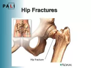
FRACTURES OF THE HIP AND ANKLE James M. Steinberg, D.O. Garden City Hospital. HIP FRACTURES. More than 250,000 hip fractures in the U.S. each year, expected to double by year 2050 Falls are the most common cause of fracture in the elderly

E N D
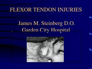
FRACTURES OF THE HIP AND ANKLE James M. Steinberg, D.O. Garden City Hospital
HIP FRACTURES • More than 250,000 hip fractures in the U.S. each year, expected to double by year 2050 • Falls are the most common cause of fracture in the elderly • High energy trauma is the most common cause in young adults • Femoral head has a very fragile blood supply
Femoral Head Fractures • Usually a result of hip dislocations, 10% of posterior hip dislocations • Most are shear or cleavage type fractures • Radiographic evaluation must include an AP and Judet views of the pelvis • Blood supply to the femoral head: -medial femoral circumflex artery (majority) -lateral femoral circumflex -artery of the ligamentum teres
Pipkin Classification • Type I: -Hip dislocation with fracture of the femoral head caudad to the fovea capitis femoris • Type II: - Hip Dislocation with fracture of the femoral head cephalad to the fovea capitis femoris
Pipkin Classification • Type III: -Type I or II injury associated with fracture of the femoral neck • Type IV: -Type I or II injury associated with fracture of the acetabular rim
Treatment of Femoral Head Fractures • Pipkin Type I: -If < 1mm step-off closed treatment, four weeks of traction and four weeks of toe-touch weight bearing -If > 1mm step-off, ORIF with small cancellous screws or herbert screws -In young patients immediate ORIF recommended to allow for early mobilization and prevention of AVN
Treatment of Femoral Head Fractures • Pipkin Type II: -For nonoperative care an anatomic reduction must be achieved -ORIF is the treatment of choice utilizing screw fixation
Treatment of Femoral Head Fractures • Pipkin Type III: -ORIF of the femoral head followed by screw fixation of the femoral neck -In young patients, emergent ORIF -Prognosis for this fracture is poor and depends on the degree of displacement of the femoral fracture
Treatment of Femoral Head Fractures • Pipkin Type IV: -Fracture must be treated in tandem with its associated acetabular fracture -Surgical approach dictated by acetabular fracture -Femoral head should be fixed internally even if nondisplaced
Complications of Femoral Head Fractures • AVN • Degenerative arthritis • Sciatic nerve palsy • Heterotopic ossification • Wound infection • Chronic instability
Femoral Neck Fractures • Low energy trauma (older patients) : -Fall onto the greater trochanter or forced external rotation of the lower extremity • High energy trauma (younger patients): -MVA or fall from significant height • Cyclical loading/stress fractures (athletes)
Evaluation of Femoral Neck Fractures • Physical exam: -Weight bearing status -Shortening and external rotation -Pain with provocative movements • Imaging: -AP in internal rotation and cross-table lateral -MRI if x-rays are negative with a high index of suspicion (first 48 hours) -Bone scan 48 hours after injury
Classification of Femoral Neck Fractures • Anatomic: -Subcapital -Transcervical -Basicervical • Pauwel: -Based on angle of fracture from horizontal -Type I: 30 degrees; Type II: 50 degrees; Type III: 70 degrees
Garden Classification of Femoral Neck Fractures • Based on degree of valgus displacement -Type I: incomplete/impacted -Type II: complete nondisplaced -Type III: complete with partial displacement (trabecular pattern does not line up) -Type IV: completely displaced (trabecular pattern in a parallel orientation)
Treatment of Femoral Neck Fractures • Fatigue/stress fractures: -Tension: in situ screw fixation -Compression: crutch ambulation • Impacted/nondisplaced fractures: -In situ fixation with three cancellous screws except in pathologic fractures, severe OA/RA and Paget’s disease (prosthetic replacement)
Treatment of Femoral Neck Fractures • Displaced fractures: ORIF and capsulotomy -25% incidence of AVN within 12 hours -30% within 12-24 hours -40% within 24-48 hours -In young patients, treat as a surgical emergency
Operative Techniques of Femoral Neck Fractures • Multiple screw fixation: -Favored technique -Threads should cross the fracture site to allow for compression -Three parallel screws yield the best fixation • Sliding screw devices -Not recommended -If used, second pin should be inserted superiority to control rotation
Hemiarthroplasty of Femoral Neck Fractures • Allows for immediate weight bearing • Indications: comminuted fractures, pathologic fractures, nonambulatory status, and neurological conditions • Contraindications: young active patients, active sepsis, and acetabular disease • Bipolar: reduces acetabular erosion (young patients) • Unipolar: less active patients
Total Hip Arthroplasty of Femoral Neck Fractures • Indications: -Contralateral hip disease -Ipsilateral acetabular metastatic disease -Preexisting degenerative disease
Complications of Femoral Neck Fractures • Nonunion • Osteonecrosis: -10% of nondisplaced fractures -27% of displaced fractures • Fixation failure • Infection • Thromboemboli
Intertrochanteric Hip Fractures • Fracture between the greater and lesser trochanters • Extracapsular fracture • Musculature produces shortening, external rotation and varus position at the fracture site: -Abductors displace the greater troch. -Iliopsoas displaces the lesser troch. -Hip flexors, extensors, and adductors pull the shaft proximally
Evaluation of Intertrochanteric Hip Fractures • Typically fractures result from a fall, direct blow to the greater troch. • Imaging: -AP and cross-table lateral -Bone scan or MRI may be useful in nondisplaced or occult fractures
Classification of Intertrochanteric Hip Fractures • Kyle: -Type I: nondisplaced, stable -Type II: displaced into varus with a small lesser troch. fragment -Type III: displaced into varus, posteromedial comminution and greater troch. fracture -Type IV: Type III with subtrochanteric extension • Other classifications: Boyd and Griffin, Evans, and Zuckerman
Treatment of Intertrochanteric Hip Fractures • Nonoperative: only for patients who are an extreme risk for surgery • Sliding hip screw (130 degrees-150 degrees) -Screw placement should be within 1cm of subchondral bone -Screw should be located slightly posterioinferior or centrally in the femoral head
Treatment of Intertrochanteric Hip Fractures • Prosthetic replacement -For patients with failed ORIF -Calcar replacement hemiarthroplasty or bipolar endoprosthesis • Cephalomedullary nails for reverse obliquity fracture pattern • Greater troch. displacement should be fixed with tension banding • Large posteriomedial fragments should be fixed with a lag screw or cerclage wires
Complications of Intertrochanteric Hip Fractures • Fixation failure • Malunion • Nonunion • Infection • Acetabular penetration • Pressure sores
Subtrochanteric Hip Fractures • Fracture between the lesser troch. and a point 5 cm distal to the lesser troch. • Closed reduction difficult because straight femoral traction does not neutralize deforming muscle forces -Proximal Fragment: Abduction (gluteus), External Rotation (short rotators), Flexion (psoas) -Distal Fragment: Varus (adductors)
Subtrochanteric Hip Fractures • Frequent site for pathological fractures, 17-35% of all subtroch. fractures • Mechanism of injury: -High energy trauma in younger patients with normal bone -Minor fall in older patients with weakened bone
Seinsheimer Classification • Type I: nondisplaced fracture or any fracture with <2mm of displacement of the fracture fragments • Type II: -A: two-part transverse femoral fracture -B: two-part spiral fracture with the lesser troch. attached to the proximal fragment -C: two-part spiral fracture with the lesser troch. attached to the distal fragment
Seinsheimer Classification • Type III: -A: three part spiral fracture in which the lesser troch. is part of the 3rd fragment -B: three part spiral fracture of the proximal third of the femur, with the 3rd part a butterfly fragment
Seinsheimer Classification • Type IV: Comminuted fracture with four or more fragments • Type V: Subtroch-intertroch fracture, any subtroch. fracture with extension into the greater troch. • Other Classifications: -Fielding: based on location of primary fracture line in relation to lesser troch. -AO: based on comminution of fracture
Nonoperative Treatment of Subtrochanteric Hip Fractures • Reserved for poor operative candidates • Skeletal traction in the 90/90 position followed by spica casting or cast bracing • Associated with increased morbidity and mortality
Operative Treatment of Subtrochanteric Hip Fractures • Choice of fixation dependent on the involvement of the trochanters -Intact greater and lesser trochs.: conventional locked IM nail -Intact greater troch., fractured lesser troch.: recon nail (2nd gen. IM device) -Fractured greater and lesser trochs. 95 degree blade plate or dynamic compression screw
Complications of Subtrochanteric Hip Fractures • Malunion • Nonunion • Loss of fixation • Technical difficulty of fixation device
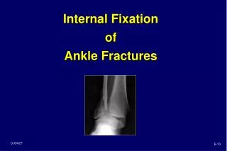
Ankle Fractures • The most common type of fracture treated by orthopedic surgeons • Only slight variation from normal is compatible with good joint function • Imaging: AP, lateral, and mortise views
Ankle Anatomy • Complex hinge joint with articulations with the fibula, tibia, and talus • Ligaments: -Deltoid ligament: superficial and deep -ATFL -PTFL -Calcaneofibular -Syndesmosis: anterior and posterior inferior tibiofibular ligaments, inferior transverse ligament, and interosseous ligament
Lauge-Hansen: Supination-Adduction • 10-20% of malleolar fractures • Stage I: transverse/avulsion fracture of the distal fibula or a rupture of the lateral collateral ligaments • Stage II: vertical fracture of the medial malleolus
Lauge-Hansen: Supination-External Rotation • Most common malleolar fracture • Stage I: disruption of the ATFL with or without an avulsion fracture at its attachment • Stage II: spiral fracture of the distal fibula • Stage III: disruption of the PTFL or a fracture of the posterior malleolus • Stage IV: transverse/avulsion fracture of the medial malleolus or a rupture of the deltoid ligament
Lauge-Hansen: Pronation-Abduction • Stage I: transverse fracture of the medial malleolus of rupture of the deltoid ligament • Stage II: rupture of the syndesmotic ligaments or an avulsion fracture at their insertions • Stage III: transverse or short oblique fracture of the distal fibula at or above the level of the syndesmosis
Lauge-Hansen: Pronation-External Rotation • Stage I: transverse fracture of the medial malleolus or rupture of the deltoid ligament • Stage II: disruption of the ATFL with or without avulsion fracture at its insertion sites • Stage III: spiral fracture of the distal fibula at or above the level of the syndesmosis • Stage IV: rupture of the PTFL or avulsion fracture of the posteriolateral tibia
Weber Classification • Based on the level of the fibular fracture • Type A: fracture below the level of the syndesmosis • Type B: oblique or spiral fracture at the level of the syndesmosis • Type C: fracture above the level of the syndesmosis
Fracture Variants • Maisonneuve fx: ankle injury with a fracture of the proximal third of the fibula, PER • Curbstone fx: avulsion fracture of the posterior tibia • LeForte-Wagstaffe fx: anterior fibular tubercle avulsion by ATFL, SER • Tillaux-Chaput fx: avulsion of anterior tibia by ATFL
Treatment of Ankle Fractures • Reduce dislocated ankles prior to x-rays • Cover open fractures with sterile, saline soap dressing, antibiotics, tetanus, etc. • Nonoperative(closed reduction): reserved for stable fracture patterns with an intact syndesmosis • Operative treatment required when closed reduction requires forced abnormal positioning of the foot, unstable fractures, open fractures, and widening of the mortise (1-2mm)
Operative Treatment of Ankle Fractures • Key to reduction is restoration of fibular length: lag screw and 1/3 tubular plate • Medial malleolus can be held with 2 cancellous screws perpendicular to the fracture line • Posterior malleolus should be fixed if there is >2mm of displacement or involvement of >25% of the articular surface • Syndesmotic screw for fibula fractures above the syndesmosis, placed 1.5-2cm above the joint line from the fibula to the tibia
Complications of Ankle Fractures • Nonunion • Malunion • Infection • Posttraumatic arthritis • Compartment Syndrome • Reflex sympathetic dystrophy • Tibiofibular synostosis
Pilon Fractures • Mechanism of injury: -Axial compression force through the talus -Shear: rotation combined with a varus or valgus stress • Etiology: mva, fall from height, direct crush injury, and sporting injuries (ski boot) • Imaging: AP, lateral, and oblique x-rays; CT scan for articular surface
Ruedi and Allgower Classification • Based on the severity of comminution and displacement of the articular surface • Type I: nondisplaced with splitting fracture lines • Type II: articular surface displaced; split fracture types • Type III: significant comminution and displacement of articular surface • Other classifications: AO, Mast, and Ovadia and Beale
Treatment of Pilon Fractures • ORIF of the fibula with 1/3 tubular plate • Reconstruction of tibial joint surface with K-wires • Bone graft metaphyseal deficits • Plate tibia (medial malleolus), cloverleaf plate • Fractures with metaphyseal comminution and severe soft-tissue injury consider external fixation
Complications of Pilon Fractures • Skin slough • Infection • Nonunion • Malunion • Posttraumatic arthritis • Joint stiffness