Download
1 / 28
390 likes | 967 Views
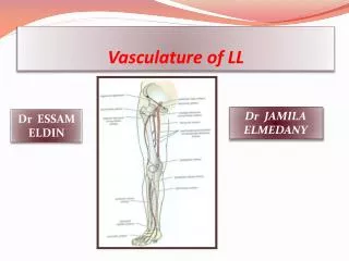
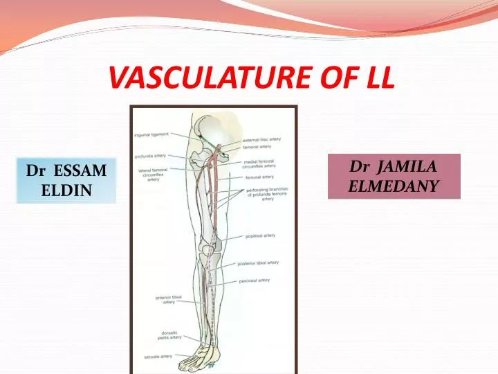
VASCULATURE OF LL. Dr JAMILA ELMEDANY. Dr ESSAM ELDIN. OBJECTIVES. At the end of the lecture, students should: List the main arteries of the lower limb. Describe their anatomy regarding: origin, course distribution & branches. List the main arterial anastomosis .

E N D
VASCULATURE OF LL Dr JAMILA ELMEDANY Dr ESSAM ELDIN
OBJECTIVES • At the end of the lecture, students should: • List the main arteries of the lower limb. • Describe their anatomy regarding: origin, course distribution & branches. • List the main arterial anastomosis. • List the sites to feel the peripheral arterial pulse. • Describe the anatomy of the veins of the lower limb regarding: differentiation into superficial& deep, origin, course & termination
FEMORAL ARTERY • It is the main arterial supply to the lower limb. • Origin: • It is the continuation of the External iliac artery. • How it enters the thigh? • Behind the inguinal ligament; midway between the anterior superior iliac spine and the symphysis pubis.
RELATIONS • Anterior: • Upper part: Skin &fascia. • Lower part: passes behind the Sartorius. • Posterior: • Psoas (separates it from the hip J),Pectineus & Adductor longus. • Medial: • Femoral vein. • Lateral : • Femoral nerve and its branches.
Femoral A. & Femoral V. At the inguinal ligament: The vein lies medial to the artery. At the apex of the femoral triangle: The vein lies posterior to the artery. At the opening in the adductor magnus: The vein lies lateral to the artery. V A
BRANCHES OF FEMORAL A. 1. Superficial Epigastric. 2. Superficial Circumflex iliac. 3. Superficial External Pudendal. 4. Deep External Pudendal. 5. Profunda femoris.
PROFUNDA FEMORIS A. It is an important, large artery. Arises from the lateral side of the femoral artery(4 cm below the inguinal ligament). It Passes medially behind the femoral vessels. Branches: Medial & Lateral circumflex femoral arteries. Three Perforating arteries. It ends by becoming the 4th perforating artery. . P
POPLITEAL ARTERY • It is the continuation of Femoral artery. • It enters the Popliteal fossa through an opening in the Adductor magnus.
RELATIONS & BRANCHES • Anterior: • Popliteal surface of the femur. • Knee joint. • Popliteus muscle. • Posterior: • Popliteal vein, Tibial nerve, skin and fascia. • Branches: • Muscular & Articular to the knee. • Termination: • At the lower border of Popliteus muscle by dividing into • Anterior and Posterior Tibial Arteries.
ANTERIOR TIBIAL ARTERY • It is the smaller of the two terminal branches of the popliteal artery. • It enters the anterior compartment of the leg through an opening in the upper part of the interosseous membrane. • It descends with the Deep Peroneal nerve. • In the upper part of its course, it lies Deep. • In the lower part, it becomes Superficial in front of the lower end of the tibia. • Branches: • Muscular& Anastomotic.
DORSALIS PEDIS ARTERY • Begins in front of ankle joint as a continuation of theAnterior Tibial artery. • It is superficial in position. • Crossed by the inferior extensor retinaculum and the first tendon of extensor digitorum brevis. • Medially: • Tendon of extensor hallucis longus. • Laterally: • Deep peroneal nerve& extensor digitorum longus.
DORSALIS PEDIS ARTERY • It Terminates by passing between the two heads of the 1st dorsal interosseous muscle. • It joins the Lateral plantar artery to complete the Plantar Arch. • Branches: • Lateral tarsal artery. • Arcuate artery. • 1st dorsal metatarsal artery.
POSTERIOR TIBIAL ARTERY Descends Deep to Soleus & Gastrocnemius. Lies on the posterior surface of TibialisPosterior muscle above and on the posterior surface of Tibia below. Its lower part is covered by Skin & Fascia only. Passes Behind Medial Malleolus , Deep to Flexor Retinaculm.
POSTERIOR TIBIAL ARTERY • Terminates by dividing into: Medial & Lateral plantar arteries. • Branches: 1. Peroneal artery: • A large artery, descends behind the fibula (the artery of the lateral compartment of the leg). • Gives : • A. Nutrient artery to the fibula. • B. Muscular branches. • C. Perforating branch to lower part of front of leg. • D. Shares in the Anastomosis around the ankle joint.
BRANCHES 2.Nutrient artery to the tibia. 3. Anastomotic branches to anastomosis around ankle joint. 4. Medial & Lateral plantar arteries.
MEDIAL PLANTAR ARTERY • The smaller of the two terminal branches of the posterior tibial artery. • Arises beneath the Flexor Retinaculum. • Gives:Muscular, Articular and Cutaneous branches. • Ends by supplying the medial side of the big toe.
LATERAL PLANTAR ARTERY • The larger of the two terminal branches of the posterior tibial artery. • At the base of the 5th metatarsal bone, it curves medially to form the Deep Plantar Arch. • Joins the Dorsalis pedis artery at the proximal end of the 1st intermetatarsal space. • Gives: • Muscular, Articular & Cutaneous branches. • The Plantar Arch gives Plantar Digital Arteries.
ARTERIAL ANASTOMOSISES TROCHANTERIC (supplies the head of femur) CRUCIATE AROUND THE KNEE
WHERE TO FEEL PERIPHERAL ARTERIAL PULSE ? • Femoral pulse: • Inferior to the inguinal ligament and midway between the anterior superior iliac spine and symphysis pubis. • Popliteal pulse: • Deep in the popliteal fossa medial to the midline. • Posterior tibial pulse: • Posteroinferior to the medial malleolus in the groove between the malleolus and the heel. • Dorsalis pedispulse • Over the tarsal bones between the tendons of extensor hallucislongusand extensor digitorum.
VEINS OF THE L.L • The veins of the lower limb are: (1)Superficial. (2) Deep.
SUPERFICIAL VEINS They are immediately under the skin in the subcutaneous tissue. Dorsal Venous arch (network): Drains most of the blood of the foot through Digital and Communicating veins. It is Drained on: Medial side by the Great Saphenous vein. Lateral side by the Small Saphenous vein
GREAT SAPHENOUS VEIN • The Longest Superficial vein of the body. • Begins from the medial end of the dorsal venous arch (as the medial marginal vein). • Ascends:In front of the Medial Malleolus accompanied by the (Saphenous nerve). • Posterior to the Medial Condyle of the femur. • Passes through the Saphenous Opening (2.5-3.25) cm below and lateral to the pubic tubercle. • Terminates in: Femoral Vein.
SMALLSAPHENOUS VEIN • Originates from the lateral end of the dorsal venous arch. • Ascends:Behind the lateral Malleolus in company with the Sural nerve. Along the middle of back of the leg. • Termination : • 1. It may join the Great Saphenous vein. • 2. Or Bifurcates: • One branch joins the Great saphenous and the other joins the Popliteal vein.
DEEP VEINS • Popliteal vein • Formed by the union of venaecomitantesaround the anterior & posterior tibial arteries. • lies posterior to popliteal artery. • Femoral vein • It enters the thigh by passing through the opening in the adductor magnus . • It leaves the thigh in the intermediate compartment of the femoral sheath. • Passes behind the inguinal ligament to become the External iliac vein
DEEP VEINS (VENAE COMITANTES) • Accompany all the major arteries and their branches. • Usually paired. • They are contained within the vascular sheath of the artery, whose pulsations help to compress and move blood in the veins..
PERFORATING VEINS • Connect the Great Saphenous veinwith the deep veins along the medial side of the calf. • Their valves only allow blood to flow from the superficial to the deep veins.
VARICOSE VEINS • Dilatation and Degeneration of the superficial veins that may be complicated by ulcers. • More common in the postero medial part of the lower limb. • Results from incompetence of the valves in the perforating veins, or within the great saphenous vein itself. • This allows the passage of high pressure blood from the deep to the superficial veins.