Download
1 / 13
150 likes | 719 Views
Meiosis – Grasshopper –predivision nuclei. These two nuclei were stained with Feulgen. Heterochromatin is visible, but chromosomes are not. DNA replication and chromosome duplication have occurred, but the latter will not be seen until later. Meiosis – Very early Prophase 1.

E N D
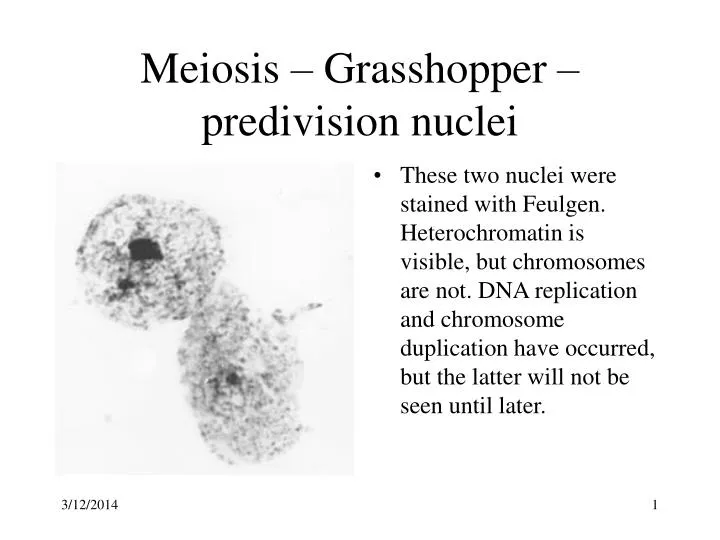
Meiosis – Grasshopper –predivision nuclei • These two nuclei were stained with Feulgen. Heterochromatin is visible, but chromosomes are not. DNA replication and chromosome duplication have occurred, but the latter will not be seen until later.
Meiosis – Very early Prophase 1 • Chromosomes are condensing and so are beginning to become visible as threads.
Meiosis – Early Prophase 1 • Homologous chromosomes have paired. The dark blob at the left is heterochromatin.
Meiosis – Mid Prophase 1 • Pairing is complete. This is the time that chiasma form, but we cannot see this through the optical microscope.
Meiosis – Late Prophase 1 • The attraction between the homologous chromosomes has given way to repulsion. Chiasmata and the result of chromosome duplication are clear.
Meiosis – Late Prophase 1 Another picture • The attraction between the homologous chromosomes has given way to repulsion. Chiasmata and the result of chromosome duplication are clear.
Meiosis – Metaphase 1 • The spindle has formed (it has not been stained), the chromosomes have attached to spindle fibers and are now aligned with the centromeres of the paired homologues on opposite side of the metaphase plate.
Meiosis – Anaphase 1 • The homologous pairs of chromosomes are now separating from each other, moving to opposite poles of the spindle.
Meiosis – Late Anaphase 1 • Homologous chromosomes are fully separated. Products of chromosome duplication and crossing over have stayed together. There are 9 duplicated chromosomes at left, 8 at the right.
Meiosis – Very Late Anaphase 1 • The heterochromatic X-chromosome appears fainter at the top of the right group.
Meiosis – Metaphase 2 • Polar view of chromo-somes arranged approximately in a circle. Chomosomes with a centromere near the middle appear to have 4 arms, while those with terminal centromeres have only 2 arms.
Meiosis- Metaphase 2 • Equatorial view, including the X-chromosome
Meiosis – Anaphase 2 • Eight separating from eight. Thus there is no X-chromosome here.