Download
1 / 18
230 likes | 461 Views
Mechanism of Breast Cancer progression. Natalia Pelaez. Breast Cancer Facts. About 39,840 women die from breast cancer each year in the US Cancer deaths are due to metastases Lung and bone metastases. Metastatic Cascade. Loss of cell-cell adhesion. 1. Cell-matrix (ECM) adhesion. 2.

E N D
Mechanism of Breast Cancer progression Natalia Pelaez
Breast Cancer Facts • About 39,840 women die from breast cancer each year in the US • Cancer deaths are due to metastases • Lung and bone metastases
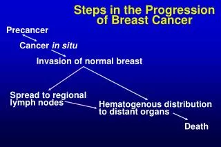
Metastatic Cascade Loss of cell-cell adhesion 1 Cell-matrix (ECM) adhesion 2 3 Degradation of ECM and migration 4 5 Extravasation and migration Intravasation 6 Proliferation
Hs 578T – Hs 578Ts(i)8 Breast Cancer model • Hs 578T cell line • Derived from a female breast carcinoma • Hs 578Ts(i)8 cell line • Was generated in the University College of Dublin, by the group of Dr. Susan McDonnell • 3 x more invasive • 2.5 x more migratory • Tumorigenic in vivo
Difference between Hs 578T – Hs 578Ts(i)8 Cell-matrix Adhesion Assay Cell-cell aggregation Assay Hs578T Hs578Ts(i)8
Woundhealing/scratch Assay Hs 578T Hs 578Ts(i)8 T=0 h T=6 h
Staining of the actin cytoskeleton Hs578T Hs578Ts(i)8
Hs 578T – Hs 578Ts(i)8 + function blocking antibodies Cell-cell aggregation Assay Cell-matrix Adhesion Assay Hs 578T Hs 578Ts(i)8 Control + function blocking antibody against cadherins
Expression levels of integrins and cadherins Cell surface expression Total expression Hs 578T Hs 578Ts(i)8 N-Cadherin kDa N-Cadherin - - 135 Integrin a5 - - 130 Integrin a5b1 Integrin b1 - - 130 Integrin aV - - 125 Integrin aVb5
Expression and activity levels of signaling molecules: Focal adhesion kinase (FAK) and src kinase • Signaling molecules involved in the migratory and invasion processes • Over-expressed or aberrantly activated Focal adhesion kinase src kinase Hs 578T Hs 578Ts(i)8 Hs 578T Hs 578Ts(i)8 FAK src - 60 - 125 FAK Y397 src Y418 - 60 - 125 FAK Y407 - 125
Fluorescent microscopy: Localization of FAK Y407and Actin Hs 578T Hs 578Ts(i)8 Actin FAK Y407
Staining for actin and FAK Y407 Overlay Hs 578T actin FAK Y407 Overlay Hs 578Ts(i)8 actin FAK Y407
Staining for integrin aVb5, actin and FAK Y407 Staining for actin and integrinaVb5 Staining for integrinaVb5 and FAK Y407 Hs 578T Hs 578Ts(i)8 Hs 578T Hs 578Ts(i)8 Red: avb5 integrin Green: FAK Y407 Red: actin Green: avb5 integrin
Conclusion • Cell-cell aggregation -> Cadherins -> cell surface and total expression of N-cadherin • Cell-matrix adhesion -> Integrins -> cell surface and total expression of integrinaVb5 and a5b1 • Migratory velocity • Signaling molecules FAK and src are more activated • Actin staining, FAK Y407 at the focal adhesion points, integrinaVb5
Future Work • Elucidate differences • Human breast cancer tissue
Acknowledgements Dr. Van Slambrouck and Dr. WimSteelant Melissa Candelaria-Lyons The School of Science of St. Thomas Univesity The Science Fellows Program My exceptional lab partners